Circulating, but not local lung, IL-5 is required
for the development of antigen-induced airways
eosinophilia.
J Wang, … , M Jordana, Z Xing
J Clin Invest.
1998;
102(6)
:1132-1141.
https://doi.org/10.1172/JCI2686
.
IL-5 is induced locally in the lung and systemically in the circulation during allergic airways
eosinophilic inflammation both in humans and experimental animals. However, the precise
role of local and systemic IL-5 in the development of allergic airways eosinophilia remains
to be elucidated. In our current study, we demonstrate that compared with their IL-5(+/+)
counterparts, IL-5(-/-) mice lacked an IL-5 response both in the lung and peripheral blood,
yet they released similar amounts of IL-4, eotaxin, and MIP-1alpha in the lung after
ovalbumin (OVA) sensitization and challenge. At cellular levels, these mice failed to
develop peripheral blood and airways eosinophilia while the responses of lymphocytes,
neutrophils, and macrophages remained similar to those in IL-5(+/+) mice. To dissect the
relative role of local and systemic IL-5 in this model, we constructed a gene transfer vector
expressing murine IL-5. Intramuscular IL-5 gene transfer to OVA-sensitized IL-5(-/-) mice led
to raised levels of IL-5 compartmentalized to the circulation and completely reconstituted
airways eosinophilia upon OVA challenge, which was associated with reconstitution of
eosinophilia in the bone marrow and peripheral blood. Significant airways eosinophilia was
observed for at least 7 d in these mice. In contrast, intranasal IL-5 gene transfer, when
rendered to give rise to a significant but compartmentalized level of transgene protein IL-5 in
the lung, was unable to reconstitute airways eosinophilia […]
Research Article
Find the latest version:
J. Clin. Invest.
© The American Society for Clinical Investigation, Inc. 0021-9738/98/09/1132/10 $2.00
Volume 102, Number 6, September 1998, 1132–1141 http://www.jci.org
Circulating, but not Local Lung, IL-5 Is Required for the Development of
Antigen-induced Airways Eosinophilia
Jun Wang,* Kay Palmer,* Jan Lˇotvall,* Sandra Milan,* Xue-Feng Lei,* Klaus I. Matthaei,‡ Jack Gauldie,* Mark D. Inman,*
Manel Jordana,* and Zhou Xing*
*Immunology and Infection Program, Department of Pathology and Molecular Medicine, McMaster University, Hamilton, Ontario, Canada L8N 3Z5; ‡Division of Biochemistry and Molecular Biology, John Curtin School of Medical Research, Australian National
University, Canberra ACT 0200, Australia
Abstract
IL-5 is induced locally in the lung and systemically in the circulation during allergic airways eosinophilic inflamma-tion both in humans and experimental animals. However, the precise role of local and systemic IL-5 in the develop-ment of allergic airways eosinophilia remains to be eluci-dated. In our current study, we demonstrate that compared with their IL-51/1 counterparts, IL-52/2 mice lacked an IL-5 response both in the lung and peripheral blood, yet they re-leased similar amounts of IL-4, eotaxin, and MIP-1a in the lung after ovalbumin (OVA) sensitization and challenge. At cellular levels, these mice failed to develop peripheral blood and airways eosinophilia while the responses of lympho-cytes, neutrophils, and macrophages remained similar to those in IL-51/1 mice. To dissect the relative role of local and systemic IL-5 in this model, we constructed a gene transfer vector expressing murine IL-5. Intramuscular IL-5 gene transfer to OVA-sensitized IL-52/2 mice led to raised levels of IL-5 compartmentalized to the circulation and completely reconstituted airways eosinophilia upon OVA challenge, which was associated with reconstitution of eosin-ophilia in the bone marrow and peripheral blood. Signifi-cant airways eosinophilia was observed for at least 7 d in these mice. In contrast, intranasal IL-5 gene transfer, when rendered to give rise to a significant but compartmentalized level of transgene protein IL-5 in the lung, was unable to re-constitute airways eosinophilia in OVA-sensitized IL-52/2 mice upon OVA-challenge, which was associated with a lack of eosinophilic responses in bone marrow and periph-eral blood. Our findings thus provide unequivocal evidence that circulating but not local lung IL-5 is critically required for the development of allergic airways eosinophilia. These findings also provide the rationale for developing strategies to target circulating IL-5 and/or its receptors in bone mar-row to effectively control asthmatic airways eosinophilia. (J. Clin. Invest. 1998. 102:1132–1141.) Key words: IL-5 • eosino-philia • asthma • IL-5 knock-out mice • gene transfer
Introduction
Airways eosinophilic inflammation is a central pathologic fea-ture of allergic asthma. The severity of asthma is correlated with the degree of airways eosinophilia (1, 2). Eosinophils con-tribute to the pathogenesis of asthma by releasing a number of inflammatory mediators and toxic products including oxygen radicals and cationic proteins that can severely damage the air-way epithelium and increase airair-way reactivity (1, 2). Thus, un-derstanding the molecular mechanisms underlying allergic air-ways eosinophilia has been a subject of intensive investigation. Eosinophils, like other types of leukocytes, originate from my-eloid precursors in the bone marrow, but different from others, they are not present in abundance in the peripheral blood un-der resting conditions. It is believed that during immune-inflammatory responses, in addition to local tissue signals, a systemic signal(s) capable of eosinophil mobilization in the bone marrow is also required for the development of allergic airways eosinophilia. Among many soluble signals, IL-5 is con-sidered to play an important role in the genesis of airways eo-sinophilia (1–4). Indeed, this cytokine has been found at raised levels both in the peripheral blood and lung tissue compart-ments in asthmatic patients (5–8). Similar findings were ob-tained from experimental models of allergic asthma where the peak circulating level of IL-5 preceded that in the lung after aerosol antigen challenge (9, 10), suggesting the presence of antigen-specific lymphocytes both within and outside the respi-ratory mucosa. IL-5 is a well recognized eosinopoietic growth factor capable of stimulating eosinophil differentiation and maturation both in vitro and in vivo (2, 3, 11–13). Further-more, IL-5 has also been shown to be an eosinophil chemoat-tractant and a potent eosinophil survival enhancer (14–17). By using transgene approaches, we and others have shown that overexpression of IL-5 locally in the lung, as opposed to sys-temic overexpression (18), induces marked airways philia (19, 20) or reconstitutes antigen-induced airways
eosino-philia in IL-52/2 mice (21), and such induction of airways
eosinophilia is always associated with systemic leakage of IL-5 and peripheral blood eosinophilia (19–21). On the other hand, systemic administration of anti–IL-5 monoclonal antibodies, which abrogates both local and systemic IL-5, has been shown to inhibit antigen-induced airways eosinophilia in experimen-tal models (22–24). While these findings support an important role of IL-5 in the pathogenesis of allergic airways eosino-philia, the precise functional role of local and systemic IL-5 re-mains to be dissected.
In our current study, we have used two transgenic tools, IL-5 gene-deficient mice and an IL-5 gene transfer vector, to inves-tigate the role of local and circulating IL-5 in the development of antigen-induced airways eosinophilia. Combined use of these genetic tools allowed us to reveal that circulating but not local lung IL-5 is critically required, via its effects on bone Address correspondence to Zhou Xing, MD, PhD, Rm. 4H19, Health
Sciences Centre, Department of Pathology, McMaster University, 1200 Main Street West, Hamilton, Ontario, Canada L8N 3Z5. Phone: 905-525-9140 ext 22471; FAX: 905-522-6750; E-mail: [email protected]. mcmaster.ca
marrow and peripheral blood eosinophils, for the development of antigen-induced airways eosinophilia. Our findings suggest that circulating but not local IL-5 or its receptors in bone mar-row represent a single, straightforward target for therapeutic strategies designed to effectively control asthmatic airways eosinophilia.
Methods
Mice and antigen-induced allergic airways inflammation.The genera-tion and characterizagenera-tion of C57BL/6 IL-5 gene knock-out (IL-52/2)
and littermate control mice (IL-51/1) have been described elsewhere
(12). Male or female mice were bred and maintained in the Level B pathogen-free facility at McMaster University Animal Quarter. Mice at the age of 8–12 wk were used. A mouse model of antigen-induced allergic airways inflammation was set up by ovalbumin (OVA)1 sensi-tization and challenge as previously described by us (10). In brief, mice were intraperitoneally sensitized twice, 5 d apart (day 217 and day 212) with 0.5 ml of a solution containing 8 mg OVA (Sigma Chemical Co., St. Louis, MO) adsorbed overnight at 48C to 4 mg of aluminum hydroxide (Aldrich Chemical Company, Inc., Milwaukee, WI) in PBS (Fig. 1). Mice were then challenged at 12 d after the sec-ond sensitization with aerosolized OVA (day 0) (Fig. 1).
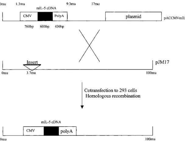
Construction and preparation of recombinant replication-deficient adenoviral vector expressing mIL-5.A 600-bp EcoRI/HindIII frag-ment of full-length murine IL-5 cDNA was isolated from pEDFM-16 (a kind gift from Dr. Alistair Ramsay, Australian National Univer-sity, Canberra, Australia) and ligated into the multicloning site of the shuttle vector pACCMV. The resultant pACCMV mIL-5 contained the mIL-5 cDNA positioned between a human cytomegalovirus pro-moter (CMV) and a SV40 splicing junction/polyA signal (polyA) in an orientation that allowed for the transcription of mIL-5 cDNA un-der control of the CMV promoter (Fig. 2). This plasmid was cotrans-fected into 293 cells along with a plasmid pJM17, which contained the entire Ad5 DNA sequences with an insert in the E1 region (25). By homologous recombination, the recombinant replication-deficient adenovirus Ad5E1pACCMVmIL-5 (AdIL-5) was rescued (Fig. 2). AdIL-5 was characterized by Southern and Northern hybridizations. The production of IL-5 was determined by ELISA with the superna-tant of A549 cells infected by AdIL-5 in vitro. Viral vectors were pu-rified, titered, and stored as previously described (26). A control viral vector Addl70-3 containing no transgene was used throughout the en-tire study.
Intramuscular or intranasal IL-5 gene transfer by AdIL-5.Mice were injected intramuscularly (i.m.) with AdIL-5 or Addl70-3 at 5 d before OVA challenge following a procedure we have previously described (27). This approach was shown to result in marked transgene expres-sion only in the muscle at the site of injection and active release of transgene protein into the circulation (27). A dose of 0.1 3 109 pfu of vector was diluted in a total of 100 ml of PBS and injected i.m. into two hind legs of mice (two injection sites per leg, 25 ml per site). In separate experiments, intranasal administration (i.n.) was performed to deliver transgene into the lung at 3 d before OVA challenge fol-lowing a procedure we have previously described (28). We have pre-viously demonstrated that this approach targets the transgene prima-rily to the bronchial epithelial cells and, to a lesser degree, to the alveolar epithelial cells and macrophages (28). A dose of 0.12 3 109 pfu of vector was diluted in a total 30 ml of PBS and delivered i.n. into ap-propriately anesthetized mice with a fine pipette tip (two administra-tions, 15 ml each).
Preparation of blood smear, serum, bronchoalveolar lavage fluid and lung tissue.The mice were sacrificed at days 1, 3, or 7 after OVA challenge, and the peripheral blood, bronchoalveolar lavage (BAL), lung, and bone marrow were collected. Blood samples were obtained by retro-orbital bleeding. Total white blood cell numbers were deter-mined after red blood cells lysis with RBC lysis buffer, and peripheral blood smears were prepared in duplicate with heparinized blood sam-ples. Differential cell types were determined on blood smears by ran-domly counting 300–500 leukocytes. Serum was prepared from the whole blood by centrifugation at 12,000 rpm in a microcentrifuge for 10 min at 48C after incubation at 378C for 30 min and stored at 2208C until cytokine assays. BAL was performed as previously described (10, 28). The mouse lung was lavaged with a total of 450 ml of PBS in two separate aliquots (250 ml and 200 ml) through a polyethylene tube (Becton Dickinson, Sparks, MD) cannulated into the mouse trachea. Approximately 350 ml of BAL was recovered by gentle massage of the lungs while retrieving. BAL fluids were then spun in a microcen-trifuge at 5,000 rpm for 5 min, and supernatants were stored in 2208C until cytokine measurements. Cell pellets were resuspended in PBS and total cell numbers counted on a hemocytometer. Cytospins were prepared by cyto-centrifugation (Shandon Inc., Pittsburgh, PA). Dif-ferential cell counts were determined on Diff-Quik-stained (Baxter, McGaw Park, IL) cytospins by randomly counting z 400 cells per slide. The lung was fixed by perfusion with 10% formalin. Lung sec-tions were stained with hematoxylin and eosin.
Preparation of bone marrow cells. The femur was surgically re-moved and the connective tissues were carefully scraped off the bone. Both ends of the femur were opened using a sharp scalpel, and the marrow was perfused by repeatedly injecting 1.5 ml of 5A culture me-dium from the distal end of the femur, using a 3-ml syringe and a 23G needle. Bone marrow cells were dispersed by repeatedly moving the perfusate with bone marrow fragments in and out of the syringe, through the needle. Cytospins of dispersed bone marrow cells were prepared on APTEX coated glass slides. The slides were fixed using Diff-Quik fixative (Dade Diagnostics of P.R. Inc., Aguada, Puerto Rico), and stained using standard eosin staining techniques. The number of mature eosinophils was estimated by differential counting of 500–2,000 cells under a microscope with oil immersion.
Cytokine and IgE measurements. Murine IL-5 was measured by using an ELISA kit (Amersham, Buckinghamshire, UK). Murine IL-4, MIP-1a, and eotaxin were measured by using ELISA kits purchased from R&D Systems (Minneapolis, MN). The sensitivity of detection of these ELISA kits was 5 pg/ml for IL-5 and 2 to 3 pg/ml for IL-4, eo-taxin, and MIP-1a. The level of OVA-specific IgE in serum was de-1. Abbreviations used in this paper: BAL, bronchoalveolar lavage;
[image:3.612.318.556.58.182.2]CMV, cytomegalovirus promoter; i.m., intramuscularly; i.n., intrana-sally; OVA, ovalbumin.
Figure 1. The mouse model of antigen-induced allergic airways
termined by using an antigen-capture ELISA method as previously described (10).
Data analysis. Wherever applicable, results and differences were statistically analyzed by using a Minitab statistical software package (Minitab; State College, PA). An unpaired t test was used and the dif-ference was considered statistically significant when P# 0.05.
Results
Lack of antigen-induced airways and peripheral blood eosino-philia in IL-52/2 mice. We characterized the difference in air-ways inflammation and peripheral blood responses between
C57BL/6 IL-52/2 and littermate control IL-51/1 mice. OVA
sensitization and aerosol challenge were carried out in these mice as previously described (10) (Fig. 1). Samples were col-lected and analyzed at days 1 and 3 after OVA challenge. We have previously demonstrated that cytokine and cellular re-sponses peaked at days 1 and 3 in mice (10, 29). The resting numbers of pulmonary macrophages in BAL fluids of naive
IL-51/1 and IL-52/2 mice were similar (Fig. 3). However, OVA
challenge induced an approximately fivefold increase in total
cell number in BAL from sensitized IL-51/1 mice and only a
threefold increase in sensitized IL-52/2 mice at day 3.
Approxi-mately 50% of these cells in IL-51/1 mouse lung were
eosino-phils. In contrast, there was only a marginally increased
num-ber of eosinophils found in BAL from IL-52/2 mice (Fig. 3).
Of note, the number of lymphocytes, neutrophils, and
mac-rophages increased to a similar degree in both IL-52/2 and
IL-51/1 mice. We then examined the inflammatory response in
the peripheral blood. An increased level of peripheral blood
eosinophilia in IL-51/1 mice was observed after second OVA
sensitization that further increased after OVA challenge (Ta-ble I). In contrast, there was a lack of peripheral blood
eosino-philia in IL-52/2 mice, thus in keeping with a lack of airways
eosinophilia. The numbers of other cell types were comparable
between IL-51/1 and IL-52/2 mice. While not surprisingly,
there was no measurable IL-5 in BAL and serum from IL-52/2
mice, and z 260 and 390 pg/ml of IL-5 were detected in serum
and BAL collected from IL-51/1 mice at day 1 after OVA
chal-lenge, respectively (Fig. 4).
Characterization of an adenoviral gene transfer vector ex-pressing murine IL-5.Having demonstrated a lack of IL-5 and eosinophilia in the peripheral blood and airways in
OVA-sen-sitized IL-52/2 but not IL-51/1 mice upon antigen challenge, we
set out to investigate the relative contribution of local and cir-culating IL-5 to the development of airways eosinophilia. A transgene approach was chosen to achieve IL-5 levels in a
se-lected compartment in IL-52/2 mice. This approach, in contrast
[image:4.612.58.362.56.293.2]to the use of recombinant protein, would allow us to achieve transient but prolonged levels of IL-5 in vivo (30). To this end, a recombinant replication-deficient adenoviral gene transfer vector expressing murine IL-5 transgene was constructed (AdIL-5; Fig. 2). AdIL-5 was characterized by HindIII restric-tion digesrestric-tion and Southern and Northern hybridizarestric-tion (data not shown). Upon infection with 10 pfu/cell of AdIL-5 but not with Addl70-3, A549 cells released 40.46 ng/ml of IL-5 in 48 h in vitro. To characterize AdIL-5 in vivo, four different doses of
Figure 2. Construction of recombinant
adenovi-rus Ad5E1pCCMVmIL-5 (AdIL-5) expressing murine IL-5. The plasmid pACCMVmIL-5 was constructed by inserting murine IL-5 cDNA into a shuttle vector pACCMV, which carried a CMV and an SV40 splicing junction/polyA signal (polyA). The recombinant adenovirus AdIL-5 was rescued by homologous recombination after cotransfecting 293 cells with pACCMVmIL-5 and a virus-rescuing vector pJM17.
Figure 3. Comparison of cellular responses in the BAL between
[image:4.612.314.558.520.681.2]AdIL-5 were given i.m. to naive C57BL/6 mice, and sera were collected at 24 h and measured for murine IL-5 by ELISA. We have previously demonstrated that by this approach, transgene is localized to the muscle with active release of transgene pro-tein into the circulation for a period of 10 to 12 d (27). The cir-culating level of IL-5 displayed a dose-dependent pattern (Fig.
5) with z 350 pg/ml being measured after delivery of a dose of
0.1 3 109 pfu of Ad5, a level similar to that detected in
IL-51/1 mice during OVA-induced immune-inflammatory
re-sponses. This dose was thus chosen for i.m. delivery in IL-52/2
mice in the following experiments. In a kinetic study, the level of IL-5 in the circulation was observed to peak at day 1, crease but still remain significant by day 5, and markedly de-cline close to background by day 8 after i.m. IL-5 gene transfer (not shown).
Reconstitution of antigen-induced airways eosinophilia by intramuscular IL-5 gene transfer in IL-52/2mice. We then
in-vestigated whether circulating IL-5, in the absence of local IL-5 in the lung, was sufficient to reconstitute antigen-induced
air-ways eosinophilia in antigen-sensitized/challenged IL-52/2
mice by using AdIL-5. Intramuscular gene transfer to naive
IL-52/2 mice of a dose of 0.1 3 109 pfu AdIL-5 led to highly
compartmentalized IL-5 levels in the circulation with 350 pg/ml measured in serum but little in BAL fluids at the peak time (Fig. 6). This dose of AdIL-5 or control vector Addl70-3 was
then injected i.m. to OVA-sensitized IL-52/2 mice at day 25
(Fig. 1). Delivery of AdIL-5 at day 25 was to ensure that the
bone marrow eosinophil progenitors be stimulated by IL-5 be-fore OVA challenge. Our previous study has suggested the in-volvement of an early IL-5 response in eosinophil responses in the bone marrow and peripheral blood before the onset of air-ways eosinophilia by OVA aerosol challenge (10). On exami-nation of cellular responses in the BAL at day 3 after OVA
challenge, we observed a lack of airways eosinophilia in IL-52/2
mice receiving no vector or i.m. Addl70-3 control vector. In contrast, airways eosinophilia was fully reconstituted in the
lung of IL-52/2 mice receiving i.m. delivery of AdIL-5,
com-pared with that in IL-51/1 mice (Fig. 7 A). The number of
other leukocyte types was similar among various groups. To examine whether such antigen-induced airways eosinophilia could persist without IL-5 present in the local lung tissue, cel-lular profiles in BAL obtained at 7 d after OVA challenge were analyzed. The number of eosinophils still remained
markedly increased in the lung of IL-52/2 mice receiving
AdIL-5 (Fig. 7 B). This level of lung eosinophilia, albeit
some-what lower, was still comparable with that in the lung of IL-51/1
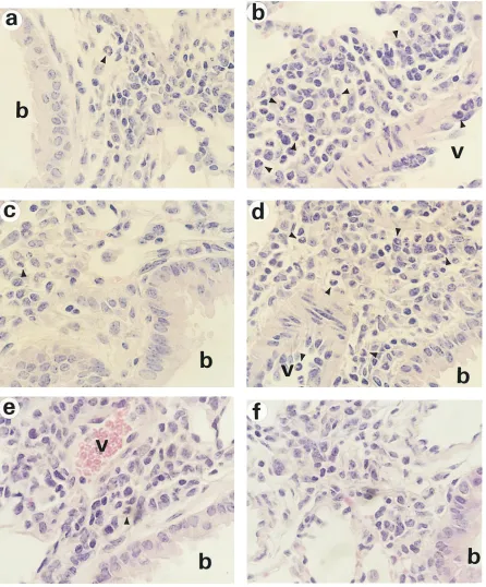
mice. We next examined histopathology of lung tissues ob-tained at day 3 after OVA challenge. In accord with BAL cyto-logic analysis, while there were similar degrees of perivascular and peribronchial accumulation of lymphocytes, neutrophils,
and monocytes in the lung of both IL-51/1 and IL-52/2 mice,
there were few eosinophils seen in the lung of IL-52/2 mice, in
sharp contrast to significant accumulation of eosinophils in the
lung of IL-51/1 mice (Fig. 8, a and b). However, intramuscular
IL-5 gene transfer but not Addl70-3 administration fully
re-constituted airways eosinophilia in lung tissues of IL-52/2 mice
(Fig. 8, c and d). Such differences were further confirmed by counting the number of eosinophils per high power field in peribronchial regions on multiple tissue sections. On average, we enumerated 3, 51, 3, and 33 eosinophils per high power field
in the lung of IL-52/2, IL-5+/+, IL-52/2 i.m. dl70-3, and IL-52/2
i.m. AdIL-5 mice, respectively.
Reconstitution of bone marrow and peripheral blood eo-sinophilia by intramuscular IL-5 gene transfer. To investigate
the mode by which circulating transgene protein IL-5
reconsti-tuted antigen-induced airways eosinophilia in IL-52/2 mice, we
examined the eosinophilic response both in the bone marrow and peripheral blood. At 3 d after OVA challenge, there was a minimal increase in eosinophil percentage in bone marrow of
IL-52/2 mice without or with i.m. Addl70-3 control vector
treatment. In contrast, the level of eosinophilia markedly
[image:5.612.57.300.60.200.2]in-creased in the bone marrow of IL-52/2 mice receiving i.m. IL-5
Figure 4. Comparison of IL-5 content in the BAL and serum
be-tween IL-52/2 and IL-5+/+ mice. BAL fluids and sera were collected 1 d after OVA challenge from IL-52/2 and IL-5+/+ mice, and IL-5 content was determined by ELISA. Data represent mean6SEM from three mice per group.
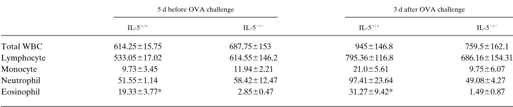
Table I. The Comparison of Cellular Responses in the Peripheral Blood of IL-51/1 and IL-52/2 Mice (3104/ml)
5 d before OVA challenge 3 d after OVA challenge
IL-51/1 IL-52/2 IL-51/1 IL-52/2
Total WBC 614.25615.75 687.756153 9456146.8 759.56162.1
Lymphocyte 533.05617.02 614.556146.2 795.366116.8 686.166154.31
Monocyte 9.7363.45 11.9462.21 21.065.61 9.7566.07
Neutrophil 51.5561.14 58.42612.47 97.41623.64 49.0864.27
Eosinophil 19.3363.77* 2.8560.47 31.2769.42* 1.4960.87
[image:5.612.56.555.599.703.2]gene transfer, similar to that in IL-51/1 mice (Fig. 9 A).
Ac-companied with eosinophilic responses in the bone marrow
was a peripheral blood eosinophilia in IL-52/2 mice receiving
IL-5 i.m. gene transfer, the level of which also increased to that
in IL-51/1 mice (Fig. 9 B). These were in contrast to the lack of
peripheral blood eosinophilia in IL-52/2 mice without or with
i.m. delivery of Addl70-3.
Similar levels of IL-4 and chemokines eotaxin and MIP-1a in the lung of IL-5+/+ and IL-52/2 mice. These results have thus
far indicated that circulating IL-5, via its effects on bone mar-row and peripheral blood eosinophils, is a systemic signal re-quired for the development of antigen-induced airways eosin-ophilia, which can occur in the absence of local levels of IL-5 in the lung. It is believed, however, that certain local tissue sig-nals are required for circulating eosinophils to migrate across the vasculature into the airways. In this regard, IL-4, in
addi-tion to its other activities, enhances eosinophil adhesion to the endothelium by upregulating VCAM-1 expression (31), and
C-C chemokines eotaxin and MIP-1a have chemotactic effects
on eosinophils (32, 33). We measured the content of IL-4,
eo-taxin, and MIP-1a in BAL fluids collected at day 1 after OVA
challenge from IL-51/1 and IL-52/2 mice. While very little IL-4
was measured in BAL from both naive IL-52/2 and IL-51/1
mice, similarly increased levels of IL-4 were measured in BAL
fluids collected from OVA-sensitized/challenged IL-51/1 mice
and IL-52/2 mice with or without i.m. delivery of AdIL-5 or
Addl70-3 (Table II). Similar induction of chemokines eotaxin
and MIP-1a was also observed in BAL fluids from both IL-51/1
mice and IL-52/2 mice under these conditions (Table II).
Simi-lar levels of immune responses were further supported by
sim-ilar anti-OVA IgE responses between IL-51/1 and IL-52/2
[image:6.612.59.299.59.259.2]mice, 137.7637.5, 219.9692.8, 161.7660.1 ng/ml serum in
Figure 7. Reconstitution of OVA-induced airways eosinophilia by
[image:6.612.314.559.63.419.2]i.m. IL-5 gene transfer in OVA-sensitized IL-52/2 mice. Cellular re-sponses were determined with BAL cell preparations by cell differen-tial counting. (A) day 3 after challenge; OVA only (n = 3), Addl70-3 i.m. (n = 5), AdIL-5 i.m. (n = 6), IL-5+/+ OVA (n = 5). (B) day 7 after challenge; Addl70-3 i.m. (n = 3), AdIL-5 i.m. (n = 4), IL-5+/+ OVA (n = 4). Results represent mean6SEM. The degree of eosinophilia between IL-52/2 AdIL-5 and IL-52/2 Addl70-3 groups is statistically significantly different (P = 0.011) but is not statistically different be-tween IL-52/2 OVA/AdIL-5 and IL-5+/+ OVA groups (P = 0.16).
Figure 5. In vivo characterization of AdIL-5. Various doses of AdIL-5
were giving i.m. to naive C57BL/6 mice and sera collected at 24 h were measured for mIL-5 by ELISA. Results represent mean values from two mice per dose.
Figure 6. Compartmentalized distribution of transgene protein IL-5
in BAL after i.m. administration of AdIL-5 (0.1 3 109 pfu) in IL-52/2
[image:6.612.57.298.529.692.2]IL-51/1, IL-52/2, and IL-52/2 i.m. AdIL-5 mice, respectively
(day 1 after OVA challenge).
Lack of reconstitution of antigen-induced airways eosino-philia following compartmentalized intrapulmonary IL-5 gene transfer in IL-52/2 mice. Our findings have thus far strongly
suggested that if only present locally in the lung but not in the
circulation, IL-5 cannot reconstitute airways eosinophilia in
IL-52/2 mice. To demonstrate this, we delivered a dose of
0.12 3 109 pfu of AdIL-5 i.n. into the lung of IL-52/2 mice at
day 23 (Fig. 1). We have previously shown that by this
ap-proach the level in BAL of transgene protein peaks at z day 4
[image:7.612.58.504.59.597.2](28); thus, delivering AdIL-5 3 d before OVA challenge would
allow a maximal level of transgene protein IL-5 present in the
lung at day 1 after OVA challenge in IL-52/2 mice. By delivering
0.12 3 109 pfu of AdIL-5, a significant level of IL-5 was
mea-sured in BAL fluids collected at day 4, which led to little spill
of IL-5 into the circulation in IL-52/2 mice (Table III). This
level of compartmentalized IL-5 transgene protein in the lung
failed to reconstitute antigen-induced airways eosinophilia as shown by BAL cellular analysis (Fig. 10). The lack of eosino-philic responses under such conditions was further supported by histologic examination (Fig. 8, e and f). In keeping with these findings, there were minimal increases in the number of eosinophils in the bone marrow and peripheral blood (data not shown).
Discussion
In this study, we demonstrated that, in contrast to their IL-51/1
[image:8.612.56.370.59.443.2]counterparts, the lack of IL-5 response both in the lung and
[image:8.612.57.299.566.663.2]Figure 9. Peripheral eosinophilic responses in IL-52/2 and IL-5+/+ mice. The level of eosinophilia in bone marrow (A) and peripheral blood (B) was analyzed from indicated groups at day 3 after OVA challenge. The number of mice used for each group was identical to that indicated for Fig. 7 A. Data represent mean6SEM. There is no statistically signifi-cant difference between IL-52/2 OVA/AdIL-5 and IL-5+/+ OVA groups.
Table II. The Content of Cytokines in BAL (pg/ml)
IL-4 Eotaxin MIP-1a
IL-52/2 OVA 495.256170.94 131.72641.02 20.2762.86
IL-52/2 OVA
Add170-3 i.m. 689.716310.29 96.62645.58 50.58620.91 IL-52/2 OVA
AdIL-5 i.m. 455.956272.21 29.6362.3 18.1466.7 IL-51/1 OVA 378.556103.58 24.3963.74 17.8364.63
BAL fluids were collected at day 1 after OVA-challenge. IL-4, eotaxin and MIP-1a were measured by specific ELISA. Data represent mean6SEM from IL-52/2 OVA (n 5 3), IL-52/2 OVA Add170-3 (n 5 3), IL-52/2 OVA AdIL-5 (n 5 3) and IL-51/1 OVA (n 5 4) mice. There is no statistically significant difference between treatments. The levels of these cytokines in BAL from naive IL-52/2 or IL-51/1 mice were under or close to the assay detection limit.
Table III. IL-5 Levels in BAL and Serum Post-i.n. AdIL-5 Delivery in IL-52/2 Mice
BAL Serum No. of mice
IL-52/2 Add170-3 0 0 4
IL-52/2 AdIL-5 49.8618 4.561.9 9
[image:8.612.314.556.639.691.2]peripheral blood in IL-52/2 mice resulted in a lack of
periph-eral and airways eosinophilia in response to antigen sensiti-zation and challenge, whereas the response of lymphocytes, neutrophils, and macrophages, and the level of IL-4 and chemokines in the lung were not markedly weakened in these mice. Circulating IL-5 by intramuscular IL-5 gene transfer to
antigen-sensitized IL-52/2 mice reconstituted eosinophilia not
only in the bone marrow and peripheral blood but also in the airway upon antigen challenge. In contrast, local compartmen-talized IL-5 achieved by intrapulmonary IL-5 gene transfer to these mice was unable to reconstitute airways eosinophilia, which was associated with a failure in reconstituting bone mar-row and peripheral blood eosinophilia. These findings indicate a critical role of circulating IL-5 in the development of anti-gen-induced airways eosinophilia and suggest that, contrary to previously thought, local lung IL-5 plays a relatively less im-portant role in the process of eosinophil accumulation in the airways.
IL-5, GM-CSF, and IL-3 are members of the hematopoietic growth factor family. Different from GM-CSF and IL-3, IL-5 has restricted biologic effects, primarily on eosinophils and eosinophil progenitors (2, 3). IL-5 has been shown to stimulate the differentiation of eosinophil progenitor cells in vitro and in systemic IL-5 transgenic mice (11, 13). In contrast to marked peripheral blood eosinophilia in IL-5 transgenic mice, GM-CSF transgenic mice developed only mild peripheral blood eosinophilia, together with markedly increased numbers of neutrophils and monocytes (34). However, IL-5 is not merely an eosinopoietic growth factor since a wealth of in vitro evi-dence has suggested that it is also an eosinophil chemoattrac-tant and survival factor (14–17). Indeed, the level of IL-5 is in-creased in the peripheral blood and local lung tissue both in asthmatic patients and experimental models of asthmatic in-flammation (5–10). And recently, IL-5, but not IL-3, receptor
expression on CD34+ progenitors and the number of
eosino-phil progenitor cells in the bone marrow have been found markedly increased in mild asthmatic patients upon local lung allergen challenge (35, 36). Thus, the role of IL-5 in the patho-genesis of allergic asthma has been thought to be mediated through its effects not only on eosinophil differentiation in the bone marrow but also on eosinophil influx and survival locally in the lung (1–4, 17). This notion appears to be supported by further experimental observations. Systemic administration of anti–IL-5 monoclonal antibodies inhibited antigen-induced
airways eosinophilia (22–24); IL-52/2 mice failed to mount
an-tigen-induced airways eosinophilia unless an IL-5 gene trans-fer vector was repeatedly intranasally delivered before and during antigen challenge (21); overexpression of IL-5 locally in the lung, either by delivering repeated large doses of recombi-nant IL-5 (37), or gene transfer vectors (19), or in lung-specific IL-5 transgenic mice (20), induced airways eosinophilia. How-ever, systemically delivered antibodies will abrogate not only circulating IL-5 but also local lung IL-5. High levels of local overexpression of IL-5 result in not only higher than physio-logic levels of IL-5 protein in the lung, but also spillovers of IL-5 into the circulation; induction of airways eosinophilia was in-variably associated with an increased number of peripheral blood eosinophils (19–21). On the other hand, since IL-5 was shown to be chemotactic to eosinophils in vitro only when present at much higher concentrations compared with classic eosinophil chemoattractants (17), it is very likely that the in-flux of eosinophils into the airways occurs as a result of eosino-phil chemotaxis to transgenic levels of IL-5 in the lung, which may not represent a physiologic functional aspect of IL-5 dur-ing immune-inflammatory responses. Hence, the relative role of local and systemic IL-5 in allergic airways eosinophilia re-mains to be clarified.
To dissect the role of local and systemic IL-5 in the patho-genesis of allergic airways eosinophilia, we first examined the cellular and cytokine responses in OVA-sensitized and chal-lenged IL-5–deficient mice. Consistent with a previous study (21), we found a lack of airways and peripheral blood
eosino-philia in IL-52/2 mice. However, in contrast to that study, we
found similar responses of other leukocyte subsets including lymphocytes, neutrophils, and macrophages both in BAL and lung tissues. Such discrepancies are likely due to differences in the protocol for sensitization and challenge. Upon examina-tion of cytokines, we found significantly increased levels of
IL-5 in BAL and peripheral blood in IL-51/1 mice but not in
IL-52/2 mice upon OVA challenge. In contrast, the level of
an-other Th2 cytokine IL-4 and CC chemokines eotaxin and
MIP-1a in the lung was similar between IL-51/1 and IL-52/2 mice,
thus likely explaining the similar extent of airways inflamma-tory responses of lymphocytes, neutrophils, and macrophages. Of interest, although not statistically significant, there ap-peared lower levels of eotaxin in the lung of mice with airways
eosinophilia (IL-51/1 OVA or IL-52/2 OVA AdIL-5 i.m.) as
compared with those in mice without airways eosinophilia. This may not reflect a lower production but rather suggests a greater consumption of eotaxin in the lung by infiltrating eosin-ophils. These findings, to our knowledge, represent the first experimental evidence that the lack of IL-5 and airways eosin-ophilia has little effect on the level of cytokines and influx of leukocytes other than eosinophils in the lung during allergic airways inflammation.
We then investigated the role of local and systemic IL-5 in allergic airways eosinophilia by using an IL-5 transgene
ap-Figure 10. Lack of reconstitution of OVA-induced airways
[image:9.612.57.300.60.213.2]proach in sensitized IL-52/2 mice. Intramuscular delivery of an
IL-5 gene transfer vector led to raised IL-5 levels compartmen-talized to the peripheral blood compartment and completely
reconstituted airways eosinophilia in IL-52/2 mice after OVA
challenge. Of importance, such a reconstitution of airways eosinophilia was associated with a restored eosinophilic re-sponse in the bone marrow and peripheral blood. The magni-tude of airways eosinophilia was identical or even slightly
higher than that seen in IL-51/1 mice at day 3 after OVA
chal-lenge (8 d after i.m. IL-5 gene transfer). It is noteworthy that
circulating levels of transgene protein IL-5 in IL-52/2 mice
dur-ing the period from OVA challenge (day 0) to death (day 3) had declined towards the baseline (5–8 d after i.m. gene trans-fer). This suggests that the eosinophil priming or mobilizing ef-fect by systemic IL-5 before OVA-aerosol challenge is of criti-cal importance in the onset of airways eosinophilia. Indeed, we observed an increase in the number of peripheral blood
eo-sinophils in OVA-sensitized IL-51/1 mice before OVA
chal-lenge. Furthermore, it suggests that high circulating levels of IL-5 that emerge and peak around day 1 after OVA challenge
in IL-51/1 mice are not required for the peak airways
eosino-philic response (day 3). On the other hand, our finding that full
reconstitution of airways eosinophilia in IL-52/2 mice by i.m.
IL-5 gene transfer took place in the absence of lung tissue IL-5, indicates that local lung IL-5 is not required for the develop-ment of a full-blown antigen-induced airways eosinophilia. Such dissociability of antigen-induced airways eosinophilia from significant tissue levels of IL-5 in the lung is further sup-ported by our observation that there was still marked airways
eosinophilia in IL-52/2 mice receiving i.m. IL-5 gene transfer
7 d after OVA challenge. This latter finding also suggests a sig-nificant role of molecules other than IL-5 in the perpetuation of airways eosinophilia during asthmatic inflammation. While it remains to be determined whether i.m. IL-5 gene transfer
concurrently reconstituted airways hyperreactivity in IL-52/2
mice, a recent study in a different model system has suggested that systemic IL-5, in the presence of specific IgE, is also in-volved in enhanced airways reactivity (38). With establishment of the critical role of systemic IL-5 in airways eosinophilia, we explored the potential mechanisms within the lung by which eosinophils migrated into the airways despite the absence of local IL-5. We found that the level of IL-4, eotaxin, and
MIP-1a was not compromised in the lung of antigen-challenged
IL-52/2 mice, thus indicating that other eosinophil-active
cyto-kines are fully capable of mounting airways eosinophilia in the absence of local IL-5. We have recently found that IL-4,
to-gether with TNFa, is required for VCAM-1 upregulation on
pulmonary vasculature, which is in turn required for the de-velopment of allergic airways eosinophilia (39). Eosinophil
chemokines including eotaxin and MIP-1a have been shown
involved in eosinophil chemotaxis in the lung during allergic airways inflammation (32, 33, 40–42). In addition, we have pre-viously shown that there is a small but significant level of GM-CSF being induced in the lung during allergic airways inflam-mation (10). It is thus possible that GM-CSF implements many biologic activities that IL-5 is capable of in this model. The re-quirement of such coordination between systemic IL-5 and lo-cal lung cytokines such as eotaxin for the onset of airways eo-sinophilia is also supported by recent findings from skin models (43–45).
Thus, we have demonstrated that circulatory but not local lung IL-5 is critically required for the development of
antigen-induced airways eosinophilia via its effect on bone marrow and peripheral blood eosinophils. On the other hand, local cyto-kine signals in the absence of IL-5 are sufficient to set up a stage for airway eosinophil accumulation to take place. While these findings shall not dismiss the possible potentiating/acti-vating activities by local lung IL-5, together with other cyto-kines, on accumulating eosinophils in asthmatic airways tissues, they provide important rationale for developing strategies to target circulating, rather than local, IL-5, or its receptors in BM to effectively control asthmatic airways eosinophilia, par-ticularly in steroid-resistant asthmatics.
Acknowledgments
We wish to thank Anna Zganiacz, Xueya Feng, Duncan Chong, Jen-nifer Wattie, Sussana Goncharova, Scott Neigh, and Bruce Vallance for their invaluable help. We also thank Dr. Todd Braciak for his ini-tial contribution to the construction of plasmids and Sara DeSilvio for her secretarial assistance.
This study is supported by the Medical Research Council (MRC) of Canada and Astra Draco AB, Sweden. M. Jordana is a Career Sci-entist of the Ontario Ministry of Health. Z. Xing is a scholar of MRC, Canada.
References
1. Seminario, M.-C., and G.J. Gleich. 1994. The role of eosinophils in the pathogenesis of asthma. Curr. Opin. Immunol. 6:860–864.
2. Bochner, B.S., B.J. Undem, and L.M. Lichtenstein. 1994. Immunological aspects of allergic asthma. Annu. Rev. Immunol. 12:295–335.
3. Anderson, G.P. 1995. Eosinophilia in genetically altered mice. Eur. Respir. Rev. 29:231–237.
4. Robinson, D.S., S.R. Durham, and A.B. Kay. 1993. Cytokines in asthma. Thorax. 48:845–853.
5. Hamid, Q., M. Azzawi, S. Ying, R. Moqbel, A.J. Wardlaw, C.J. Corrigan, B. Bradley, S.R. Durham, J.V. Collins, P.K. Jeffery, et al. 1991. Expression of mRNA for interleukin-5 in mucosal bronchial biopsies from asthma. J. Clin. In-vest. 87:1541–1546.
6. Robinson, D.S., Q. Hamid, S. Ying, A. Tsicopoulos, J. Barkans, A.M. Bentley, C. Corrigan, S.R. Durham, and A.B. Kay. 1992. Predominant TH2-like bronchoalveolar T-lymphocyte population in atopic asthma. N. Engl. J. Med. 30:298–304.
7. Walker, C., E. Bode, L. Boer, T.T. Hansel, K. Blaser, J.C. Virchow, Jr. 1992. Allergic asthmatics have distinct patterns of T-cell activation and cyto-kine production in peripheral blood and bronchoalveolar lavage. Am. Rev. Respir. Dis. 146:109–115.
8. Alexander, A.G., J. Barkans, R. Moqbel, N.C. Barnes, A.B. Kay, and C.J. Corrigan. 1994. Serum interleukin-5 concentrations in atopic and non-atopic patients with glucocorticoid-dependent chronic severe asthma. Thorax. 49:1231–1233.
9. Yamaguchi, S., H. Nagai, H. Tanaka, M. Tsujimoto, and N. Tsuruoka. 1994. Time course study for antigen-induced airway hyperreactivity and the ef-fect of soluble IL-5 receptor. Life Sci. 54:471–475.
10. Ohkawara, Y., X.-F. Lei, M.R. Stampfli, J.S. Marshall, Z. Xing, and M. Jordana. 1997. Cytokine and eosinophil responses in the lung, peripheral blood, and bone marrow compartments in a murine model of allergen-induced airways inflammation. Am. J. Respir. Cell Mol. Biol. 16:510–520.
11. Clutterbuck, E.J., and C.J. Sanderson. 1988. Human eosinophil hemato-poiesis studies in vitro by means of murine eosinophil differentiation factor (IL-5): production of functionally active eosinophils from normal human bone mar-row. Blood. 71:646–651.
12. Kopf, M., F. Brombacher, P.D. Hodgkin, A.J. Ramsay, E.A. Milbourne, W.J. Dai, K.S. Ovington, C.A. Behm, G. Kohler, I.G. Young, et al. 1996. IL-5 deficient mice have a developmental defect in CD5+ B-1 cells and lack
eosino-philia but have normal antibody and cytotoxic T cell responses. Immunity. 4:15–24. 13. Lee, N.A., M.P. McGarry, K.A. Larson, M.A. Horton, A.B. Kristensen, and J.J. Lee. 1997. Expression of IL-5 in thymocytes/T cells leads to the devel-opment of a massive eosinophilia, extramedullary eosinophilopoiesis, and unique histopathologies. J. Immunol. 158:1332–1344.
14. Yamaguchi, Y., Y. Hayashi, Y. Sugama, Y. Miura, T. Kasahara, S. Kita-mura, M. Torisu, S. Mita, A. Tominaga, K. Takatsu, et al. 1988. Highly purified murine interleukin-5 (IL-5) stimulates eosinophil function and prolongs in vitro survival. IL-5 as an eosinophil chemotactic factor. J. Exp. Med. 167:1737–1742.
Mantovani. 1989. Recombinant human interleukin 5 is a selective eosinophil chemoattractant. Eur. J. Immunol. 19:701–705.
16. Tai, P.C., L. Sun, and C.J.F. Spry. 1991. Effects of IL-5, granulocyte/ macrophage colony-stimulating factor (GM-CSF) and IL-3 on the survival of human blood eosinophils in vitro. Clin. Exp. Immunol. 85:312–316.
17. Resnick, M.R., and P.F. Weller. 1993. Mechanisms of eosinophil recruit-ment. Am. J. Respir. Cell Mol. Biol. 8:349–355.
18. Dent, L.A., M. Strath, A.L. Mellor, and C.J. Sanderson. 1990. Eosino-philia in transgenic mice expressing interleukin-5. J. Exp. Med. 172:1425–1431.
19. Xing, Z., Y. Ohkawara, T. Braciak, G.M. Tremblay, P.J. Sime, M. Jor-dana, and J. Gauldie. 1996. Disparate functional consequences of transient overexpression of GM-CSF and IL-5 in the lung. Am. J. Respir. Crit. Care Med. 153:A793.
20. Lee, J.J., M.P. McGarry, S.C. Farmer, K.L. Denzler, K.A. Larson, P.E. Carrigan, I.E. Brenneise, M.A. Horton, A. Haczku, E.W. Gelfand, et al. 1997. Interleukin-5 expression in the lung epithelium of transgenic mice leads to pul-monary changes pathognomonic of asthma. J. Exp. Med. 185:2143–2154.
21. Foster, P.S., S.P. Hogan, A.J. Ramsay, K.I. Matthaei, and I.G. Young. 1996. Interleukin 5 deficiency abolished eosinophilia, airways hyperreactivity, and lung damage in a mouse asthma model. J. Exp. Med. 183:195–201.
22. Coffman, R.L., B.W. Seymour, S. Hudak, J. Jackson, and D. Rennick. 1989. Antibody to interleukin-5 inhibits helminth-induced eosinophilia in mice. Science. 245:308–310.
23. Mauser, P.J., A.M. Pitman, X. Fernandez, S.K. Foran, G.K. Adams III, W. Kreutner, R.W. Egan, and R.W. Chapman. 1995. Effects of an antibody to interleukin-5 in a monkey model of asthma. Am. J. Respir. Crit. Care Med. 152: 467–472.
24. Kung, T.T., D.M. Stelts, J.A. Zurcher, G.K. Adams III, R.W. Egan, W. Kreutner, A.S. Watnick, H. Jones, and R.W. Chapman. 1995. Involvement of IL-5 in a murine model of allergic pulmonary inflammation: prophylactic and therapeutic effect of an anti-IL-5 antibody. Am. J. Respir. Cell Mol. Biol. 13: 360–365.
25. McGrory, W.J., D.S. Bautista, and F.L. Graham. 1988. A simple tech-nique for the rescue of early region I mutations into infectious human adenovi-rus type 5. Virology. 163:614–617.
26. Graham, F.L., and L. Prevec. 1991. Gene transfer and expression proto-cols. In Methods in Molecular Biology. E.J. Murry and J.M. Walker, editors. Humana Press Inc., Clifton, NJ. 109–127.
27. Xing, Z., Y. Ohkawara, M. Jordana, F.L. Graham, and J. Gauldie. 1997. Adenoviral vector-mediated IL-10 expression in vivo: intramuscular gene transfer inhibits cytokine responses in endotoxemia. Gene Ther. 4:140–149.
28. Lei, X.-F., Y. Ohkawara, M.R. Stampfli, J. Gauldie, K. Croitoru, M.
Jordana, and Z. Xing. 1998. Compartmentalized transgene expression of granu-locyte-macrophage colony-stimulating factor (GM-CSF) in mouse lung en-hances allergic airways inflammation. Clin. Exp. Immunol. 113:157–165.
29. Stämpfli, M.R., S.A. Ritz, G.S. Neigh, P.J. Sime, X.-F. Lei, Z. Xing, K. Croitoru, and M. Jordana. 1998. Intramuscular viral infection inhibits antigen-induced airways inflammation in mice. Clin. Exp. Allergy. In press.
30. Xing, Z., T. Braciak, Y. Ohkawara, J.-M. Sallenave, R. Foley, P.J. Sime, M. Jordana, F.L. Graham, and J. Gauldie. 1996. Gene transfer for cytokine functional studies in the lung: the multifunctional role of GM-CSF in pulmo-nary inflammation. J. Leukoc. Biol. 59:481–488.
31. Schleimer, R.P., S.A. Sterbinsky, J. Kaiser, C.A. Bickel, D.A. Klunk, K. Tomioka, W. Newman, F.W. Luscinskas, M.A. Gimbrone, Jr., B.W. McIntyre, et al. 1992. IL-4 induced adherence of human eosinophils and basophils but not neutrophils to endothelium. Association with expression of VCAM-1. J.
Immu-nol. 148:1086–1092.
32. Gonzalo, J.-A., G.-Q. Jia, V. Aguirre, D. Friend, A.J. Coyle, N.A. Jen-kins, G-s. Lin, H. Katz, A. Lichtman, N. Copeland, et al. 1996. Mouse eotaxin expression parallels eosinophil accumulation during lung allergic inflammation but it is not restricted to a Th2-type response. Immunity. 4:1–14.
33. Lukacs, N.W., R.M. Strieter, K. Warmington, P. Lincoln, S.W. Chensue, and S.L. Kunkel. 1997. Differential recruitment of leukocyte populations and alteration of airways hyperreactivity by C-C family chemokines in allergic air-ways inflammation. J. Immunol. 158:4398–4404.
34. Lang, R.A., D. Metcalf, R.A. Cuthbertson, I. Lyons, E. Stanley, A. Kelso, G. Kannourakis, D.J. Williamson, G.K. Klintworth, T.J. Gonda, et al. 1987. Transgenic mice expressing a hemopoietic growth factor gene (GM-CSF) develop accumulations of macrophages, blindness, and a fatal syndrome of tis-sue damage. Cell. 51:675–686.
35. Sehmi, R., L.J. Wood, R. Watson, R. Foley, Q. Hamid, P.M. O’Byrne, and J.A. Denburg. 1997. Allergen-induced increases in IL-5 receptor a-subunit
expression on bone marrow-derived CD34+ cells from asthmatic subjects. J.
Clin. Invest. 100:2466–2475.
36. Wood, L.J., M.D. Inman, R.M. Watson, R. Foley, J.A. Denburg, and P.M. O’Byrne. 1998. Changes in bone marrow inflammatory cell progenitors af-ter inhaled allergen in asthmatic subjects. Am. J. Respir. Crit. Care Med. 157: 99–105.
37. Van Oosterhout, A.J.M., D. Fattah, I. Van Ark, G. Hofman, T.L. Buck-ley, and F.P. Nijkamp. 1995. Eosinophil infiltration precedes development of airway hyperreactivity and mucosal exudation after intranasal administration of interleukin-5 to mice. J. Allergy Clin. Immunol. 96:104–112.
38. Hamelmann, E., A. Oshiba, J. Schwarze, K. Bradley, J. Loader, G.L. Larsen, and E.W. Gelfand. 1997. Allergen-specific IgE and IL-5 are essential for the development of airways hyperresponsiveness. Am. J. Respir. Cell Mol. Biol. 16:674–682.
39. Lei, X.-F., Y. Ohlawara, M.R. Stampfli, C. Mastruzzo, R.A. Marr, D. Snider, Z. Xing, and M. Jordana. 1998. Disruption of antigen-induced inflam-matory responses in CD40 ligand knock-out mice. J. Clin. Invest. 101:1342– 1353.
40. Lamkhioued, B., P.M. Renzi, S. Abi-Younes, E.A. Garcia-Zepada, Z. Allakhverdi, O. Ghaffar, M.D. Rothenberg, A.D. Luster, and Q. Hamid. 1997. Increased expression of eotaxin in bronchoalveolar lavage and airways of asth-matics contributes to the chemotaxis of eosinophils to the site of inflammation. J. Immunol. 159:4593–4601.
41. Rothenberg, M.E., J.A. Maclean, E. Pearlman, A.D. Luster, and P. Le-der. 1997. Targeted disruption of the chemokine eotaxin partially reduces anti-gen-induced tissue eosinophilia. J. Exp. Med. 185:785–790.
42. Maclean, J.A., R. Ownbey, and A.D. Luster. 1996. T cell-dependent regulation of eotaxin in antigen-induced pulmonary eosinophilia. J. Exp. Med. 184:1461–1469.
43. Rothenberg, M.E., R. Ownbey, P.D. Mehlhop, P.M. Loiselle, M. Van de Rijn, J.V. Bonvebtre, H.C. Oettgen, P. Leder, and A.D. Luster. 1996. Eotaxin triggers eosinophil-selective chemotaxis and calcium flux via a distinct receptor and induces pulmonary eosinophilia in the presence of interleukin-5 in mice. Mol. Med. 2:334–348.
44. Mould, A.W., K.I. Matthaei, I.G. Young, and P.S. Foster. 1997. Rela-tionship between interleukin-5 and eotaxin in regulating blood and tissue eosin-ophilia in mice. J. Clin. Invest. 99:1064–1071.