Induction of circulating myelin basic protein
and proteolipid protein-specific transforming
growth factor-beta1-secreting Th3 T cells by
oral administration of myelin in multiple
sclerosis patients.
H Fukaura, … , H L Weiner, D A Hafler
J Clin Invest.
1996;
98(1)
:70-77.
https://doi.org/10.1172/JCI118779
.
Oral administration of antigen is a long recognized method of inducing systemic immune
tolerance. In animals with experimental autoimmune disease, a major mechanism of oral
tolerance triggered by oral administration of antigen involves the induction of regulatory T
cells that mediate active suppression by secreting the cytokine TGF-beta 1. Multiple
sclerosis (MS) is a presumed T cell-mediated Th1 type autoimmune disease. Here, we
investigated whether in MS patients oral myelin treatment, containing both myelin basic
protein (MBP) and proteolipid protein (PLP), induced antigen specific MBP or PLP reactive
T cells that either secreted IL4, TGF-beta1, or alternatively did Th1 type sensitization occur
as measured by IFN-gamma secretion. Specifically, 4,860 short-term T cell lines were
generated to either MBP, PLP, or tetanus toxoid (TT) from 34 relapsing-remitting MS
patients: 17 orally treated with bovine myelin daily for a minimum of 2 yr as compared to 17
nontreated patients. We found a marked increase in the relative frequencies of both MBP
and PLP specific TGF-beta1-secreting T cell lines in the myelin treated MS patients as
compared to non-treated MS patients (MBP P < 0.001, PLP P < 0.003). In contrast, no
change in the frequency of MBP or PLP specific IFN-gamma or TT specific TGF-beta1
secreting T cells were observed. These results suggest that the oral administration of
antigens generates antigen specific […]
Research Article
Find the latest version:
J. Clin. Invest.
© The American Society for Clinical Investigation, Inc. 0021-9738/96/07/70/08 $2.00
Volume 98, Number 1, July 1996, 70–77
Induction of Circulating Myelin Basic Protein and Proteolipid Protein-specific
Transforming Growth Factor-
b
1–secreting Th3 T Cells by Oral Administration of
Myelin in Multiple Sclerosis Patients
Hikoaki Fukaura, Sally C. Kent, Matthew J. Pietrusewicz, Samia J. Khoury, Howard L. Weiner, and David A. Hafler Center for Neurologic Diseases, Department of Neurology, Brigham and Women’s Hospital and Harvard Medical School, Boston, Massachusetts
Abstract
Oral administration of antigen is a long recognized method of inducing systemic immune tolerance. In animals with ex-perimental autoimmune disease, a major mechanism of oral tolerance triggered by oral administration of antigen in-volves the induction of regulatory T cells that mediate ac-tive suppression by secreting the cytokine TGF-b1. Multiple sclerosis (MS) is a presumed T cell–mediated Th1 type au-toimmune disease. Here, we investigated whether in MS pa-tients oral myelin treatment, containing both myelin basic protein (MBP) and proteolipid protein (PLP), induced anti-gen specific MBP or PLP reactive T cells that either secreted IL-4, TGF-b1, or alternatively did Th1 type sensitization occur as measured by IFN-g secretion. Specifically, 4,860 short-term T cell lines were generated to either MBP, PLP, or tetanus toxoid (TT) from 34 relapsing-remitting MS pa-tients: 17 orally treated with bovine myelin daily for a mini-mum of 2 yr as compared to 17 nontreated patients. We found a marked increase in the relative frequencies of both MBP and PLP specific TGF-b1–secreting T cell lines in the myelin treated MS patients as compared to non-treated MS patients (MBP P, 0.001, PLP P, 0.003). In contrast, no
change in the frequency of MBP or PLP specific IFN-g or TT specific TGF-b1 secreting T cells were observed. These results suggest that the oral administration of antigens gen-erates antigen specific TGF-b1 secreting Th3 cells of pre-sumed mucosal origin that represent a distinct lineage of T cells. Since antigen-specific TGF-b1 secreting cells localize to the target organ and then suppress inflammation in the local microenvironment, oral tolerization with self antigens may provide a therapeutic approach for the treatment of cell-mediated autoimmune disease which does not depend upon knowledge of the antigen specificity of the original T cell clone triggering the autoimmune cascade. (J. Clin. In-vest. 1996. 98:70–77.) Key words: demyelination • oral
toler-ance • autoantigen • regulatory cytokines
Introduction
Multiple sclerosis (MS)1 is a chronic inflammatory disease characterized by lymphocytic infiltration and demyelination in the central nervous system (CNS) thought to be initiated by Th1 type T cells recognizing myelin components of the CNS (1, 2). Alterations in regulation of immune responses are found in patients with the disease, though these defects are not well defined (1, 2). Experimental autoimmune encephalomy-elitis (EAE), a model for a cell mediated Th1 type autoim-mune CNS disease, shows pathologic similarities to MS and is induced by activated T cells recognizing myelin basic protein (MBP) (3), proteolipid protein (PLP) (4) or myelin-oligoden-drocyte glycoprotein (MOG) (5). In the EAE model, acti-vated, CD41 myelin reactive T cells secreting the cytokines
in-terleukin-2 (IL-2), interferon-gamma (IFN-g), tumor necrosis factor-a, and lymphotoxin migrate into the CNS and initiate a cascade of events that can lead to clinical paralysis in the ani-mals. Spontaneous recovery is associated with secretion of IL-4, IL-10, and transforming growth factor-beta 1 (TGF-b1) (6).
The oral administration of antigen is a long recognized method of inducing tolerance by suppressing systemic T cell mediated immune responses (7, 8). More recently, oral toler-ance has been used to suppress experimental autoimmune dis-eases, including EAE (9, 10), collagen (11, 12), and adjuvant-induced arthritis (13), uveitis (14), and spontaneous diabetes in the nonobese mouse (15). Three distinct mechanisms have been elucidated for the systemic-antigen–specific immune sup-pression associated with oral tolerance. In the first two mecha-nisms, feeding high doses of antigen induces either deletion (16) or anergy (17, 18) of antigen-specific cells presumably by providing a strong T cell receptor signal to antigen specific T cells. In contrast, feeding multiple low doses of antigen induces regulatory T cells that mediate suppression by secreting the cy-tokines IL-4, IL-10, and TGF-b1 (19–21). Specifically, T cell clones isolated from the mesenteric lymph nodes of mice orally tolerized with low doses of MBP were structurally iden-tical to Th1 encephalitogenic CD41 clones in T cell receptor
usage, MHC class II restriction and epitope recognition while secreting IL-4, IL-10, and TGF-b1 (20). These clones sup-pressed ongoing EAE induced by either MBP or PLP. This in-dicates that antigen-specific regulatory T cells generated in the gut can migrate to the target organ and suppress ongoing in-flammatory reactions in an antigenic nonspecific fashion, a phenomena termed bystander suppression (19, 20).
Address correspondence to David A. Hafler, M.D., Center for Neu-rologic Diseases, 221 Longwood Ave., Boston, MA 02115. FAX: 617-732-7787; E-mail: [email protected]
Received for publication 27 November 1995 and accepted in re-vised form 4 April 1996.
These investigations in experimental models of autoim-mune diseases have led to a series of phase I/II double-blind clinical trials of oral tolerance in subjects with MS, rheumatoid arthritis, and uveitis. We reported previously no adverse toxic-ity or side effects in a pilot double-blind trial involving 30 re-lapsing-remitting MS patients receiving daily capsules of bo-vine myelin containing both MBP and PLP proteins for 1 yr. The frequency of T cells reactive with MBP from myelin-treated MS individuals were reduced as compared to nonmyelin treated control MS individuals. Clinically, there was a ten-dency for the treated group to have fewer exacerbations, espe-cially in the subgroup of DR2-males (22). A statistically significant clinical effect after oral administration of type II collagen has been shown in the subjects with rheumatoid ar-thritis (23). The clinical efficacy of oral myelin tolerization in subjects with MS is presently under phase III investigation in a multicenter, 504 patient, randomized, double-blind clinical trial (24).
Based on the identification of antigen-reactive TGF-b1 se-creting T cells in mice that were orally tolerized to MBP, we wished to determine whether the oral administration of myelin to MS patients induced antigen-specific MBP or PLP reactive T cells that either secreted IL-4, TGF-b1, or alternatively did Th1 type sensitization occur as measured by IFN-g secretion. As MS patients are known to have poorly defined defects in the generation of suppression (1, 2), it was particularly impor-tant to determine whether the prolonged oral administration of myelin autoantigens would result in the desired TGF-b1 se-creting cells or instead would such treatment provoke un-wanted Th1 type response to the orally administered myelin antigens. Despite numerous studies of oral tolerance in ani-mals, such questions can only be addressed in patients with the disease who are given oral autoantigens over prolonged peri-ods of time. Our results demonstrate no Th1 sensitization after prolonged oral antigen administration in MS patients; instead, MBP and PLP reactive TGF-b1 secreting cells were induced.
Methods
Patients. 17 relapsing-remitting MS patients from the continuation study of the phase I/II oral myelin trial were examined (24). These patients had been orally dosed with 300 mg bovine myelin (My-loralTM) daily for at least 2 yr. The preparation contained
z 7.5 mg of
MBP and 15 mg of PLP and was supplied to patients by AutoIm-mune, Inc. (Lexington, MA). The open label clinical results of the continuation study have been reported separately (24). 17 relapsing-remitting MS patients who did not receive bovine myelin were exam-ined as controls. The average disease duration for oral myelin-treated group was 11.864.3 yr (age: 37.864.8) and for control was 11.668.7 yr (age: 37.165.7). MS patients were not treated with immunosuppressive drugs or BetaseronTM in the past or with steroids within 3 mo of blood
sampling. These investigations were approved by the human subjects committee of the Brigham and Women’s Hospital (Boston, MA).
Antigens. Human MBP was purified from the white matter of the human brain by the method previously described and was provided by AutoImmune, Inc. (25). Bovine PLP was purified as previously de-scribed (26). Tetanus toxoid (TT) was obtained in purified form from the Massachusetts Public Health Laboratory (Boston, MA).
Antigen-specific T cell lines. Peripheral blood mononuclear cells (PBMC) were isolated from heparinized venous blood by Ficoll-Hypaque density gradient, washed twice with HBSS, counted, and re-suspended in media containing 10% autologous serum (collected from each patient and heat-inactivated; this serum was used through-out the experiment for each patient) in RPMI 1640, 10 mM Hepes
buffer, 2 mM l-glutamine, and 100 U/100 mg per ml
penicillin/strepto-mycin. All media and components were purchased from BioWhit-taker (Walkersville, MD). PBMC from each patient were frozen in 10% DMSO/FBS (from Sigma Chemical Co., St. Louis, MO and Bio-Whittaker, respectively) at 2708C and were used as antigen-present-ing cells for the remainder of the assay. PBMC from each patient were pulsed with antigen; 1.2 3 107 PBMC in 1.2 ml were pulsed with
either MBP (50 mg/ml), bovine PLP (50 mg/ml), or TT (12 Lf/ml) for 2 h at 378C, washed in media twice with 10% autologous serum and then seeded in 96-well U-bottom plates (CoStar Corp., Cambridge, MA) at 2 3 105 cells/well (with total vol of 200 ml/well). On day 7, each well
was restimulated with the primary antigen as used at day 0. Autolo-gous PBMC were pulsed with antigen by incubating 107 PBMC in 1
ml of media with antigen at a concentration of 100 mg/ml (MBP or PLP) or 10 Lf/ml (TT) for 2 h at 378C, washed twice in media, and then irradiated with 5,000 rad. 2 3 105 antigen pulsed PBMC were
added to each well. On day 9, 100 ml media was removed from each well and 100 ml of media with IL-2 (T cell supernatant derived from Phytohemagglutinin-P stimulated human PBMC, final concentration per well, 5% vol/vol) (Collaborative Biomedical Products, Bedford, MA) and rIL-4 (2.5 U/ml, final concentration/well) (Boehringer Mannheim, GmbH, Mannheim, Germany) was added to the wells.
On day 14, a split well assay was performed. Each well was split into four wells: two received 105 autologous, antigen-pulsed, washed,
and irradiated cells each, and two received 105 autologous,
non–anti-gen-pulsed, washed, and irradiated cells each. All antigen pulsing, washing, and irradiation was performed as described above for the day 7 antigen pulse. Final well volume was 200 ml. Supernatants were collected after 24 h for cytokine measurement of IL-4 and IFN-g by ELISA. The lines were cultured for an additional 72 h in 150 ml/well of serum free media (X-Vivo 20; BioWhittaker), and supernatants were then collected for measurement of TGF-b1. Each cell line was then pulsed with 1 mCi/well of 3H-thymidine during the last 18 h of
culture and subsequently harvested by an automated cell harvester (Beta plate 1295-004; Wallac, Gaithersburg, MD). 3H-thymidine
up-take was measured in a beta scintillation counter (Beta plate 1205; Wallac). As previously described, antigen-reactive lines were defined by exhibiting both a stimulation index of greater than 3 and a DCPM of . 500. The relative frequency of antigen-reactive lines were calcu-lated by dividing the numbers of wells positive for reactivity, as de-fined above, by the total number of lines generated after stimulation with that antigen (22). Each well in the 96-well plate yielded a growth positive well under these culture conditions that could be examined for antigen reactivity.
Cytokine assays. Cytokines produced by T cell lines were as-sayed by ELISA. To measure IL-4, a capture ELISA method was used as follows: Immulon 4 microtiter plates (Dynatech, Chantilly, VA) were coated with capture mAb (Pharmingen, San Diego, CA) at 1 mg/ml diluted in 0.1 M NaHCO3 (pH 8.2), and incubated at 48C
overnight. Plates were then blocked with 3% BSA (Kirkegaard and Perry Labs, Gaithersburg, MD) in PBS for 2 h at room temperature (RT) and supernatant (100 ml of culture supernatant) and standards of IL-4 (R&D Systems, Gaithersburg, MD) (21–1,667 pg/ml) were added and incubated at 48C overnight. Plates were washed and de-tecting biotinylated mAb (Pharmingen) were added to plates at 0.5
IFN-g standard was purchased from GIBCO BRL, Gaithersburg, MD. Human TGF-b1 was measured by a TGF-b1 ELISA kit (Promega Corp., Madison, WI). Cytokine production from each line, performed in duplicate, was calculated by subtracting antigen-negative cytokine production from antigen-positive cytokine production. A line was considered positive if the antigen-specific cytokine secretion was . 50 pg/ml of IL-4 and . 100 pg/ml of IFN-g or TGF-b1. The lowest mea-surable concentration for each assay was 5 pg/ml for IL-4, 10 pg/ml for IFN-g, and 16 pg/ml for TGF-b1.
Statistics analyses. A nonpaired two-tailed Student’s t test was used to perform the statistical analyses.
Results
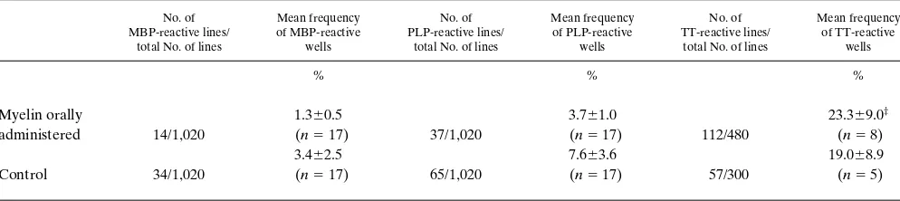
Frequency of antigen-specific T cell lines and correlation of pro-liferation with cytokine production. The frequency of antigen-reactive T cell lines from myelin-treated and non–myelin-treated MS patients’ PBMCs was determined by 3H-thymidine incorporation. A T cell line was considered positive when the stimulation index was . 3.0 and the delta CPM was . 500. A comparison of the frequencies in responses of the two groups to individual antigens is shown in Table I. Although there was a lower frequency of MBP- and PLP-reactive T cells in the my-elin-treated group as compared to the nontreated group in re-sponse to MBP and PLP, this difference did not reach statisti-cal significance in this cross-sectional analysis. The frequency of TT reactive lines were similar in both myelin-treated and nontreated groups.
Reactivity of human T cells is usually measured by 3 H-thy-midine incorporation. Thus it was important to know if this measure of T cell proliferation correlated with cytokine secre-tion. This was examined by screening several hundred lines (both negative and positive for antigen-induced 3H-thymidine incorporation) for secretion of IL-4, IFN-g, and TGF-b1 by ELISA. Although a correlation between thymidine uptake and IFN-g secretion was found (correlation coefficient r 5
0.60, P 5 0.0004), there was no correlation between stimula-tion index and cytokine secrestimula-tion for IL-4 and TGF-b1. Thus, we measured cytokine secretion from all wells whether or not there was antigen-specific 3H-thymidine incorporation.
The relative frequency of cytokine-secreting lines. The rel-ative frequency of cytokine-secreting lines in response to MBP, PLP, and TT in T cell lines from oral myelin-treated and non-treated patients was calculated for individual subjects, and the mean relative frequency for each group was calculated (Fig. 1). There was an increase in TGF-b1–secreting lines recognizing
MBP in the oral myelin-treated as compared to nontreated pa-tients (myelin treated 9.962, nontreated 1.360.5, P , 0.001) (Fig. 1 A). We also observed an increase in the relative fre-quency of IL-4–secreting lines reactive to MBP in the oral my-elin-treated MS patients, but this did not reach statistical sig-nificance (myelin-treated 6.562.7, nontreated 1.360.7, P ,
0.099). No difference in the relative frequency of IFN-g secret-ing lines was observed (myelin treated 5.062.5, nontreated 6.461.8). In response to PLP, there was also a significant in-crease in the relative frequency of TGF-b1–secreting T cell lines in the myelin-treated group (myelin treated 9.562.2, nontreated 1.160.7, P , 0.003) while there were no differences observed for IL-4 secreting lines (myelin treated 3.161.9, non-treated 261.6) (Fig. 1 B). There was a trend for lower IFN-g
secretion from the oral myelin-treated group in response to PLP, but this did not achieve statistical significance (myelin treated 6.162.5, nontreated 13.767.4, P , 0.264). The fre-quency of TGF-b1 secreting lines from individual subjects is shown in Fig. 2.
To examine whether the frequency of TGF-b1 secreting lines from the oral myelin-treated group was specific for mye-lin antigens, we measured cytokine secretion from T cell mye-lines using TT as a control antigen. Both oral myelin-treated and non–myelin treated groups showed similar relative frequencies of TGF-b1, IL-4, and IFN-g cell lines in response to TT
(TGF-b1; myelin treated 1.961.2, nontreated 2.061.2, IL-4; myelin treated 1.060.5, nontreated 0, IFN-g; myelin treated 39.8612.1, nontreated 65.866.3) (Fig. 1 C).
Th1- or Th2-, Th3-like cytokine secretion patterns. We ana-lyzed individual T cell lines to determine whether high TGF-b1 secretion was predominantly associated with IL-4 or IFN-g
secretion. The majority of MS patients, whether or not receiv-ing oral myelin, did not exhibit high relative frequencies of IL-4–secreting T cells. However, two individuals with rela-tively high frequencies of IL-4–secreting T cells were observed in the oral myelin-treated group. In both individuals, the IL-4– secreting T cells predominantly secreted TGF-b1 and not IFN-g. The data for one of the two patients as compared to the non– myelin-treated MS patient analyzed in the same experiment is shown in Fig. 3. Thus, the majority of TGF-b1–secreting T cells did not secrete IL-4 and are consistent with a distinct CD41 Th3 cell type (20).
[image:4.612.58.555.592.703.2]Correlation between the relative frequencies of MBP vs. PLP TGF-b1–secreting T cells. The frequency of both MBP and PLP reactive TGF-b1–secreting T cells varied among MS
Table I. Relative Frequency of Antigen-reactive T Cell Lines in MS Patients*
No. of MBP-reactive lines/
total No. of lines
Mean frequency of MBP-reactive
wells
No. of PLP-reactive lines/
total No. of lines
Mean frequency of PLP-reactive
wells
No. of TT-reactive lines/
total No. of lines
Mean frequency of TT-reactive wells % % % Myelin orally administered 14/1,020
1.360.5
(n5 17) 37/1,020
3.761.0
(n5 17) 112/480
23.369.0‡
(n5 8)
Control 34/1,020
3.462.5
(n5 17) 65/1,020
7.663.6
(n5 17) 57/300
19.068.9 (n5 5)
*PBMC were plated directly into 96-well plates and stimulated with antigens as described in Methods. Each well was restimulated in the presence of
antigen with APC on day 7 and for reactivity by a split well assay on day 14. ‡The values are expressed as the percentage of antigen reactive lines out
Figure 1. Relative frequency of antigen-reactive T cell lines secreting IL-4, IFN-g, or TGF-b1 from MS patients either receiving myelin orally or controls. The difference in cytokine secretion on day 14 of T cell lines stimulated by antigen-pulsed APCs were compared to T cell lines stimu-lated with APCs alone, as described in Methods. Culture supernatants were collected 24 hours after antigenic stimulation for an IL-4 and IFN-g
ELISA. Serum-free media was added to each well, and after an additional 72 h, culture supernatant was collected for a TGF-b1 ELISA. A line was considered positive if the antigen-specific cytokine secretion was . 50 pg/ml of IL-4 and . 100 pg/ml of IFN-g or TGF-b1. The relative fre-quencies of cytokine-secreting lines with antigenic stimulation were calculated for each patient, 17 myelin-treated and 17 nontreated patients and the average values, 6standard errors, are shown here. Significant P values for TGF-b1 secretion are highlighted in bold. (A) MBP stimula-tion. TGF-b1, myelin treated 9.962, nontreated 1.360.5, P, 0.001; IL-4, myelin-treated 6.562.7, nontreated 1.360.7, P, 0.099; IFN-g, myelin treated 5.062.5, nontreated 6.461.8. (B) PLP stimulation. TGF-b1, myelin treated 9.562.2, nontreated 1.160.7, P, 0.003; IL-4, myelin treated
3.161.9, nontreated 261.6; IFN-g, myelin treated 6.162.5, nontreated 13.767.4, P, 0.264. (C) TT stimulation. TGF-b1, myelin treated 1.961.2, nontreated 2.061.2; IL-4, myelin treated 1.060.5, nontreated 0, IFN-g, myelin treated 39.8612.1, nontreated 65.866.3, all values not signifi-cantly different for TT stimulation.
Figure 2. Distribution of relative frequencies of TGF-b1–producing lines in response to antigen. PBMC were stimulated with antigens (MBP, PLP, and TT) and cultured as described. 24 h after antigen stimulation on day 14, culture media were replaced with serum-free media, and cells were cultured for an additional 72 h. TGF-b1 secretion in supernatant was measured by ELISA. Each data point represents a patient sample and indicates percentage of positive cytokine-producing lines out of total generated lines. (A) MBP, (B) PLP, (C) TT. There were significantly more TGF-b1–secreting lines generated from treated patients than nontreated patients (P, 0.001 and P, 0.003 for MBP and PLP-reactive T cell
[image:5.612.59.556.475.672.2]patients orally treated with myelin. This led us to examine whether there was a correlation between the relative frequen-cies of MBP vs. PLP TGF-b1–secreting T cells among individ-ual subjects. As is shown in Fig. 4, there was a highly signifi-cant correlation in individual patients between the frequency of MBP vs. PLP TGF-b1–secreting T cells.
Discussion
[image:6.612.61.556.65.428.2]The present study was carried out to determine whether the prolonged oral administration of myelin in MS patients alters the frequency or cytokine secretion patterns of MBP or PLP
Figure 3. TGF-b1 secretion is associated with IL-4, not IFN-g–secreting T cells. Cytokine production by MBP-specific T cell lines generated from one of two oral myelin-treated patients generating significant numbers of IL-4–secreting T cells and one concurrently examined nontreated patient. (A) IL-4 versus IFN-g; (B) IL-4 versus TGF-b1; (C) IFN-g versus TGF-b1. Cytokine production in supernatant from single T cell lines in response to MBP is represented by each data point. In this representative patient set, T cell lines from a nontreated patient secreted IFN-g or small amounts of TGF-b1 and no IL-4; lines generated from an oral myelin-treated patient secreted IL-4 or TGF-b1 and IL-4, but not IFN-g and TGF-b1.
[image:6.612.57.299.510.735.2]reactive T cells. Here, we demonstrate an increase in the fre-quency of MBP and PLP but not TT reactive TGF-b 1–secret-ing T cell lines in myelin-treated MS patients as compared to non–myelin-treated MS patients. These data, showing cyto-kine deviation with antigen-specific immunotherapy in sub-jects with autoimmune disease, have not been previously re-ported and are consistent with our observation that in mice receiving oral MBP, there is an increase in MBP-specific TGF-b1 secreting T cells.
A number of studies have defined mechanisms by which oral tolerization occurs in rodents. Investigations of the EAE model have shown that one of the primary mechanisms in-volves the generation of active suppression, and this is medi-ated by T cells that secrete TGF-b1 and suppress the immune response (27). Furthermore, T cell clones isolated from the mesenteric lymph nodes of mice orally tolerized with MBP se-creting IL-4, IL-10, and TGF-b1 suppressed ongoing EAE in-duced by either MBP or PLP (20). The regulatory properties of these T cell clones were abrogated when mice were given anti-TGF-b1 antibodies, suggesting a critical role for local se-cretion of TGF-b1 in suppressing the immune response. Simi-lar cytokine patterns were observed in MBP T cell receptor transgenic mice orally tolerized with low doses of MBP (21). The importance of cytokines in regulating the inflammatory CNS response in EAE has also been suggested by immuno-pathologic examination of cytokine secretion in the CNS. Ani-mals with acute EAE exhibit perivascular infiltration with acti-vated mononuclear cells secreting the inflammatory cytokines IL-l, IL-2, TNF-a, IFN-g, IL-6, and IL-8. In contrast, animals with EAE that had been orally tolerized with MBP exhibit a marked reduction of the perivascular infiltrate, with down reg-ulation of inflammatory cytokines and increased amounts of TGF-b1 and IL-4 (6). These results suggest that the suppres-sion of EAE, by oral tolerization and natural recovery, is re-lated to regulatory cells that secrete inhibitory cytokines at the target organ.
We examined TGF-b1 secretion from T cell lines using ir-radiated whole mononuclear cells as antigen-presenting cells (APC). The secretion of TGF-b1 by T cell lines cultured with the APC in the absence of antigen was compared to that of lines stimulated with APC pulsed with antigen. No TGF-b1 could be detected in supernatants at 48 or 96 h after the addi-tion of antigen to the culture of either fresh PBMC or Mo1-positive macrophages from treated subjects, indicating that macrophages or other APCs did not directly secrete TGF-b1 without T cell help (data not shown). However, as macro-phages and other APCs are capable of secreting TGF-b1, we cannot rule out that T cells, activated by antigen, signaled APC to secrete TGF-b1. Further analysis of the TGF-b1–secreting cell population is limited both by the unavailability of reagents to perform intracytoplasmic staining for this cytokine, and the low cell numbers hampering isolation of T cell populations at the early time points when cytokines are measured. While it is clear that the T cell lines are stimulated by antigen and thus presumably are secreting TGF-b1, it should be noted that the identity of the cytokine secreting cells has not been formally demonstrated.
TGF-b1 has a number of properties consistent with a role in regulating the immune response. It is potent in inhibiting T and B cell entry into S/G2/M cell cycle and thus inhibiting pro-liferation. Paradoxically, TGF-b1 also can attract macrophages to sites of inflammation while inducing upregulation of IgA
se-cretion by activated B lymphocytes. TGF-b1 can also induce fibrin deposition and is involved in scar formation (28, 29). Thus the combined physiologic action of TGF-b1 may be to down-regulate inflammatory T cell responses while inducing tissue repair.
The oral administration of antigen is a well known method in experimental animals of inducing tolerance to prevent sys-temic immune reactions (7, 8, 30). However, there are few in-vestigations in humans regarding the mechanism of oral toler-ization. The immune responses in normal individuals orally administered 50 mg KLH daily for 2 wk over a 3-wk period de-creased subsequent cell-mediated immune responses when subjects were injected with KLH, although antibody responses were not affected (31). The mechanism for this suppression has not been elucidated. Myelin antigens have also been given by other routes in attempts to tolerize human T cell responses. Porcine MBP was previously administered to patients with MS with subsequent examination of immune sensitization (32). These studies showed that with parenteral administration of MBP, subsequent delayed type hypersensitivity responses were observed, presumably representing a Th1 type response.
In our study, we measured responses to human rather than bovine MBP while we orally administered bovine myelin. Ex-amination of one and not both MBP antigens was restricted by the amount of blood we could obtain from the patients. Though the immunodominant epitope (residues 84–102) of MBP are identical between human and bovine MBP (1, 2), there are sequence differences in other parts of the protein. We believe examination of the response to human MBP is of greater clinical importance as, ultimately, we are interested in generating TGF-b1–secreting T cell clones that recognize hu-man proteins in the CNS. This is not an issue with PLP as the sequence between human and bovine PLP is almost identical.
We previously reported that the frequency of MBP-reac-tive T cells was significantly decreased in a longitudinal study investigating frequencies before and after initiating oral ad-ministration of myelin in patients with MS (22). While a simi-lar trend was observed in the present study, the data did not reach statistical significance. We believe the lack of statistical significance in the present cross-sectional study is related to the variability in the frequency of MBP reactive T cells among different patients with MS as compared to the serial measure-ments we performed in our first investigation. A longitudinal study of the frequency and cytokine secretion patterns of MS patients is in progress. Although the patients were being fol-lowed in an open label, nonrandomized fashion, we attempted to determine whether there was a correlation between fre-quency of TGF-b1–secreting cells and response to therapy by arbitrarily dividing patients into responders and nonresponders and performing a chi-square statistical analysis. No correlation was found in this small number of patients studied. As men-tioned above, this will be examined in a larger study powered to address this experimental question.
Thus, as mentioned above, a longitudinal dosing trial is in progress to address this question.
As expected from treating an outbred group of MS pa-tients, there was a distribution in the range of both MBP and PLP reactive TGF-b1–secreting T cells. This may have been due to differences among individual patients in their ability to recognize these myelin antigens, or alternatively, individual patients may have differential abilities to generate TGF-b1– secreting T cells. This question was addressed by determining whether there was a correlation among individual patients in the frequencies of MBP vs. PLP reactive TGF-b1–secreting T cells. Indeed we found a highly significant correlation in indi-vidual MS patients between the frequency of these two mye-lin-reactive T cell populations. As MBP and PLP-reactive TGF-b1–secreting T cells were found in the same patients, these data suggest the later hypothesis that individual patients, similar to different mice strains, may have a differential ability to generate different cytokine secreting populations of T cells.
While MBP and PLP are the most abundant proteins in the central nervous system myelin and may represent major tar-gets of the putative cell-mediated autoimmune response, other antigens such as MOG (5), and S100 beta (33), are also candi-date antigens to induce effector T cells which may be of critical importance in the pathogenesis of MS. Moreover, Sercarz and co-workers have defined the concept of immunodominant and cryptic antigens in mice based on recognition of peptides after primary in vivo stimulation with either whole protein antigen or peptides, respectively (34). Thus while the T cell response may be initially focused on immunodominant epitopes, with time there is spreading to other cryptic epitopes or other tissue antigens recognized by T cells. This concept is supported by studies of patients with MS where an increased frequency of activated T cells is found to both MBP and PLP in the same patients (35). Together, these data indicate that while toleriza-tion to a single protein epitope or antigen in animals may ef-fectively ameliorate experimental models of autoimmunity early in the course of the disease, immune therapy for T cell– mediated chronic human autoimmune disease such as MS or type I diabetes will require approaches that suppress ongoing immune responses to a multitude of organ-associated self anti-gens. The use of oral myelin tolerization to generate MBP and PLP-reactive T cells in humans that have the ability to migrate into the central nervous system and nonspecifically suppress ongoing autoimmune responses at the target organ by secret-ing TGF-b1 represents an antigen-specific immunotherapy ca-pable of suppressing autoimmune responses to multiple self antigens at the target organ. Moreover, these data show that autoantigen-specific T cells secreting TGF-b1 can be gener-ated in humans with a putative autoimmune disease.
In summary, oral administration of myelin to MS patients generates an increased frequency of MBP and PLP-specific TGF-b1–secreting T cells. These results are consistent with the cytokine secretion profile of regulatory T cells in animals after oral tolerization with MBP (19–21, 27). Such antigen-specific regulatory T cells, upon homing to the central nervous system, would be predicted to suppress ongoing autoimmune immune responses in an antigen nonspecific fashion by the secretion of TGF-b1, a mechanism which has been termed bystander sup-pression. T cells induced by mucosal stimulation with antigen which secrete TGF-b1 appear to represent a distinct lineage of T cells (Th3) (36). Our data demonstrated that oral tolerization with self antigens in a human autoimmune disease may
pro-vide a therapeutic approach to the treatment of autoimmune disease which does not depend upon knowledge of the antigen specificity of the original T cell clone triggering the autoim-mune cascade.
Acknowledgments
This work was supported by National Institutes of Health grants RO1-NS24247 (D.A. Hafler), RO1-NS-29352 (H.L. Weiner), Pro-gram Project Grant AR 43220 (D.A. Hafler and H.L. Weiner), and grants from The National Multiple Sclerosis Society (H.L. Weiner, D.A. Hafler), and AutoImmune Inc., Lexington, MA. Drs. Hafler and Weiner have a financial interest in AutoImmune, Inc.
References
1. McFarlin, D.E., and H.F. McFarland. 1982. Multiple sclerosis. N. Engl. J. Med. 307:1183–1188.
2. Hafler, D.A., and H.L. Weiner. 1995. Immunologic mechanisms and therapy in multiple sclerosis. Immunol. Reviews. 144:75–107.
3. Zamvil, S., P. Nelson, J. Trotter, D. Mitchell, R. Knobler, R. Fritz, and L. Steinman. 1985. T cell clones specific for myelin basic protein induce chronic re-lapsing paralysis and demyelination. Nature (Lond.). 317:355–358.
4. Tuohy, V.K., R.A. Sobel, and M.B. Lees. 1988. Myelin proteolipid pro-tein-induced experimental allergic encephalomyelitis. Variations of disease ex-pression in different strains of mice. J. Immunol. 140:1868–1873.
5. Linington, C., T. Berger, L. Perry, S. Weerth, D. Hinze-Selch, Y. Zhang, H.C. Lu, H. Lassmann, and H. Wekerle. 1993. T cells specific for the myelin oli-godendrocyte glycoprotein mediate an unusual autoimmune inflammatory re-sponse in the central nervous system. Eur. J. Immunol. 23:1364–1372.
6. Khoury, S.J., W.W. Hancock, and H.L. Weiner. 1992. Oral tolerance to myelin basic protein and natural recovery from experimental autoimmune en-cephalomyelitis are associated with downregulation of inflammatory cytokines and differential upregulation of transforming growth factor-b, interleukin-4, and prostaglandin E expression in the brain. J. Exp. Med. 176:1355–1364.
7. Chase, M.W. 1946. Inhibition of experimental drug allergy by prior feed-ing of the sensitizfeed-ing agent. Proc. Soc. Exp. Biol. Med. 61:257–259.
8. Mowat, A.M. 1987. The regulation of immune responses to dietary pro-tein antigens. Immunol. Today. 8:93–98.
9. Higgins, P.J., and H.L. Weiner. 1988. Suppression of experimental au-toimmune encephalomyelitis by oral administration of myelin basic protein and its fragments. J. Immunol. 140:440–445.
10. Bitar, D.M., and C.C. Whitacre. 1988. Suppression of experimental au-toimmune encephalomyelitis by the oral administration of myelin basic protein. Cell. Immunol. 112:364–370.
11. Thompson, H.S., and N.A. Staines. 1986. Gastric administration of type II collagen delays the onset and severity of collagen-induced arthritis in rats. Clin. Exp. Immunol. 64:581–586.
12. Nagler-Anderson, C., L.A. Bober, M.E. Robinson, G.W. Siskind, and G.J. Thorbecke. 1986. Suppression of type II collagen-induced arthritis by in-tragastric administration of soluble type II collagen. Proc. Natl. Acad. Sci. USA. 83:7443–7446.
13. Zhang, Z.J., C.S.Y. Lee, O. Lider, and H.L. Weiner. 1990. Suppression of adjuvant arthritis in Lewis rats by oral administration of type II collagen. J. Immunol. 145:2489–2493.
14. Nussenblatt, R.B., R.R. Caspi, R. Mahdi, C.-C. Chan, F. Roberge, O. Lider, and H.L. Weiner. 1990. Inhibition of S-antigen induced experimental au-toimmune uveoretinitis by oral induction of tolerance with S-antigen. J. Immu-nol. 144:1689–1695.
15. Zhang, Z.J., L. Davidson, G. Eisenbarth, and H.L. Weiner. 1991. Sup-pression of diabetes in nonobese diabetic mice by oral administration of por-cine insulin. Proc. Natl. Acad. Sci. USA. 88:10252–10256.
16. Chen, Y., J.-I. Inobe, R. Marks, P. Gonnella, V.K. Kuchroo, and H.L. Weiner. 1995. Peripheral deletion of antigen-reactive T cells in oral tolerance. Nature (Lond.). 376:177–180.
17. Whitacre, C.C., I.E. Gienapp, C.G. Orosz, and D.M. Bitar. 1991. Oral tolerance in experimental autoimmune encephalomyelitis. III. Evidence for clonal anergy. J. Immunol. 147:2155–2163.
18. Friedman, A., and H.L. Weiner. 1994. Induction of anergy or active sup-pression following oral tolerance is determined by antigen dosage. Proc. Natl. Acad. Sci. USA. 91:6688–6692.
19. Miller, A., O. Lider, and H.L. Weiner. 1991. Antigen-driven bystander suppression following oral administration of antigens. J. Exp. Med. 174:791– 798.
20. Chen, Y., V.K. Kuchroo, J.-I. Inobe, D.A. Hafler, and H.L. Weiner. 1994. Regulatory T cell clones induced by oral tolerance: suppression of au-toimmune encephalomyelitis. Science (Wash. DC). 265:1237–1240.
H.L. Weiner. 1996. Oral tolerance in myelin basic protein T-cell receptor trans-genic mice: suppression of autoimmune encephalomyelitis and dose-dependent induction of regulatory cells. Proc. Natl. Acad. Sci. USA. 93:388–391.
22. Weiner, H.L., G.A. Mackin, M. Matsui, E.J. Orav, S.J. Khoury, D.M. Dawson, and D.A. Hafler. 1993. Double-blind pilot trial of oral tolerization with myelin antigens in multiple sclerosis. Science (Wash. DC). 259:1321–1324.
23. Trentham, D.E., R.A. Dynesius-Trentham, E.J. Orav, D. Combitchi, C. Lorenzo, K.L. Sewall, D.A. Hafler, and H.L. Weiner. 1993. Effects of oral ad-ministration of type II collagen on rheumatoid arthritis. Science (Wash. DC). 261:1727–1730.
24. Hohol, M.J., S.J. Khoury, S.L. Cook, E.J. Orav, D.A. Hafler, and H.L. Weiner. 1996. Three year open protocol continuation study of oral tolerization with myelin antigens in multiple sclerosis and design of a phase III pivotal trial. Annu. NY Acad. Sci. 778:243–250.
25. Chou, F.C.-H., C.-H.J. Chou, R. Shapira, and R.F. Kibler. 1976. Basis of microheterogeneity of myelin basic protein. J. Biol. Chem. 251:2671–2679.
26. Bizzozero, O.A., G. Zuniga, and M.B. Lees. 1991. Fatty acid composi-tion of human myelin proteolipid protein in peroxisomal disorders. J. Neuro-chem. 56:872–878.
27. Miller, A., O. Lider, A.B. Roberts, M.B. Sporn, and H.L. Weiner. 1992. Suppressor T cells generated by oral tolerization to myelin basic protein sup-press both in vitro and in vivo immune responses by the release of transforming growth factor b after antigen-specific triggering. Proc. Natl. Acad. Sci. USA. 89: 421–425.
28. Lebman, D.A., F.D. Lee, and R.L. Coffman. 1990. Mechanism for trans-forming growth factor b and IL-2 enhancement of IgA expression in li-popolysaccharide-stimulated B cell cultures. J. Immunol. 144:952–959.
29. Wahl, S.M., N. McCarthy-Francis, and S.E. Mergenhagen. 1989. Inflam-matory and immunomodulatory roles of TGFb. Immunol. Today. 10:258–261.
30. Weiner, H.L., A. Friedman, A. Miller, S.J. Khoury, A. Al-Sabbagh, L. Santos, M. Sayegh, R.B. Nussenblatt, D.E. Trentham, and D.A. Hafler. 1994. Oral tolerance: immunologic mechanisms and treatment of animal and human organ-specific autoimmune disease by oral administration of autoantigens. Annu. Rev. Immunol. 12:809–837.
31. Husby, S., J. Mestecky, Z. Moldoveanu, S. Holland, and C.O. Elson. 1994. Oral tolerance in humans: T cell but not B cell tolerance after antigen feeding. J. Immunol. 152:4663–4670.
32. Salk, R.J.S. 1983. A study of myelin basic protein as a therapeutic probe in patients with multiple sclerosis. In Multiple Sclerosis. J.F. Hallpike, C.W.M. Adams, and W.W. Tourtellotte, editors. Chapman and Hall, London. 621–630.
33. Kojima, K., T. Berger, H. Lassman, D. Hinze-Selch, Y. Zhang, J. Gehr-mann, K. Reske, H. Wekerle, and C. Linington. 1994. Experimental autoim-mune panencephalitis and uvoretinitis transferred to the Lewis rat by T lym-phocytes specific for the S100-beta molecule, a calcium binding protein of astroglia. J. Exp. Med. 180:817–829.
34. Lehmann, P.V., T. Forsthuber, A. Miller, and E.E. Sercarz. 1992. Spreading of T-cell autoimmunity to cryptic determinants of an autoantigen. Nature (Lond.). 358:155–157.
35. Zhang, J., S. Markovic-Plese, B. Lacet, J. Raus, H.L. Weiner, and D.A. Hafler. 1994. Increased frequency of interleukin-2 responsive T cells specific for myelin basic protein and proteolipid protein in peripheral blood and cere-brospinal fluid of patients with multiple sclerosis. J. Exp. Med. 179:973–984.