the Linear Array Assay for HPV Screening in an Asian Population
Oscar Gee-Wan Wong,aC. K. Lo,aJoanne N. K. Chow,aObe K. L. Tsun,aElaine Szeto,aStephanie S. Liu,bHextan Y. S. Ngan,band Annie N. Y. Cheunga
Departments of Pathologya
and Obstetrics & Gynaecology,b
Queen Mary Hospital, The University of Hong Kong, Hong Kong Special Administrative Region, China
High-risk human papillomavirus (HR-HPV) DNA detection in cervical cytology samples is useful for primary screening of cervi-cal cancer and for triage of patients with equivocervi-cal cytologicervi-cal findings. The GenoFlow HPV array test (GF assay; Diagcor Biosci-ence Inc., Hong Kong) was recently developed to detect 33 HPV genotypes by a “flowthrough” hybridization technology. In this study, we assessed the analytical sensitivity and reproducibility of the GF assay and compared its genotyping results with those of the Linear Array (LA) assay (Roche Molecular Diagnostics, Indianapolis, IN), using 400 archived liquid-based cytology sam-ples representing the full range of cytology findings. Genotyping findings of the GF and LA assays were concordant or compati-ble for 93.44% of tested samples, with a good ( ⴝ0.797) to very good ( ⴝ0.812) strength of agreement for assay-common and oncogenic HPV types, respectively. The two assays showed good ( ⴝ0.635) agreement in detecting infections with multiple HPV genotypes. The lowest detection limits of the GF assay for HPV16 and HPV18 were 25 copies and 20 copies, respectively. Repeat testing of 60 samples by use of two different lots of the GF assay revealed no discordant results, suggesting good repro-ducibility of the assay. Both assays achieved approximately 80% and 100% sensitivity for identifying cases of atypical squamous cells of undetermined significance (ASC-US) and low-grade squamous intraepithelial lesions (LSIL) with subsequent detection of LSILⴙand high-grade squamous intraepithelial lesions or higher (HSILⴙ) in 2 years, respectively. Among ASC-US samples, the GF assay achieved the highest specificity (23.08%) for indicating subsequent identification of HSIL compared with the LA (19.23%) and Hybrid Capture 2 (HC2) (8.97%) assays. The GF and LA assays showed significant discrepancy in detecting HPV genotypes 11, 26, 39, 52, and 66. More sensitive detection of HPV52 by GF assay offers an advantage in regions where HPV52 is more prevalent. The sensitivity of the GF assay for detecting patients with HSILⴙwas noninferior to that of the LA assay.
H
uman papillomavirus (HPV) is an established essential etio-logical factor for cervical cancer (31). Among the estimated ⬎200 HPV types, around 15 anogenital types are associated with cervical cancers and are termed high-risk HPV (HR-HPV) (4, 21). HR-HPV DNA testing has been proposed as a tool for primary cancer screening (29) or as an adjunct test for the triage of patients with the equivocal cytology finding of atypical squamous cells of undetermined significance (ASC-US) (23). For these purposes, HR-HPV cocktail tests such as the Hybrid Capture 2 (HC2) test and the Amplicor test are usually employed. These tests detect the presence of HR-HPV but do not distinguish the genotype present. However, it has been demonstrated that specific identification of HPV16 and HPV18 can highlight patients at the greatest risk of developing cervical intraepithelial neoplasia 3 or above (CIN3⫹) (6, 15, 28). HPV genotyping tests such as the Linear Array (LA) test (Roche) may hence provide more specific information.For each new HPV detection test, it is important to validate the clinical performance by using established tests as benchmarks (20). The HC2 test (Qiagen, Gaithersburg, MD [previously Di-gene]) is the first U.S. FDA-approved HR-HPV DNA test. It is a signal-amplified cocktail assay that detects 13 common HR-HPV types (types 16, 18, 31, 33, 35, 39, 45, 51, 52, 56, 58, 59, and 68). It has been validated extensively in many large studies (3, 19). The Linear Array genotyping test (Roche) is a reverse line blot assay for detection and genotyping of 37 types of HPV (10, 11). The LA test has been the standard for assessing new HPV genotyping tests (12, 18, 26).
The GenoFlow human papillomavirus array test (GF test) is a new HPV assay, developed by Diagcor Bioscience Inc., that is ca-pable of genotyping 33 types of HPV (27). The GF test is a reverse
dot blot assay utilizing a modified PGMY primer set to amplify the L1 region of HPV. PCR products are hybridized to probes spotted on a membrane by a rapid flowthrough hybridization process. In addition to the 33 HPV genotyping probe spots, a universal probe spot is available for detecting HPV genotypes outside the panel, as well as some HPV variants.
This study aimed to investigate the concordance in genotyping results of the GF and LA tests, as well as their clinical sensitivity and specificity, for a selected cohort of patients with different cy-tology diagnoses. The analytical sensitivity and interlot reproduc-ibility of the GF test were also evaluated.
MATERIALS AND METHODS
Cervical cytology specimen and sample preparation.Four hundred cer-vical cytology samples were retrieved from the archive of the Cercer-vical Cytology Laboratory, Queen Mary Hospital. The specimens were ran-domly selected from consecutive samples reported in 2009. The ages of the patients ranged from 17 to 76 years, and the median age was 38 years. The cytological diagnoses of the samples were as follows: normal (20 samples), ASC-US (120 samples), ASC– cannot exclude high-grade squamous intra-epithelial lesions (HSIL) (ASC-H) (40 samples), atypical glandular cells
Received2 October 2011Returned for modification15 November 2011 Accepted3 February 2012
Published ahead of print15 February 2012
Address correspondence to Annie N. Y. Cheung, [email protected].
Supplemental material for this article may be found athttp://jcm.asm.org/.
Copyright © 2012, American Society for Microbiology. All Rights Reserved.
doi:10.1128/JCM.05933-11
on May 16, 2020 by guest
http://jcm.asm.org/
(AGC) (30 samples), low-grade squamous intraepithelial lesions (LSIL) (100 samples), HSIL (60 samples), and squamous cell carci-noma (SCC) (30 samples). All of the ASC-US samples had been tested for HR-HPV by the Digene HC2 HPV test. The follow-up cytology/ colposcopic biopsy findings obtained within 2 years were retrieved. The use of archival human cytology specimens for research purposes was approved by the local institutional ethics committee (HKU/HA HKW IRB no. UW-10-301).
Input DNA for the GF assay was extracted from 1 ml of each sample by use of a Qiagen blood minikit according to the “Blood and Body Fluid Spin Protocol” of the kit instructions. DNA concentration was deter-mined by spectrophotometry, but the same volume (5l) of extracted DNA was used in each PCR for GF assay. Input DNA for the LA assay was extracted using the reagents and protocol included in the kit according to the manufacturer’s instructions.
HPV genotyping by GF and LA assays.DNA samples were tested for HPV by the GF and LA assays according to instructions of the manufac-turers. Briefly, for the GF assay, DNA extracted from a sample by use of a QIAamp blood minikit (Qiagen) was amplified by use of a biotin-labeled primer mix. PCR was performed in a reaction volume of 25l containing 5l of DNA template, 19.25l of the master mixture provided, and 0.75
l of DNATaqpolymerase (5 U/l) in a Perkin-Elmer GeneAmp PCR system 9700 apparatus (Applied Biosystems). The reaction mixture was first incubated at 95°C for 9 min and then went through 43 cycles of 20 s of denaturation at 95°C, 30 s of annealing at 55°C, and 30 s of elongation at 72°C, followed by a final extension for 5 min at 72°C. The amplified products were subsequently denatured and then hybridized to probes prespotted on a membrane through a flowthrough hybridization process. After a stringent wash, hybridized DNA was detected with streptavi-din-alkaline phosphatase followed by colorimetric development using nitroblue tetrazolium–5-bromo-4-chloro-3-indolylphosphate (NBT-BCIP). The results were interpreted by direct visualization.
The LA test uses a biotinylated PGMY09/11 primer set to amplify a 450-bp region of the L1 gene and is capable of detecting 37 HPV geno-types, including 15 HR types. DNA was amplified by PCR in a Perkin-Elmer GeneAmp PCR system 9700 apparatus (Applied Biosystems). The denatured PCR product was then hybridized to an array strip containing immobilized oligonucleotide probes. The results were interpreted by us-ing the reference guide and readus-ing the matchus-ing individual types down the length of the strip.
Interpretation of genotyping results.The results of GF tests were interpreted according to the manufacturer’s instructions. A valid HPV-positive result must include visible signals at the “universal,” “HC” (hy-bridization control), and “AC” (amplification control) probe spots. A valid negative result must include signals at the HC and AC probe spots. An HPV of unknown genotype was present when only the universal, HC, and AC probe spots were positive. Some HPV types share the same probe spots and cannot be distinguished by GF assay, e.g., HPV66 and HPV68, HPV54 and HPV55, and HPV40 and HPV61. Both genotypes were inter-preted as present if a signal was seen in the probe spot shared by two HPV genotypes.
The results of LA tests were read according to the manufacturer’s instructions. The results were interpreted by using the reference guide and reading the matching individual types down the length of the strip. A result is valid only when at least one of the globin (low and high) signal bands is visible. The LA assay offers no separate detection for HPV52. Instead, a signal band for HPV types 33, 35, 52, and 58 and three separate bands for HPV33, HPV35, and HPV58 are present. HPV52 positivity is established only if the HPV33/35/52/58 signal is present and the HPV33, HPV35, and HPV58 signals are absent.
Assessment of analytical sensitivity and reproducibility.Plasmids containing the full-length genomes of HPV16 and HPV18 were used to determine the analytical sensitivity of the GF assay (18). The plasmids were serially diluted to 1⫻104, 5⫻103, 1⫻103, 5⫻102, 1⫻102, 5⫻101,
1⫻101, and 5 copies/l. Each dilution also contained 32 ng/l genomic
DNA of C33A, an HPV-negative cervical cancer cell line. The smallest amount of input HPV DNA tested was 5 copies. Each dilution was tested three times with the GF assay, and the lowest copy number that could be detected every time defined the analytical sensitivity of the assay.
Sixty samples were chosen for repeat HPV testing by GF assay to assess the reproducibility of the assay. Among the 60 samples, there were 20 ASC-US, 20 LSIL, and 20 HSIL. The samples were tested with a different lot of the GF assay.
Discrepancy analysis with L1 PCR sequencing and Cobas 4800 HPV test.Cases showing discordant results and discrepancy in terms of HR-HPV positivity were chosen for further analysis. DNAs extracted from the samples were amplified by both PGMY09/11 and MY09/11 primer sets in separate PCRs. The absence of an amplification product suggested that there was no detectable HPV DNA. PCR products were sequenced, and resultant sequences were matched with known HPV sequences. In addi-tion, the samples were subjected to the Cobas 4800 HPV test, a qualitative
in vitrotest for the detection of 14 HR-HPV types (5). The test specifically identifies (types) HPV16 and HPV18 and at the same time indicates the presence or absence of the rest of the high-risk types (types 31, 33, 35, 39, 45, 51, 52, 56, 58, 59, 66, and 68).
Data analysis and statistics.A kappa value of 0 indicates no ment better than chance, and a kappa value of 1 indicates perfect agree-ment. Kappa values of 0 to 0.20, 0.21 to 0.40, 0.41 to 0.60, 0.61 to 0.80, and
ⱖ0.81 indicate a poor, fair, moderate, good, and very good strength of agreement, respectively (2). The nonparametric McNemar test was used to analyze the complementarities of the detection methods and to deter-mine if the results obtained by the two methods were significantly differ-ent. A test of noninferiority was calculated as previously described (20).P
values of⬍0.05 were considered statistically significant.
RESULTS
HPV genotyping results of GF and LA assays are mostly concor-dant or compatible.To compare the HPV genotyping results of the GF and LA assays, we performed two independent HPV tests on 400 liquid-based cervical cytological samples, using both as-says. The two assays are capable of (Table 1) detecting different but largely overlapping sets of HPV genotypes (Table 1). In this report, “assay-common HPV genotypes” refers to those HPV ge-notypes detectable by both assays. The 400 samples were selected to encompass various cytological diagnoses, including normal, ASC-US, ASC-H, AGC, LSIL, HSIL, and SCC. LA testing of all 400 samples yielded either HPV-negative or HPV-positive results, whereas the amplification control signal could not be visualized for four samples tested by the GF assay, meaning that there was either insufficient DNA to be amplified or a PCR inhibitor was present in the extracted DNA. Therefore, comparable results of the GF and LA assays were obtained for a total of 396 samples.
In comparing results from the GF and LA assays, “concordant” means that the HPV genotypes identified by the GF and LA assays are exactly the same or that both assays yielded HPV-negative results. A pair of results is considered “compatible” if there is at least one assay-common HPV genotype in the results of the GF and LA assays. To-tally disagreeing results are considered “discordant.”
As shown in Table 2, the GF and LA assays gave rise to either concordant or compatible results for 93.44% (52.78% plus 40.66%) of the samples. Remarkably, for serious lesions (HSIL and SCC), the results of the GF and LA assays were highly consis-tent, giving concordant results for more than 70% of the samples, with no discordant results (Table 2). This difference reached sta-tistical significance (chi-square test;P⬍0.0001).
GF and LA assays highly agree on samples positive for HR-HPV.We transformed the HPV genotyping results into
on May 16, 2020 by guest
http://jcm.asm.org/
HPV-positive and HR-HPV-negative results by using the follow-ing criteria: if a sample was positive for at least 1 of the 15 HR-HPVs, i.e., types 16, 18, 31, 33, 35, 39, 45, 51, 52, 56, 58, 59, 68, 73, and 82, then it was considered HR-HPV positive; otherwise, it was considered HR-HPV negative. The two assays showed remarkable agreement for HR-HPV-positive samples, reaching an absolute agreement of 93.69%. Cohen’svalue was 0.812, meaning that the two assays showed very good agreement (Table 3). When
the criterion was changed to assay-common genotypes, absolute agreement was 95.45%, and Cohen’s value was 0.797. When only HSIL and SCC cases were considered, Cohen’svalues were 1.000 (very good) and 0.794 (good) for assay-common HPV types and oncogenic HPV types, respectively (see Table S1 in the sup-plemental material).
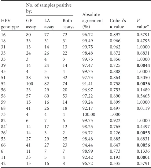
Interassay agreement of individual HPV genotypes.When individual HPV genotypes were examined, the GF and LA assays agreed for most genotypes (Table 4). Most importantly, the abso-lute interassay agreement values for HPV types 16 and 18 were 96.72% and 99.49%, respectively (Table 4). Cohen’svalues for these two HPV vaccine-covered oncogenic HPV types were 0.897 and 0.966, respectively (Table 4). However, we also noted a statisti-cally significant difference in identifying a few genotypes between the two assays (Table 4). Notably, the GF and LA assays differed signifi-cantly in detecting the HR-HPVs HPV39 and HPV52 (P⫽0.0044 andP⫽0.0036, respectively) (Table 4). The two assays also differed significantly in detecting HPV11, one of the most common causes of warts (Table 4). Notably, every genotype detectable by the GF assay was detected at least once within our cohort. The rarest type was HPV72, which was detected in only one sample. The frequency of detection of each HPV type assayed by the GF assay is presented in Table S2 in the supplemental material.
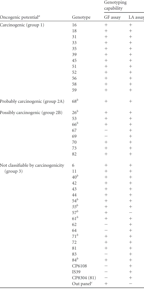
[image:3.585.41.284.81.569.2]Single- and multiple-infection detection of GF and LA as-says.One of the advantages of line blot or dot blot assays such as TABLE 1HPV genotypes recognized by GF and LA assays
Oncogenic potentiala Genotype
Genotyping capability
GF assay LA assay Carcinogenic (group 1) 16 ⫹ ⫹
18 ⫹ ⫹
31 ⫹ ⫹
33 ⫹ ⫹
35 ⫹ ⫹
39 ⫹ ⫹
45 ⫹ ⫹
51 ⫹ ⫹
52 ⫹ ⫹
56 ⫹ ⫹
58 ⫹ ⫹
59 ⫹ ⫹
Probably carcinogenic (group 2A) 68b ⫹ ⫹
Possibly carcinogenic (group 2B) 26b ⫹ ⫹
53 ⫹ ⫹
66b ⫹ ⫹
67 ⫺ ⫹
69 ⫺ ⫹
70 ⫹ ⫹
73 ⫹ ⫹
82 ⫹ ⫹
Not classifiable by carcinogenicity (group 3)
6 ⫹ ⫹
11 ⫹ ⫹
40b ⫹ ⫹
42 ⫹ ⫹
43 ⫹ ⫹
44 ⫹ ⫹
54b ⫹ ⫹
55b ⫹ ⫹
57b ⫹ ⫺
61b ⫹ ⫹
62 ⫺ ⫹
64 ⫺ ⫹
71b ⫹ ⫹
72 ⫹ ⫹
81 ⫹ ⫹
83 ⫺ ⫹
84b ⫹ ⫹
CP6108 ⫺ ⫹
IS39 ⫺ ⫹
CP8304 (81) ⫺ ⫹ Out panelc ⫹ ⫺ a
According to reference 4.
bHPV66 and HPV68, HPV54 and HPV55, HPV40 and HPV61, HPV57 and HPV71,
and HPV84 and HPV26 share probe spots on the GF membrane.
cThe GF assay provides a universal probe for detection of HPV types outside the panel.
[image:3.585.299.545.97.229.2]According to the manufacturer, types 54 (new subtype), 74 (new subtype), CP8304 (81), 87, and 89 have been detected successfully in clinical samples.
TABLE 2Concordant/compatible HPV detection (assay-common genotypes) in relation to cytological classification between GF and LA assays
Cytology finding
No. (%) of samples with the indicated result between GF and LA assays
Total no. of samples Concordant Compatible Discordant
[image:3.585.299.542.566.652.2]Normal 13 (65.00) 6 (30.00) 1 (5.00) 20 ASC-H 20 (50.00) 19 (47.50) 1 (2.50) 40 ASC-US 59 (50.43) 48 (41.03) 10 (8.55) 117 AGC 16 (53.33) 9 (30.00) 5 (16.67) 30 LSIL 38 (38.00) 54 (54.00) 8 (8.00) 100 HSIL 43 (72.88) 16 (27.12) 0 (0.00) 59 SCC 21 (70.00) 9 (30.00) 0 (0.00) 30 Total 210 (52.78) 161 (40.66) 25 (6.57) 396
TABLE 3HPV positivity agreement of GF and LA assaysa
GF assay result
No. of samples with LA assay result
Assay-common genotypesb Oncogenic genotypesc
Positive Negative Total Positive Negative Total Positive 336 7 343 299 13 324 Negative 11 42 53 12 72 84 Total 347 49 396 311 85 396
a
The absolute agreement levels between assays for assay-common genotypes and oncogenic genotypes were 95.45% and 93.69%, respectively. Cohen’svalues for assay-common genotypes and oncogenic genotypes were 0.797 and 0.812, respectively.P
values determined by the McNemar test for assay-common genotypes and oncogenic genotypes were 0.4795 and 1.0000, respectively.
bAssay-common genotypes are HPV genotypes detectable by both assays.
c
Oncogenic genotypes are types 16, 18, 31, 33, 35, 39, 45, 51, 52, 56, 58, 59, 68, 73, and 82.
on May 16, 2020 by guest
http://jcm.asm.org/
the GF and LA assays is their ability to detect multiple infections. We categorized the genotyping results of the GF and LA assays into single-infection, multiple-infection, and negative results. The GF and LA assays showed good agreement on the number of HPV types present ( ⫽0.635;P⬍0.01 by chi-square test) (Table 5). Notably, more multiple-infection cases were identified by the GF assay (190 cases) than by the LA assay (154 cases).
Discrepancy analysis of HR-HPV discordant cases.Among the 396 cases compared, 25 cases showed discordant GF and LA assay results. In other words, for 25 cases, the results from the GF and LA assays were completely different. Among the 25 discordant cases, 13 disagreed on their detection of HR-HPV. These cases were further evaluated by PCR and sequencing using PGMY09/11 and MY09/11 primer sets, as well as by the Cobas 4800 HPV test, a cocktail HR-HPV test capable of indicating the presence of HPV16, HPV18, or non-HPV16/18 HR-HPV or the absence of HR-HPV. We could not detect any HPV by the PCR method in 9 of the cases, which agreed with the GF assay results in 5 of the cases (Table 6). Among the 4 PCR-positive cases, 3 agreed with the LA assay (one of the genotypes suggested by the test) and 1 agreed with the GF assay. Taken together, the results show that 5 cases matched by PCR and LA assay, 6 cases matched by PCR and GF assay, and in 2 cases the PCR results did not agree with either assay (Table 6). In 8 cases, the results of the Cobas 4800 system agreed with the LA assay, and in 5 cases the Cobas 4800 system results agreed with the GF assay (Table 6).
Analytical sensitivity and interlot reproducibility of GF as-say.To assess the analytical sensitivity of the GF assay, serial dilu-tions of two plasmids, harboring the full genomes of HPV16 and
HPV18, were prepared (10,000 copies/l to 5 copies/l). Each dilution also contained the same amount (32 ng/l) of genomic DNA of C33A, an HPV-negative cervical cancer cell line. The low-est detection limits of HPV16 and HPV18 DNAs were found to be 25 copies and 20 copies, respectively.
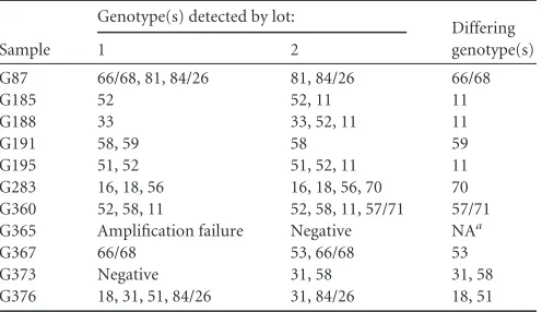
To assess the interlot reproducibility of the GF assay, 60 sam-ples were selected for repeat testing with a different lot of the test. Of these 60 samples, 49 (81.67%) yielded exactly the same HPV genotyping results. Among the 11 samples which yielded different genotyping results, it was most common to see that one of the tests detected one additional low-risk HPV type (Table 7). One sample failed to be amplified in one of the tests and hence could not be compared (Table 7).
Sensitivity and specificity of GF and LA assays for triage of ASC-US cases in comparison to HC2 test.Either cytology or his-tology follow-up data were available for 119 of the ASC-US cases, and all of the ASC-US samples had been tested for HR-HPV by HC2 assay. To compare our results with those of the HC2 test, HPV types 16, 18, 31, 33, 35, 39, 45, 51, 52, 56, 58, 59, and 68 were used to define HR-HPV. When the GF and LA assays were used to identify cases that would progress to LSIL/CIN1⫹, their sensitiv-ities were 81.82% and 87.88%, respectively, which are lower than that of the HC2 test (96.97%).
All three tests could identify all cases that would progress to HSIL/CIN2⫹(sensitivity⫽100%). However, both the GF and LA assays achieved higher specificities than that of the HC2 test (23.08% for GF assay, 19.23% for LA assay, and 8.97% for HC2 test).
ASC-H and AGC are rare but nonetheless significant cytology findings (14, 22). In total, there were 138 cases of ASC-US, ASC-H, or AGC with available follow-up data. We calculated the sensitivity and specificity of using the GF and LA assays for pre-dicting disease on follow-up. When ASC-H and AGC cases were included, the sensitivity and specificity of detecting cases that would progress to LSIL/CIN1⫹were 80.00% and 36.67%, respec-tively, for the GF assay and 80.56% and 40%, respecrespec-tively, for the LA assay. The sensitivity and specificity of detecting cases that would progress to HSIL/CIN2⫹were 84.00% and 25.45%, re-spectively, for the GF assay and 88.00% and 26.55%, rere-spectively, for the LA assay.
In summary, we found that the GF assay and LA assay per-formed very similarly for triage of cases with equivocal cytology findings.
[image:4.585.39.286.86.360.2]Sensitivity of GF assay is noninferior to that of LA assay for detecting HSILⴙ.A test of noninferiority was performed using data from the HSIL and SCC cases (20). To assess the noninferi-ority of the sensitivity (i.e., relative sensitivity of no lower than TABLE 5Single and multiple HPV infections identified by GF and LA assaysa
GF assay result
No. of samples with LA assay result Single
HPV type
Multiple
HPV types Negative Total Single HPV type 131 17 6 154 Multiple HPV types 50 136 4 190
Negative 10 1 41 52
Total 191 154 51 396
a
Cohen’svalue for the number of HPV types detected was 0.635. TABLE 4Interassay agreements for individual HPV (assay-common)
genotypes detected by GF and LA assays
HPV genotype
No. of samples positive by:
Absolute agreement (%)
Cohen’s
value
P
valuea
GF assay
LA assay
Both assays
16 80 77 72 96.72 0.897 0.5791 18 33 31 31 99.49 0.966 0.4795 31 13 14 13 99.75 0.962 1.0000 33 24 26 22 98.48 0.872 0.6831 35 3 4 3 99.75 0.856 1.0000 39 14 24 14 97.47 0.725 0.0044
45 4 5 4 99.75 0.888 1.0000 51 38 35 32 97.73 0.864 0.5050 52 100 82 74 91.41 0.758 0.0036
56 23 29 20 96.97 0.753 0.1489 58 57 60 53 97.22 0.890 0.5465 59 15 16 14 99.24 0.899 1.0000 68 41 26 18 92.17 0.497 0.0119 73 4 4 4 100.00 1.000
82 6 7 6 99.75 0.922 1.0000 84b 14 17 12 98.25 0.765 0.4497
26b 14 3 2 96.72 0.226 0.0055
53 27 29 25 98.48 0.885 0.6831 66 41 27 23 94.44 0.647 0.0056
6 11 7 7 98.99 0.773 0.1336 11 33 5 4 92.42 0.193 0.0001
42 13 16 8 96.72 0.535 0.5791
aDetermined by the McNemar test. Values in bold show significant results.
b
HPV84 and HPV26 share the same probe spot in the GF assay.
on May 16, 2020 by guest
http://jcm.asm.org/
[image:4.585.302.544.88.178.2]90%), the GF and LA test results were tabulated and the noninfe-riority scoreTwas calculated (Table 8). The null hypothesis was rejected (T⫽3.24893;P⫽0.00058), and hence the sensitivity of the GF assay was not inferior to the sensitivity of the LA assay. The test score for the noninferiority of specificity calculated based on cases with diagnoses lower than HSIL, however, did not reach statistical significance (T⫽0.91698;P⫽0.17958), and hence the null hypothesis (i.e., the relative specificity was lower than 98%) was not rejected.
DISCUSSION
The Diagcor GF HPV test provides a faster alternative to the LA test, as reported previously (27). In practice, the time required for us to test similar numbers of samples (⬇25) by the GF assay is approximately 25% shorter than that required for the LA assay. This is partly attributable to the adaption of flowthrough hybrid-ization technology, which significantly shortened the hybridiza-tion step of the GF assay (27). However, before it is adopted for clinical or research use, the performance of the GF assay needs to be characterized. The GF test has been compared to the LA assay only in a study with a limited number of samples (27). Our study provides a more comprehensive comparison of the GF and LA
assays. Generally, we found the results from the two tests to be highly compatible (Table 2). When the tests were used as tools for detecting the presence of HR-HPV without detailing the individ-ual types present, the tests highly agreed with each other (Table 3). Therefore, when used for triage of ASC-US cases, the two tests achieved similar sensitivities and specificities. At the individual HPV type level, the two tests agreed for most (13/15 types) HR-HPV types, being significantly different only in detecting HVP39 and HPV52 (Table 4).
[image:5.585.41.549.77.241.2]HPV tests capable of identifying individual HPV types are use-ful tools for epidemiological research (1, 9, 13, 16). In addition, identification of specific oncogenic HPV types may bear implica-tions for the management of HPV-positive women. In a recent study involving more than 40,000 patients, detection of HPV16, HPV18, or both had a better sensitivity and similar positive pre-dictive value (PPV) for detection of CIN3 or worse than for ASC-US or worse alone among HPV-positive women (6, 30). Our previous study also found that detection of HPV16 and HPV18 improved the sensitivity of identifying HPV-positive ASC-US TABLE 6Discrepancy analysis for cases showing discordant results and discrepant HR-HPV statuses in the two tests
Sample
Genotype(s) detected bya: PCR and sequencingb Cobas 4800 HPV test
LA assay GF assay Result Matchc Result Matchc
G14 16 Negative Negative GF assay 16 LA assay G53 16, 42, 62 81 62 LA assay HR-HPV negative GF assay G88 68 43/44 44 GF assay HR-HPV negative GF assay G116 67 51 Negative XX HR-HPV negative LA assay G117 Negative 52 Negative LA assay HR-HPV negative LA assay G222 33 Negative 33 LA assay Non-type 16/18 HR-HPV LA assay G263 56 Negative Negative GF assay HR-HPV negative GF assay G272 68 Negative Negative GF assay Non-type 16/18 HR-HPV LA assay G311 51 Negative Negative GF assay Non-type 16/18 HR-HPV LA assay G334 62, 69 51, 52 Negative XX Non-type 16/18 HR-HPV GF assay G373 31, 58 Negative 58 LA assay 18, non-type16/18 HR-HPV LA assay G391 Negative 52 Negative LA assay HR-HPV negative LA assay G394 39 Negative Negative GF assay HR-HPV negative GF assay
aUnderlining indicates HR-HPV types.
b
PCR was done with both PGMY09/11 and MY09/11 primer sets.
cXX, sequencing result does not match either GF or LA assay result.
TABLE 7Samples yielding different results between two different lots of GF assays
Sample
Genotype(s) detected by lot:
Differing genotype(s)
1 2
G87 66/68, 81, 84/26 81, 84/26 66/68
G185 52 52, 11 11
G188 33 33, 52, 11 11
G191 58, 59 58 59
G195 51, 52 51, 52, 11 11 G283 16, 18, 56 16, 18, 56, 70 70 G360 52, 58, 11 52, 58, 11, 57/71 57/71 G365 Amplification failure Negative NAa
G367 66/68 53, 66/68 53 G373 Negative 31, 58 31, 58 G376 18, 31, 51, 84/26 31, 84/26 18, 51
[image:5.585.297.542.539.706.2]aNA, not available.
TABLE 8Comparison of GF and LA test results by test of noninferiority
Case type and GF test result
No. of cases with LA test result HR-HPV
positive
HR-HPV
negative Total HSIL and SCC casesa
HR-HPV positive 85 1 86 HR-HPV negative 0 3 3
Total 85 4 89
Normal, ASC-US, ASC-H, AGC, and LSIL casesb
HR-HPV positive 214 12 226 HR-HPV negative 12 69 81
Total 226 81 307
a␦ ⫽
0.90,T⫽3.24893, andP⫽0.00058.
b␦ ⫽0.98,T⫽0.91698, andP⫽0.17958.
on May 16, 2020 by guest
http://jcm.asm.org/
[image:5.585.41.289.572.715.2]cases that will progress to HSIL or worse in an Asian screening population (28). It remains to be tested whether detection of any other HPV types could similarly highlight patients at particular risk of disease progression.
It is particularly reassuring that the absolute interassay (GF assay versus LA assay) agreement levels for HPV types 16 and 18 were 96.72% and 99.49%, respectively (Table 4). Cohen’svalues for these two HPV vaccine-covered oncogenic HPV types were 0.897 and 0.966, respectively, reflecting a higher strength of agree-ment than when assay-common ( ⫽0.797) or oncogenic ( ⫽ 0.812) HPV types were compared as a group.
Four samples failed to yield results when they were tested by GF assay because no amplification control (AC) signal was present in their blots. Three of them were free of any HPV signals, but one of them was actually positive for HPV43/44. One of the failed sam-ples was verified to be HPV negative upon repeat GF testing (Table 7). It is likely that insufficient DNA was present in the samples for amplification of the-globin control. However, the four samples were able to yield readable results in the LA test. This may indicate that the sensitivity of the AC control PCR or probe of the GF assay needs to be optimized.
We evaluated the interlot reproducibility of the GF assay by testing 60 samples twice with two different lots of the test. Approx-imately 80% (49/60 samples) of the samples yielded exactly the same HPV genotyping results on repeat GF testing. This figure was similar to that previously reported for the LA assay (83%) (24). Therefore, we concluded that the interlot reproducibility of the GF assay is adequate. Among the 11 cases which yielded differ-ent results when tested with two differdiffer-ent lots of GF tests, most of the differences involved low-risk HPV types (Table 7). How-ever, in a few cases (4/11 cases), HR-HPV was detected in only one of the two GF tests involving different lots of kits. Such interlot variability, though it may have clinical implications, is difficult to avoid.
Sensitivity in detecting HPV52 may be an important difference between the LA and GF tests. Unlike all other HPV types, there is no separate, individual probe band for detecting HPV52 in the LA test. HPV52 shares one probe band with HPV types 33, 35, and 58, while these types also have their own separate probe bands. Ac-cording to the manufacturer’s instructions, when a signal is visible on the HPV52/33/35/58 probe band, HPV52 positivity can be confirmed only if no signal could be observed on the independent HPV33/35/58 probe bands. In other words, the LA test cannot detect HPV52 in samples with multiple infections with HPV52 and HPV33/35/58. The GF test is free of this limitation, and in-deed, significantly more HPV52-positive cases were detected by GF assay than by LA assay (Table 4). Such a difference may have clinical implications in Asia, and South China in particular, where HPV52 is more prevalent than in Western countries. In the last decade, HPV52 was the third most commonly found HPV type in SCC in Hong Kong, accounting for 14.7% of HPV infections, after only HPV16 and HPV18 (7). We also found that HPV52 was actually the most common high-risk type found in women residing in the Guangzhou region of China in a recent study (17). Moreover, HPV52 prevalence exceeded that of HPV16 in patients with normal cytology and ranked second in patients with ASC-US or worse cytol-ogy in Japan (25). In contrast, the prevalence of HPV52 was found to be 2.7% in studies carried out mostly in South America and Europe (21). These findings suggest that the GF assay may provide a reliable
tool for the identification of an HPV type potentially important in the management of Asian patients.
On the other hand, we noticed that several pairs of HPV types could not be distinguished unequivocally by the GF test due to probe spot sharing. Most of the types in question were low-risk HPVs, except for HPV66 and HPV68. HPV68 was among the first HPV types to be established as carcinogenic by epidemiological data (21). Recently, HPV66 was classified as carcinogenic (8). Since the carcinogenicity of HPV types may differ by an order of magnitude, we think that it is more desirable to develop separate probe spots for these two HPV types.
ACKNOWLEDGMENT
This study was supported by research funds from the Diagcor Bioscience Incorporation Ltd.
REFERENCES
1.Alsbeih G, et al.2011. Prevalence and genotypes’ distribution of human papillomavirus in invasive cervical cancer in Saudi Arabia. Gynecol. On-col.121:522–526.
2.Altman DG.1991. Practical statistics for medical research. Chapman and Hall, London, United Kingdom.
3.Arbyn M, et al.2006. Chapter 9. Clinical applications of HPV testing: a summary of meta-analyses. Vaccine24(Suppl 3):S78 –S89.
4.Bouvard V, et al.2009. A review of human carcinogens. B. Biological agents. Lancet Oncol.10:321–322.
5.Castle PE, et al.2009. Evaluation of a prototype real-time PCR assay for carcinogenic human papillomavirus (HPV) detection and simultaneous HPV genotype 16 (HPV16) and HPV18 genotyping. J. Clin. Microbiol.
47:3344 –3347.
6.Castle PE, et al.2011. Performance of carcinogenic human papillomavi-rus (HPV) testing and HPV16 or HPV18 genotyping for cervical cancer screening of women aged 25 years and older: a subanalysis of the ATHENA study. Lancet Oncol.12:880 – 890.
7.Chan PK, et al.2009. Distribution of human papillomavirus types in cervical cancers in Hong Kong: current situation and changes over the last decades. Int. J. Cancer125:1671–1677.
8.Cogliano V, et al.2005. Carcinogenicity of human papillomaviruses. Lancet Oncol.6:204.
9.De Francesco MA, et al. 2010. Prevaccination distribution of human papillomavirus types in Italian women with high-risk lesions and cervical neoplasia. Intervirology53:417– 425.
10. Gravitt PE, et al.2000. Improved amplification of genital human papil-lomaviruses. J. Clin. Microbiol.38:357–361.
11. Gravitt PE, Peyton CL, Apple RJ, Wheeler CM.1998. Genotyping of 27 human papillomavirus types by using L1 consensus PCR products by a single-hybridization, reverse line blot detection method. J. Clin. Micro-biol.36:3020 –3027.
12. Halfon P, et al.2010. Comparison of the clinical performance of carci-nogenic HPV typing of the Linear Array and Papillocheck HPV-screening assay. J. Clin. Virol.47:38 – 42.
13. Howell-Jones R, et al.2010. Multi-site study of HPV type-specific prev-alence in women with cervical cancer, intraepithelial neoplasia and nor-mal cytology, in England. Br. J. Cancer103:209 –216.
14. Iavazzo C, et al.2008. The histological outcome of glandular dyskaryo-sis—AGUS—reported in Papanicolaou smears. J. BUON13:97–100. 15. Khan MJ, et al.2005. The elevated 10-year risk of cervical precancer and
cancer in women with human papillomavirus (HPV) type 16 or 18 and the possible utility of type-specific HPV testing in clinical practice. J. Natl. Cancer Inst.97:1072–1079.
16. Kreimer AR, et al.2011. The epidemiology of oral HPV infection among a multinational sample of healthy men. Cancer Epidemiol. Biomarkers Prev.20:172–182.
17. Liu SS, et al.2011. Prevalence and risk factors of human papillomavirus (HPV) infection in southern Chinese women—a population-based study. PLoS One6:e19244.
18. Liu SS, Leung RC, Chan KK, Cheung AN, Ngan HY.2009. Evaluation of a newly developed GenoArray human papillomavirus (HPV) genotyp-ing assay by comparison with Roche Linear Array HPV genotypgenotyp-ing assay. J. Clin. Microbiol.48:756 –764.
on May 16, 2020 by guest
http://jcm.asm.org/
19. Mayrand MH, et al.2007. Human papillomavirus DNA versus Papani-colaou screening tests for cervical cancer. N. Engl. J. Med.357:1579 –1588. 20. Meijer CJ, et al.2009. Guidelines for human papillomavirus DNA test requirements for primary cervical cancer screening in women 30 years and older. Int. J. Cancer124:516 –520.
21. Munoz N, et al.2003. Epidemiologic classification of human papillo-mavirus types associated with cervical cancer. N. Engl. J. Med.348: 518 –527.
22. Sherman ME, Castle PE, Solomon D. 2006. Cervical cytology of atypical squamous cells cannot exclude high-grade squamous intraepi-thelial lesion (ASC-H): characteristics and histologic outcomes. Can-cer108:298 –305.
23. Sherman ME, Schiffman M, Cox JT.2002. Effects of age and human papil-loma viral load on colposcopy triage: data from the randomized Atypical Squamous Cells of Undetermined Significance/Low-Grade Squamous Intra-epithelial Lesion Triage Study (ALTS). J. Natl. Cancer Inst.94:102–107. 24. Steinau M, Swan DC, Unger ER.2008. Type-specific reproducibility of
the Roche linear array HPV genotyping test. J. Clin. Virol.42:412– 414. 25. Takehara K, et al.2011. Human papillomavirus types 52 and 58 are
prevalent in uterine cervical squamous lesions from Japanese women. Pathol. Res. Int.2011:246936.
26. van Hamont D, van Ham MA, Bakkers JM, Massuger LF, Melchers WJ.
2006. Evaluation of the SPF10-INNO LiPA human papillomavirus (HPV) genotyping test and the Roche linear array HPV genotyping test. J. Clin. Microbiol.44:3122–3129.
27. Wong FK, Ching JC, Chow JK.2010. Comparison of the DiagCor Geno-Flow human papillomavirus array test and Roche linear array HPV geno-typing test. Open Virol. J.4:169 –174.
28. Wong OG, Lo CK, Szeto E, Cheung AN. 2011. Efficacy of Abbott RealTime high risk HPV test in evaluation of atypical squamous cells of undetermined significance from an Asian screening population. J. Clin. Virol.51:136 –138.
29. Wright TC, Jr.2007. Cervical cancer screening in the 21st century: is it time to retire the PAP smear? Clin. Obstet. Gynecol.50:313–323. 30. Wright TC, Jr, et al.2011. Evaluation of HPV-16 and HPV-18 genotyping
for the triage of women with high-risk HPV⫹cytology-negative results. Am. J. Clin. Pathol.136:578 –586.
31. zur Hausen H.2002. Papillomaviruses and cancer: from basic studies to clinical application. Nat. Rev. Cancer2:342–350.