Ionization–Time of Flight Mass Spectrometry System for Rapid
Identification of Bacteria in Routine Clinical Microbiology
Damien Dubois,a,bMarion Grare,a,bMarie-Françoise Prere,a,bChristine Segonds,a,bNicole Marty,a,band Eric Oswalda,b Laboratoire de Bactériologie-Hygiène, Institut Fédératif de Biologie, Centre Hospitalo-Universitaire, Toulouse, France,a
and Université Paul Sabatier-Toulouse III, Toulouse, Franceb
Rapid and cost-effective matrix-assisted laser desorption ionization–time of flight mass spectrometry (MALDI-TOF MS)-based systems will replace conventional phenotypic methods for routine identification of bacteria. We report here the first evaluation of the new MALDI-TOF MS-based Vitek MS system in a large clinical microbiology laboratory. This system uses an original spectrum classifier algorithm and a specific database designed for the identification of clinically relevant species. We have tested 767 routine clinical isolates representative of 50 genera and 124 species. Vitek MS-based identifications were performed by means of a single deposit on a MALDI disposable target without any prior extraction step and compared with reference identifi-cations obtained mainly with the VITEK2 phenotypic system; if the identifiidentifi-cations were discordant, molecular techniques pro-vided reference identifications. The Vitek MS system propro-vided 96.2% correct identifications to the species level (86.7%), to the genus level (8.2%), or within a range of species belonging to different genera (1.3%). Conversely, 1.3% of isolates were misidenti-fied and 2.5% were unidentimisidenti-fied, partly because the species was not included in the database; a second deposit provided a success-ful identification for 0.8% of isolates unidentified with the first deposit. The Vitek MS system is a simple, convenient, and accu-rate method for routine bacterial identification with a single deposit, considering the high bacterial diversity studied and as evidenced by the low prevalence of species without correct identification. In addition to a second deposit in uncommon cases, expanding the spectral database is expected to further enhance performances.
D
ue to the dramatic increase of bacterial resistance and to the ecological cost of broad-spectrum antimicrobial therapies, rapid and accurate identification (ID) of bacteria is essential for the appropriate management of infections. Conventional identi-fication methods require at least 4 to 12 h, and molecular methods are not suitable for large-scale routine identification.Nearly 40 years ago, chemists proposed to identify bacterial cultures via the detection of small organic molecules using mass spectrometry (2). More than 10 years later, matrix-assisted laser desorption ionization–time of flight mass spectrometry (MALDI-TOF MS) allowed the detection of intact larger biomolecules, such as proteins, and was further developed for microbial ID in routine clinical laboratories (13). During the analysis process, proteins are ionized without fragmentation by the coordinated action of the laser and the small organic acids of the matrix and separated on the basis of their mass-to-charge ratios, a process which results in a characteristic mass spectral profile. Microbial ID is based on the comparison of the protein spectrum generated from intact whole bacterial cells to a database of species-specific reference protein profiles using a particular algorithm.
In the mid-1990s, different groups developed their own librar-ies of bacterial reference mass spectra and software for bacterial identification and taxonomic classification. After a decade of op-timization of the method parameters, like the reproducibility of mass spectral profiles at different locations, the robustness to ac-count for variations and variability in culture conditions, the ap-plication to the majority of clinically relevant bacteria, and an automated mass spectral analysis, the MALDI-TOF MS-based bacterial ID became suitable for routine use in applied laborato-ries (11). Commercial, user-friendly devices containing different algorithms for the classification of bacterial protein mass patterns
and associated with databases including several thousand bacterial reference entries for bacterial ID, represented mainly by the Bio-typer (Bruker Daltonics, Germany), Vitek MS RUO (formerly Saramis) and Vitek MS (bioMérieux, France), and Andromas (Andromas SAS, France) systems, were available (8,19). Numer-ous studies reported the fast, easy-to-use, cost-effective, and, thus, high-throughput performances of these MALDI-TOF MS systems for bacterial ID in clinical laboratories using duplicate deposits on a MALDI target (4,5,7,9,15,20,24).
The objective of the present study was to evaluate the perfor-mances and technical practicability of the Vitek MS system (bio-Mérieux), a recently commercialized MALDI-TOF-based method using an original spectrum classifier algorithm (i.e., comparison of the presence and the absence of specific peaks between the ob-tained spectrum and the typical spectrum of each claimed species, previously determined with 10 different reference strains, using an analysis mass range from 3,000 to 17,000 Da) and a database of 586 species (including 508 bacterial and 78 fungal taxa). In con-trast to most previous studies that analyzed the ID performances of other MALDI-TOF-based systems with two deposits or even protein extraction (5,7,15,20,24), the Vitek MS was assessed here
Received6 February 2012Returned for modification29 February 2012
Accepted4 May 2012
Published ahead of print16 May 2012
Address correspondence to Damien Dubois, [email protected].
Copyright © 2012, American Society for Microbiology. All Rights Reserved.
doi:10.1128/JCM.00343-12
on May 16, 2020 by guest
http://jcm.asm.org/
using a single deposit without any prior extraction step from bac-terial colonies.
(This work was presented in part at the 51st Interscience Con-ference on Antimicrobial Agents and Chemotherapy [ICAAC], Chicago, IL, 2011.)
MATERIALS AND METHODS
Bacterial isolates.In order to capture the clinically significant broad bac-terial diversity that was encountered in our large medical laboratory’s routine, bacterial isolates, including no more than 30 consecutive isolates per species (any surplus isolates tested for a given species were kept for performance analysis), were prospectively recovered over a 6-week period from various clinical specimens (such as blood, urine, stool, pus, biopsy specimens, cerebrospinal fluid, respiratory tract, wound specimens, and swabs from any site of the body) and different medical departments. Iso-late duplicates (i.e., from the same patient) were discarded.
The isolates were recovered after the laboratory’s routine ID and pu-rity control on an appropriate agar plate (5% sheep blood agar, chocolate agar, or buffered charcoal-yeast extract [BCYE] agar medium; bio-Mérieux) and under appropriate atmosphere (aerobic, microaerophilic, or anaerobic incubation) after 24 h to 72 h of incubation at 35°C.
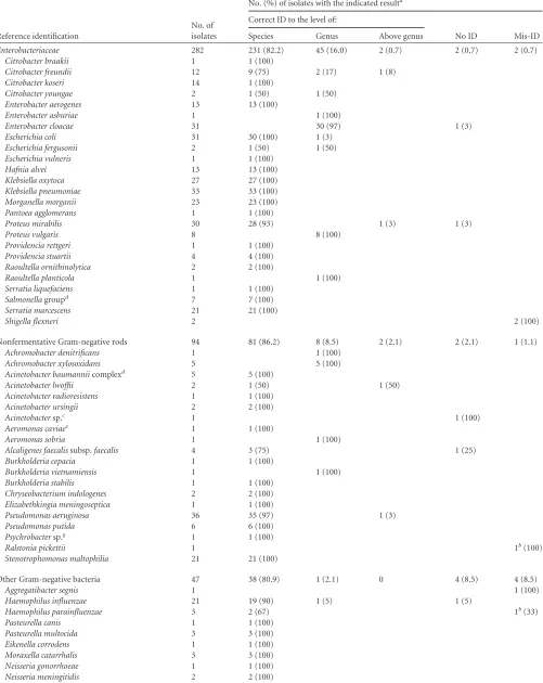
The 767 isolates included in the study encompassed 282 Enterobacte-riaceae, 94 nonfermentative Gram-negative rods, 47 other Gram-negative bacteria, 127 staphylococci and related species, 177 streptococci and re-lated species, 30 anaerobes, and 10 other Gram-positive rods (Table 1).
Reference identification and results management.Isolates were si-multaneously identified by the Vitek MS system and, as reference meth-ods, by the conventional Vitek2 system using the GP, GN, NH, or ANC card (bioMérieux) if applicable or otherwise by genomic methods. Vitek2 IDs were performed according to the recommendations of the manufac-turer, including complementary tests if required.
When the Vitek MS system proposed as a single choice or in a multiple choice the Vitek2 IDs to the species level, no further investigation was performed. In the case of discordant results between the Vitek MS and Vitek2 methods, low-discrimination results with Vitek2, or “no ID” re-sults obtained with the Vitek2 or Vitek MS methods, genomic IDs were performed and then considered to be the reference ID.
Genomic IDs were performed using the sequencing-based Mastermix 16S Complete kit (Molzym GmbH) targeting the 5=partial 16S rRNA gene as the first line. If the ID remained inconclusive, sequencing-based IDs using thesodAgene for coagulase-negative staphylococci, streptococci, and enterococci, therecAgene for theBurkholderia cepaciacomplex, and the 3=partial 16S rRNA gene for other taxa were implemented (16,17,21). Moreover, the PCR- and hybridization-based system GenoType EHEC (Hain LifeScience), detecting theipaHgene, and a serotyping method were used to confirmShigellaisolates. An optochin susceptibility test for
Streptococcus pneumoniaeisolates and a species-specific PCR targeting the
crgAgene forNeisseria meningitidisisolates were also used (23).
MALDI-TOF MS. (i)Technical training.Prior to the assessment ini-tiation, the four operators involved were trained for sample and slide preparation by performing three slides of 48 deposits with duplicate de-posits per isolate during three independent days (one slide per day). Mu-coid and rough isolates were included only in the third slide performed by each operator. A proficiency test using 16 strains with single deposits was passed by each operator.
(ii) Plate preparation.The disposable plate preparation was per-formed with the Vitek MS Preparation Station software to link sample information to the Vitek MS spectrophotometer using the single-use FlexiMass MALDI target plates, supplied in a 48-well microscope slide format, divided into three acquisition groups of 16 spots, and inoculated by picking an overnight culture with a 1-l disposable loop and by smear-ing the specimen directly onto the plate (mostly one colony/deposit). The preparations were overlaid with 1l of matrix solution (saturated solu-tion of␣-cyano-4-hydroxycinnamic acid in 50% acetonitrile and 2.5% trifluoroacetic acid) and air dehydrated for 1 to 2 min at room
tempera-ture. As recommended by the manufacturer, theEscherichia coliATCC 8739 strain, used as a calibrator and internal ID control, was inoculated on the calibration spots of each acquisition group (small spot in the middle of each acquisition group). Each bacterial isolate had been tested with a unique deposit.
(iii) Generation of mass spectra.Mass spectra were generated with a Vitek MS Axima Assurance mass spectrometer (bioMérieux) in positive linear mode at a laser frequency of 50 Hz with an acceleration voltage of 20 kV and an extraction delay time of 200 ns. For each spectrum, 500 shots in 5-shot steps from different positions of the target spot (automatic mode) were collected by the mass spectrometer operating in conjunction with the Acquisition Station software (Vitek MS version 1.0.0). Measured mass spectra ranged from 2,000 to 20,000 Da.
(iv) MS identification.For each bacterial sample, mass fingerprints were processed by the compute engine and the advanced spectrum clas-sifier (ASC) algorithm associated with the Vitek MS system, which then automatically identifies the organism by comparing the characteristics of the spectrum obtained (presence and absence of specific peaks) with those of the typical spectrum of each claimed species.
The ASC algorithm is a supervised learning method that analyzes spec-tral data and recognizes patterns used to build the knowledge base. It can be classified in the “nonprobabilistic linear classifier” family. In most cases, one pattern in the knowledge base corresponds to one species. Sometimes, when two (or more) species cannot be separated efficiently with MALDI-TOF technology, only one pattern is created by using all the spectra collected for both species. On the other hand, when spectra col-lected for one species are so variable due to strain variability or culture conditions like incubation time and culture medium, several patterns are created for one species. For each species integrated in the knowledge base, the variability of the spectra is evaluated with clustering application and interspectra distance calculation.
The spectral database was built by the manufacturer as follows. Ten isolates belonging to the same species were carefully selected to take into account diversity in clinical specimen origins, geographic origins (differ-ent countries), and year of isolation. All isolates were previously charac-terized by phenotypic and/or molecular methods. The spectral database was built through an experimental plan design, including several culture media, several media suppliers, different incubation times ranging from 18 h to 24 h to up to 72 h, and several mass spectrometers. Masses from 2 to 20 kDa were collected, and the analysis focused on the 3- to 17-kDa mass range.
The ASC algorithm compared the generated spectra to the expected spectrum of each organism or organism group of the database to provide identification. A percent probability, or confidence value, which repre-sents the similarity in terms of presence/absence of specific peaks between the generated spectrum and the database spectra, was calculated by the algorithm. A perfect match between the spectrum and the unique spec-trum of a single organism or organism group provided a confidence value of 99.9% (“good ID”). When a perfect match was not obtained, it was still possible for the spectrum to be sufficiently close to that of a reference spectrum such that a clear decision was provided about the organism ID (“good ID,” confidence value of⬎60 to 99.8%). If a unique ID pattern was not recognized, a list of possible organisms was given (“low discrimina-tion” [LD], confidence value of⬎60%) or the strain was determined to be outside the scope of the database (“no ID”). The range of percent proba-bilities in the single-choice case was 60 to 99%. Values closer to 99.9% indicate a closer match to the typical pattern for the given organism. When the confidence value obtained was below 60, the organism was considered nonidentified.
The overall correct ID was defined as including the following levels: (i) correct ID to the species level, when the system proposed the reference species ID as a single choice or with low discrimination to the subspecies level (with any level of confidence), (ii) correct ID to the genus level, when the system proposed the reference species ID among a set of low-discrim-ination results including species of the same genera, and (iii) correct ID
on May 16, 2020 by guest
http://jcm.asm.org/
TABLE 1Valid Vitek MS results of 767 bacterial isolates using a single deposit and no protein extraction step
Reference identification
No. of isolates
No. (%) of isolates with the indicated resulta
Correct ID to the level of:
No ID Mis-ID Species Genus Above genus
Enterobacteriaceae 282 231 (82.2) 45 (16.0) 2 (0.7) 2 (0.7) 2 (0.7)
Citrobacter braakii 1 1 (100)
Citrobacter freundii 12 9 (75) 2 (17) 1 (8)
Citrobacter koseri 14 1 (100)
Citrobacter youngae 2 1 (50) 1 (50)
Enterobacter aerogenes 13 13 (100)
Enterobacter asburiae 1 1 (100)
Enterobacter cloacae 31 30 (97) 1 (3)
Escherichia coli 31 30 (100) 1 (3)
Escherichia fergusonii 2 1 (50) 1 (50)
Escherichia vulneris 1 1 (100)
Hafnia alvei 13 13 (100)
Klebsiella oxytoca 27 27 (100)
Klebsiella pneumoniae 33 33 (100)
Morganella morganii 23 23 (100)
Pantoea agglomerans 1 1 (100)
Proteus mirabilis 30 28 (93) 1 (3) 1 (3)
Proteus vulgaris 8 8 (100)
Providencia rettgeri 1 1 (100)
Providencia stuartii 4 4 (100)
Raoultella ornithinolytica 2 2 (100)
Raoultella planticola 1 1 (100)
Serratia liquefaciens 1 1 (100)
Salmonellagroupd 7 7 (100)
Serratia marcescens 21 21 (100)
Shigella flexneri 2 2 (100)
Nonfermentative Gram-negative rods 94 81 (86.2) 8 (8.5) 2 (2.1) 2 (2.1) 1 (1.1)
Achromobacter denitrificans 1 1 (100)
Achromobacter xylosoxidans 5 5 (100)
Acinetobacter baumanniicomplexd 5 5 (100)
Acinetobacter lwoffii 2 1 (50) 1 (50)
Acinetobacter radioresistens 1 1 (100)
Acinetobacter ursingii 2 2 (100)
Acinetobactersp.c 1 1 (100)
Aeromonas caviaee 1 1 (100)
Aeromonas sobria 1 1 (100)
Alcaligenes faecalissubsp.faecalis 4 3 (75) 1 (25)
Burkholderia cepacia 1 1 (100)
Burkholderia vietnamiensis 1 1 (100)
Burkholderia stabilis 1 1 (100)
Chryseobacterium indologenes 2 2 (100)
Elizabethkingia meningoseptica 1 1 (100)
Pseudomonas aeruginosa 36 35 (97) 1 (3)
Pseudomonas putida 6 6 (100)
Psychrobactersp.g 1 1 (100)
Ralstonia pickettii 1 1b(100)
Stenotrophomonas maltophilia 21 21 (100)
Other Gram-negative bacteria 47 38 (80.9) 1 (2.1) 0 4 (8.5) 4 (8.5)
Aggregatibacter segnis 1 1 (100)
Haemophilus influenzae 21 19 (90) 1 (5) 1 (5)
Haemophilus parainfluenzae 3 2 (67) 1b(33)
Pasteurella canis 1 1 (100)
Pasteurella multocida 3 3 (100)
Eikenella corrodens 1 1 (100)
Moraxella catarrhalis 3 3 (100)
Neisseria gonorrhoeae 1 1 (100)
Neisseria meningitidis 2 2 (100)
(Continued on following page)
on May 16, 2020 by guest
http://jcm.asm.org/
TABLE 1(Continued)
Reference identification
No. of isolates
No. (%) of isolates with the indicated resulta
Correct ID to the level of:
No ID Mis-ID Species Genus Above genus
Neisseria subflava/flavescens/perflava 1 1 (100)
Neisseria mucosa 1 1 (100)
Campylobacter fetussubsp.fetusf 1 1 (100)
Campylobacter jejunisubsp.jejuni 3 2 (67) 1 (33)
Helicobacter pylori 3 3 (100)
Legionella pneumophila 2 2 (100)
Staphylococci and related species 127 117 (92.1) 3 (2.4) 3 (2.4) 4 (3.1) 0
Staphylococcus aureus 36 35 (97) 1 (3)
Staphylococcus capitis 9 9 (100)
Staphylococcus caprae 2 2 (100)
Staphylococcus carnosussubsp.carnosus 1 1 (100)
Staphylococcus cohniissp.cohniif 1 1 (100)
Staphylococcus epidermidis 36 35 (97) 1 (3)
Staphylococcus haemolyticus 3 2 (67) 1 (33)
Staphylococcus hominissubsp.hominis 5 5 (100)
Staphylococcus intermedius 3 3 (100)
Staphylococcus lugdunensis 9 9 (100)
Staphylococcus pseudintermedius 1 1 (100)
Staphylococcus saprophyticus 5 5 (100)
Staphylococcus schleiferi 1 1 (100)
Staphylococcus sciuri 1 1 (100)
Staphylococcus simulans 4 4 (100)
Staphylococcus warneri 6 4 (67) 2 (33)
Staphylococcus xylosus 1 1 (100)
Staphylococcus condimentic 1 1 (100)
Staphylococcus pasteuric 1 1 (100)
Micrococcus luteus/lylae 1 1 (100)
Enterococcus avium 8 5 (63) 1 (13) 2 (25)
Enterococcus casseliflavus 1 1 (100)
Enterococcus durans 1 1 (100)
Enterococcus faecalis 38 3 (100)
Enterococcus faecium 17 16 (94) 1 (6)
Enterococcus gallinarum 1 1 (100)
Streptococcus agalactiae 32 32 (100)
Streptococcus pyogenes 8 8 (100)
Streptococcus dysgalactiaesubsp.equisimilisf 8 8 (100) Streptococcus dysgalactiaesubsp.dysgalactiaef 1 1 (100)
Streptococcus canisc 1 1b(100)
Streptococcus anginosus 14 14 (100)
Streptococcus constellatus 9 8 (89) 1 (11)
Streptococcus intermedius 2 2 (100)
Streptococcus pneumoniae 19 19 (100)
Streptococcus mitis/oralis 9 9 (100)
Streptococcus gallolyticussubsp.pasteurianusf 2 2 (100)
Streptococcus parasanguinis 2 2 (100)
Streptococcus vestibularis 1 1 (100)
Streptococcus australis 1 1 (100)
Granulicatella adiacens 1 1 (100)
Vagococcus fluvialis 1 1 (100)
Anaerobes 30 25 (83) 0 1 (3) 4 (13) 0
Anaerococcus hydrogenalisc 1 1 (100)
Bacteroides vulgatus 1 1 (100)
Bacteroides fragilis 8 8 (100)
Bacteroides ovatus 1 1 (100)
Bacteroides uniformis 2 2 (100)
Clostridium difficile 5 5 (100)
Clostridium perfringensh 2 2 (100)
(Continued on following page)
on May 16, 2020 by guest
http://jcm.asm.org/
above the genus level, when the system proposed the reference species ID among a set of low-discrimination results including species of different genera.
When a human error or a poor-quality deposit occurred (including the warning messages “bad spectrum,” “not enough peaks,” “too many peaks,” and “too much background noise” or in the case of calibration/ control failure), the incriminated isolates or all the isolates of the incrim-inated acquisition group had been retested with a single deposit and the second result had been taken into account for the analysis. For informa-tive purposes, samples with “no ID” or “mis-ID” first-spot results were secondarily retested with a single spot.
Calculation of global assessment indices.For the MALDI-TOF-based identification method, positive predictive values to the genus level and to the species level, considering isolates with correct IDs to the genus level true positives and isolates with correct IDs to the species level true positives, respectively, were calculated. Misidentified isolates were consid-ered false positives. Negative predictive value, considering isolates with an absence of ID and belonging to species not included in the database true negatives and isolates with an absence of ID and belonging to species included in the database false negatives, was calculated.
RESULTS
Technical practicability.During the study, one out of 48 spot calibrations performed failed and 2.2% of all generated spectra were uninterpretable. These latter errors corresponded to seven “too many/not enough peaks” and 10 “bad spectrum” warning messages and were not associated with any particular taxonomic
group or colonial characteristics. Fifteen out of these 17 isolates were correctly identified to the species level with a second deposit. OneStaphylococcus epidermidisisolate and oneKlebsiella oxytoca
isolate again generated a bad spectrum and then were discarded from the analysis.
Global identification performances.During the study period, 767 isolates were analyzed by the Vitek MS system and, in parallel, by the conventional Vitek2 system (760 isolates, 99.1%) or di-rectly by genotypic methods (7 isolates, 0.9%, forLegionella pneu-mophila,Helicobacter pylori, andBacillus cereus/thuringiensis/my-coidesspecies). Implementation of DNA-based ID methods to manage discrepancies or to obtain a more accurate reference ID (to the species or subspecies level) was performed for 79 (10.3%) isolates. Reference IDs proposed by the Vitek MS as a single choice (SC), whatever the confidence value, or included in a multiple-choice result (LD with up to four proposed species) were considered overall correct IDs. Among the 767 isolates, including 124 species and 50 genera, 738 (96.2%) isolates were correctly identified by the MALDI-TOF MS system as defined previously (Table 1). No IDs and discor-dant results (mis-IDs) were obtained for 2.5% and 1.3% of the iso-lates, respectively.
[image:5.585.41.550.84.304.2](i) Correct identifications.A correct ID to the species or sub-species level was obtained for 86.7% (n⫽665) of the isolates, with confidence values of 99.9%, 90.0% to 99.8%, and 80.0% to 89.9% for 97.6%, 2.0%, and 0.3% of these isolates, respectively. A correct
TABLE 1(Continued)
Reference identification
No. of isolates
No. (%) of isolates with the indicated resulta
Correct ID to the level of:
No ID Mis-ID Species Genus Above genus
Clostridium celerecrescensc 1 1 (100)
Finegoldia magna 1 1 (100)
Prevotella intermedia 2 2 (100)
Prevotella nanceiensisc 1 1 (100)
Prevotella nigrescensc 1 1 (100)
Propionibacterium acnes 2 2 (100)
Propionibacterium avidum 1 1 (100)
Veillonella parvula 1 1 (100)
Other Gram-positive rods 10 4 (40) 3 (30) 0 2 (20) 1 (10)
Corynebacterium amycolatum 1 1 (100)
Corynebacterium striatum 4 4 (100)
Corynebacterium fastidiosum/segmentosumc 1 1 (100)
Corynebacterium macginleyic 1 1 (100)
Lactobacillus rhamnosus 1 1 (100)
Bacillus cereus/thuringiensis/mycoidesd 2 2 (100)
Total 767 665 (86.7) 63 (8.2) 10 (1.3) 19 (2.5) 10 (1.3)
aID, identification; mis-ID, misidentification; species, correct identification at the species level (single choice or low discrimination at the subspecies level); genus, correct
identification at the genus level (low discrimination at the species level); above genus, correct identification proposed among a set of low-discrimination results including species of different genera.
b
LD discrepancies (sets of low-discrimination results not including the reference species).
cSpecies absent from the Vitek MS database. Using sequencing-based genomic methods and according to nucleotide public databases, theAcinetobactersp. isolate corresponded to
an unnamedAcinetobacterspecies.
dThe species group is the final Vitek MS identification. The subspecies or species included in each species group are as follows: for theSalmonellagroup,S.entericasubsp.enterica,
S. entericaserovar Enteritidis,S. entericaserovar Paratyphi B,S. entericaserovar Paratyphi C,S. entericaserovar Typhimurium, andSalmonellaspp.; for theAcinetobacter baumannii complex,A. baumannii,A. calcoaceticus,Acinetobactergenomospecies 3, andAcinetobactergenomospecies TU13.
e
TheAeromonas hydrophila/caviae species group is displayed as a species group result by the Vitek MS.
fThe Vitek MS did not differentiate between the subspeciesS. cohniisubsp.cohniiandS. cohniisubsp.urealyticum,S. dysgalactiaesubsp.dysgalactiaeandS. dysgalactiaesubsp.
equisimilis,S. gallolyticussubsp.pasteurianusandS. gallolyticussubsp.gallolyticus, andC. fetussubsp.fetusandC. fetussubsp.venerealis.
gThis isolate was identified asPsychrobacter phenylpyruvicusby the Vitek MS system, but thePsychrobacterspecies was undeterminable by sequencing. h
These isolates do not possess the epsilon toxin gene.
on May 16, 2020 by guest
http://jcm.asm.org/
ID to the genus level only, that is, the correct species ID was in-cluded in a multiple-choice result of species from the same genus, was obtained for 8.2% (n⫽63) of the isolates. These LD results to the species level proved to be recurrent in 79.3% of cases, includ-ing species complexes such asEnterobacter cloacae/asburiae(n⫽
31),Proteus vulgaris/penneri(n⫽8),Achromobacter xylosoxidans/
denitrificans(n⫽6),Bacillus cereus/thuringiensis/mycoides(n⫽
2), andStaphylococcus intermedius/pseudintermedius(n⫽3). LD results above the genus level, that is, with the correct ID proposed among species of different genera, were obtained for 1.3% (n⫽
10) of the isolates (Table 2), some of which also seem to be recur-rent, like theStaphylococcus warneri/Prevotella buccalisLD result for some S. warneriisolates. An identical and high confidence value was mostly obtained for each proposed species in the case of an LD result to the species level or above the genus level. In the few cases in which a confidence value difference occurred, it argued either for or against the correct species ID.
(ii) Incorrect identifications.Ten isolates were misidentified by the Vitek MS system, six of them with the correct or closed
genus (Table 3). TwoShigellaisolates were misidentified asE. coli, even after retest. OneAggregatibacter segnisisolate (a spe-cies formerly belonging to theHaemophilusgenus), misidenti-fied asHaemophilus influenzae, was correctly identified with a second deposit, whereas oneNeisseria mucosaisolate was mis-identified as its closely related speciesNeisseria subflavatwice. Belonging to species absent from the database, one Streptococ-cus australisisolate was misidentified asStreptococcus parasan-guinis, even after one retest, and oneStreptococcus canisisolate gave an LD result between other species of the “pyogenic” group and a correct “no ID” result with a second deposit.
OneCampylobacter jejuniisolate and oneLactobacillus rham-nosusisolate were misidentified as the distant speciesCitrobacter braakiiandPropionibacterium avidum, respectively, but were cor-rectly identified with a second deposit. With an LD result between species of distant genera, the misidentifiedHaemophilus parain-fluenzaeandRalstonia pickettiiisolates were correctly identified to the species level with a second deposit.
[image:6.585.38.547.79.258.2](iii) No identifications.The Vitek MS system gave an absence
TABLE 2Correct identifications proposed by the Vitek MS among multiple-choice results including species of different generaa
Reference ID
Proposed ID results (confidence value [%])
1st choice 2nd choice 3rd choice 4th choice
Acinetobacter lwoffii Acinetobacter lwoffii(99.9) Mycobacterium tuberculosis
(99.9)
Mycobacterium bovis(99.9)
Citrobacter freundii Citrobacter freundii(99.0) Citrobacter braakii(99.9) Citrobacter koseri(92.4) Haemophilus influenzae
(71.5)
Enterococcus avium Enterococcus avium(99.9) Clostridium butyricum(96.6)
Enterococcus avium Enterococcus avium(99.9) Clostridium butyricum(85.9) Bacillus atrophaeus(78.8)
Micrococcus luteus Micrococcus luteus/lylae(99.9) Bacillus thuringiensis(99.9) Bacillus mycoides(79.1) Bacillus cereus(79.1)
Propionibacterium avidum Propionibacterium avidum(96.8) Clostridium butyricum(87.7)
Proteus mirabilis Proteus mirabilis(99.9) Streptococcus constellatus(71.3)
Pseudomonas aeruginosa Pseudomonas oryzihabitans
(99.4)
Peptoniphilus indolicus(99.2) Pseudomonas aeruginosa(82.0)
Staphylococcus epidermidis Staphylococcus epidermidis(99.9) Leuconostoc pseudomesenteroides
(99.7)
Staphylococcus warnerib Staphylococcus warneri(99.9) Prevotella buccalis(99.9) aID, identification.
b
2 isolates.
TABLE 3Misidentifications of the Vitek MS systema
Reference ID
Proposed IDs (confidence value [%]) for first spot as single or multiple choice
ID result for second spot 1st choice 2nd choice 3rd choice
Aggregatibacter segnis Haemophilus influenzae(99.4) Aggregatibacter segnis(99.9)
Campylobacter jejuni Citrobacter braakii(84.4) Campylobacter jejuni(99.9)
Haemophilus parainfluenzae Haemophilus haemolyticus
(98.8)
Enterobacter aerogenes(93.5) Haemophilus parainfluenzae
(99.9)
Lactobacillus rhamnosus Propionibacterium avidum
(99.3)
Lactobacillus rhamnosus(99.9)
Neisseria mucosa Neisseria subflava(99.9) Neisseria subflava(99.9)
Ralstonia pickettii Prevotella melaninogenica(99.9) Staphylococcus saprophyticus
(99.9)
Chryseobacterium gleum
(96.2)
Ralstonia pickettii(99.9)
Shigella flexnerib Escherichia coli(99.9) E. coli(99.9)
Streptococcus australis Streptococcus parasanguinis
(99.9)
Streptococcus parasanguinis(99.9)
Streptococcus canis Streptococcus dysgalactiaesubsp.
dysgalactiae(99.9)
Streptococcus dysgalactiaesubsp.
equisimilis(99.9)
Streptococcus equisubsp.
equi(99.9)
No ID
a
ID, identification.
b2 isolates.
on May 16, 2020 by guest
http://jcm.asm.org/
[image:6.585.44.545.536.708.2]of ID for 19 (2.5%) isolates that were tested again using one de-posit for informative purposes (Table 1). Nine isolates belonging to nine species were not included in the database of the MALDI-TOF MS system, including oneAcinetobactersp. (unnamed spe-cies in public nucleotide databases), two staphylococci, two co-rynebacteria, and four anaerobe isolates, for which the system gave the same “no ID” answer after reading a second deposit. With the analysis of an additional deposit, six out of 10 isolates with species included in the database were correctly identified. One
Staphylococcus haemolyticus isolate among three tested in the study and threeHelicobacter pyloriisolates again gave a “no ID” result despite the additional retest.
(iv) Global assessment indices.According to the criteria de-tailed in Materials and Methods, the positive predictive values to the genus level and to the species level of the Vitek MS system were 98.6 and 98.5, respectively, and the negative predictive value was 47.4.
Analysis of theStreptococcaceaegroup.Considering the or-ganism groups largely tested, the Vitek MS gave a good ID overall to the genus level with a single deposit for 98.2% of the Enterobac-teriaceae, 94.7% of the nonfermentative Gram-negative rods, 94.5% of the staphylococci, and 97.2% of the streptococci and related isolates (Table 1), with some species of the last group re-ported to be difficult to discriminate using MALDI-TOF MS sys-tems. Focusing on the 177 isolates belonging to the Streptococ-caceaefamily that were tested, 93.9% of the enterococci (62 out of 66 isolates, including six species) and 98% of the pyogenic strep-tococci (49 out of 50 isolates, including four species) were cor-rectly identified to the species level with a single deposit (Table 1). Of note, the LD results to the subspecies level seem to be recurrent forStreptococcus dysgalactiaesubsp.dysgalactiaeandS. dysgalac-tiaesubsp.equisimilis(n⫽9), probably due to the resolution limit of the settled system. Among the milleri group streptococci, 24 out of 25 isolates were correctly identified to the species level and one LD result was obtained for aStreptococcus constellatusisolate with
Streptococcus anginosus, whereas five isolates were identified as other alpha- or nonhemolytic streptococci by the Vitek2 system. The Vitek MS system correctly identified to the species level all 19
S. pneumoniaeisolates and 15 out of 17 alpha- or nonhemolytic
Streptococcaceaeisolates, including nineStreptococcus mitis/oralis
isolates. OneS. australisisolate, a species not included in the Vitek MS database, was misidentified asS. parasanguinis, and one Strep-tococcus vestibularisisolate was not discriminated from the closed
Streptococcus salivariussubsp.salivariusspecies. In contrast, the Vitek2 system misidentified two alpha-hemolytic streptococci isolates as milleri group streptococci.
DISCUSSION
The introduction of the high-tech MALDI-TOF MS technology in clinical laboratories is reducing the time required while improving the accuracy of bacterial identification. Without an intensive training background of the operators, the technical ownership of the Vitek MS system is straightforward and fast, as previously mentioned (5). However, the operator must remain vigilant in routine practice during sample preparation because of reduced interspot distances (especially for spots near theE. colicalibrant spot) that can mix two bacterial deposits, particularly during the matrix application step, as happened during the training period.
Many authors evaluating other MALDI-TOF MS-based sys-tems have previously reported the use of a formic acid-based
pro-tein extraction using a bacterial lysis step or directly on the bacte-rial smear before matrix application to be needed, mainly for Gram-positive bacteria (1,5,9,15,24). In this study, the use of the Vitek MS system generated a low frequency of unusable spectra without formic acid use that is compatible and convenient for routine practice. Only oneStaphylococcus epidermidisisolate and oneKlebsiella oxytocaisolate twice generated bad spectra; in rou-tine practice, these two isolates should have then been managed using a formic acid extraction step. These good performances of Vitek MS spectral acquisition for both negative and Gram-positive isolates may be due to the efficient displacement raster of the laser on the deposit. The laser scans the entire sample, and the instrument acquires good-quality subspectra from each 5-shot step. When 100 good-quality subspectra are not reached during the first large screening of the deposit, the laser goes back onto sample areas giving good-quality subspectra to obtain sufficient data (30 is the minimum acceptable number of subspectra) before the average spectrum data are analyzed.
LD results between species of different genera, accounting for 14 (1.8%) of the tested isolates, were not reported in the literature for other MALDI-TOF MS-based ID systems. The basis of this phenomenon may lie in the spectrum classifier algorithm that takes into account the absence and the presence of species-specific peaks. Although most of the correct species IDs included in such LD results can be found out with growth conditions or by simple and immediate tests (catalase, Gram staining, pigmentation), the correct species was proposed for only 11 out of 14 isolates with these LDs; a correct result (species ID or “no ID”) was obtained for the three other isolates using a second spot. As a consequence and according to our subsequent experience, these few LD results should not be removed by complementary tests, but the isolates should be retested in order to obtain a single choice. Moreover, whatever the level of the LD results, confidence values do not appear to be reliable to determine the right species. Nevertheless, recurrent LD results to the species level have mostly no impact on isolate management and bioclinical interpretation, since species show similar pathogenicities and antibiotics susceptibility pat-terns (for example,E. cloacae/asburiae) and/or can be discrimi-nated by simple and immediate tests (for example, indole test for
P. vulgaris/penneri). For other LD results to the species level, an additional deposit should also be performed, as it mostly provided the correct species ID during our subsequent experience.
The mis-ID ofShigellaisolates as E. coli was previously re-ported with other MALDI-TOF MS-based ID systems (4–6,15, 20). These results are not surprising since the genusShigella be-longed genetically to theE. colispecies and was kept in this way to differentiate these “specificE. coli” isolates with a particular viru-lence toward humans (12). This point reflects the resolution limits of the MALDI-TOF MS-based ID method currently used for rou-tine bacterial IDs. These mis-IDs are a major drawback from a clinical point of view, particularly for stool sample analysis, that needs to be overcome by conventional phenotypic testing. Re-garding the other major mis-IDs (Campylobacter jejuni, Lactoba-cillus rhamnosus, andAggregatibacter segnisisolates), one cannot rule out a technical mistake or an undetected mixed culture gen-erating two superimposed spectra that were wrongly inter-preted as a unique spectrum and, thus, as a third species. These major incorrect results could have been amended in routine practice according to Gram staining, growth conditions, colo-nial features, and oxidase/catalase tests. These few but
on May 16, 2020 by guest
http://jcm.asm.org/
theless critical mis-IDs confirmed that, like any identification system, experienced laboratory personnel have to manage Vi-tek MS results and take into account bacterial and clinical data, as highlighted by other authors using different MALDI-TOF MS-based systems (4,15,22).
Nine isolates belonging to nine species not included in the Vitek MS database twice obtained the same “no ID” answer, high-lighting the specificity of the algorithm used in these bacterial groups. Two out of fourCorynebacteriumspecies and four out of 15 anaerobic species collected in our routine practice were not identified. The lack of species diversity of corynebacteria and an-aerobes in the currently available database should be addressed for overall routine use. As noted by other authors, our results empha-size the widespread ignorance and failure to correctly identify an-aerobic species by biochemical methods in medical bacteriology (10, 14, 25, 26). Moreover, from microbiologist views and for clinical purposes, the ID to the genus level of many anaerobes is usually sufficient, but that is unfortunately not achievable with the original algorithm of the tested system.
With the analysis of an additional deposit, six out of 10 isolates with species included in the database were correctly identified. As mentioned for another MALDI-TOF MS-based bacterial ID sys-tem, these results may indicate that deposition of an excessive amount of bacteria during sample spot preparation can lead to a loss of accuracy of the Vitek MS system, providing quality spec-trum warning messages or “no ID” results (5). As a consequence, all isolates with a “no ID” result given by the Vitek MS using one deposit should be retested in routine practice. The absence of IDs for oneS. haemolyticusisolate and all threeH. pyloriisolates tested twice may be due to a lack of species diversity among the 10 ref-erence spectra embedded in the database or the need for a prior protein extraction.
The “good ID” performances for the largely tested Enterobac-teriaceae, nonfermentative Gram-negative rod, staphylococci, and streptococci groups using only one deposit and no extraction step were reported previously in only one study (4). In addition to the spectral acquisition step, the good results obtained with the Vitek MS system may be due to the database building using 10 different reference strains for each species and generating different spectra for each strain under different culture conditions in order to set its typical spectrum. A previous report has noted that including at least 10 strains per species in the database with many replicates per strain is a prerequisite to obtain an accurate MALDI-TOF MS ID (20). By allowing quick and reliable IDs ofStreptococcaceae iso-lates, the Vitek MS showed better ID performances for milleri group streptococci and other alpha- or nonhemolytic Streptococ-caceaethan did the Vitek2 system. In contrast to previous studies using other MALDI-TOF MS systems and although our results need to be confirmed on a larger number of isolates, the Vitek MS appears to particularly discriminateS. pneumoniae, an undeniable pathogenic species, from other alpha-hemolytic streptococci (3,4, 6,7,15,18,20,22,24).
In conclusion, the Vitek MS system allows a fast and reliable acquisition of bacterial ID for most bacterial species isolated rou-tinely in a medical laboratory with only one deposit of crude bac-teria and without any extraction step. The remarkable perfor-mance of the Vitek MS system may be due to its novel laser displacement mode, original algorithm, and quality of the data-base building. It is worth noting that the analysis of the presence and the absence of specific peaks by the spectrum classifier
algo-rithm of the Vitek MS is double edged. When the tested species is missing from the database, the algorithm gives a clear absence of ID in most cases, a result which is overall what is expected for an ID system. However, due to the limited species diversity of the database for some taxa groups, as for some anaerobe taxa, the absence of ID results to the genus level is unfortunate and requires the use of a conventional identification system. For routine pur-poses, in addition to the isolates that generated a spectrum of poor quality, we suggested a retest with one to two deposits for isolates that give “no ID” and “no recurrent” LD results with a first de-posit, as false “no ID” results can be obtained with deposits of poor quality and the confidence values are indicators with weak useful-ness in cases of LD, respectively. Considering the higher bacterial diversity included in this study than in routine practice and the low prevalence of species without correct identification, the per-formances should be even better for routine activity in clinical laboratories. However, expanding the spectral database is war-ranted, particularly for anaerobic, coryneform, and some highly pathogenic bacteria, in order to use almost exclusively this system for isolate IDs in routine medical practice.
ACKNOWLEDGMENTS
Funding for this study was provided by bioMérieux.
We thank the clinical microbiology laboratory technologists for their help in collecting isolates for this study and Françoise Bertin, Brigitte Gras-Potier, Brigitte Rigonnet, Véronique Tudal, and Emmanuel Cartron for helpful technical assistance.
REFERENCES
1.Alatoom AA, Cunningham SA, Ihde SM, Mandrekar J, Patel R.2011. Comparison of direct colony method versus extraction method for iden-tification of Gram-positive cocci by use of Bruker Biotyper matrix-assisted laser desorption ionization–time of flight mass spectrometry. J. Clin. Mi-crobiol.49:2868 –2873.
2.Anhalt JP, Fenselau C.1975. Identification of bacteria using mass spec-trometry. Ann. Chem.47:219 –225.
3.Benagli C, Rossi V, Dolina M, Tonolla M, Petrini O. 2011. Matrix-assisted laser desorption ionization-time of flight mass spectrometry for the identification of clinically relevant bacteria. PLoS One6:e16424. doi: 10.1371/journal.pone.0016424.
4.Bille E, et al.2011. MALDI-TOF MS Andromas strategy for the routine identification of bacteria, mycobacteria, yeasts,Aspergillusspp. and posi-tive blood cultures. Clin. Microbiol. Infect. doi:10.1111/j.1469-0691.2011.03688.x.
5.Bizzini A, Durussel C, Bille J, Greub G, Prod’hom G.2010. Performance of matrix-assisted laser desorption ionization–time of flight mass spec-trometry for identification of bacterial strains routinely isolated in a clin-ical microbiology laboratory. J. Clin. Microbiol.48:1549 –1554. 6.Blondiaux N, Gaillot O, Courcol RJ.2010. MALDI-TOF mass
spectrom-etry to identify clinical bacterial isolates: evaluation in a teaching hospital in Lille. Pathol. Biol. (Paris)58:55–57. (In French.)
7.Cherkaoui A, et al.2010. Comparison of two matrix-assisted laser de-sorption ionization–time of flight mass spectrometry methods with con-ventional phenotypic identification for routine identification of bacteria to the species level. J. Clin. Microbiol.48:1169 –1175.
8.Emonet S, Shah HN, Cherkaoui A, Schrenzel J.2010. Application and use of various mass spectrometry methods in clinical microbiology. Clin. Microbiol. Infect.16:1604 –1613.
9.Haigh J, Degun A, Eydmann M, Millar M, Wilks M.2011. Improved performance of bacterium and yeast identification by a commercial ma-trix-assisted laser desorption ionization–time of flight mass spectrometry system in the clinical microbiology laboratory. J. Clin. Microbiol.49:3441. 10. Justesen US, et al.2011. Species identification of clinical isolates of an-aerobic bacteria: a comparison of two matrix-assisted laser desorption ionization–time of flight mass spectrometry systems. J. Clin. Microbiol. 49:4314 – 4318.
11. Kallow W, Erhard M, Shah HN, Raptakis E, Welker M.2010.
on May 16, 2020 by guest
http://jcm.asm.org/
TOF MS for microbial identification: years of experimental development to an established protocol, p 255–276.InShah HN, Gharbia SE (ed), Mass spectrometry for microbial proteomics. John Wiley & Sons, Ltd, Chich-ester, United Kingdom.
12. Kaper JB, Nataro JP, Mobley HL.2004. PathogenicEscherichia coli. Nat. Rev. Microbiol.2:123–140.
13. Karas M, Hillenkamp F.1988. Laser desorption ionization of proteins with molecular masses exceeding 10,000 daltons. Anal. Chem.60:2299 – 2301.
14. La Scola B, Fournier PE, Raoult D.2011. Burden of emerging anaerobes in the MALDI-TOF and 16S rRNA gene sequencing era. Anaerobe17: 106 –112.
15. Neville SA, et al.2011. Utility of matrix-assisted laser desorption ioniza-tion–time of flight mass spectrometry following introduction for routine laboratory bacterial identification. J. Clin. Microbiol.49:2980 –2984. 16. Poyart C, Quesne G, Boumaila C, Trieu-Cuot P. 2001. Rapid and
accurate species-level identification of coagulase-negative staphylococci by using thesodAgene as a target. J. Clin. Microbiol.39:4296 – 4301. 17. Prere MF, Fayet OA.2011. A specific polymerase chain reaction test for
the identification of Streptococcus pneumoniae. Diagn. Microbiol. Infect. Dis.70:45–53.
18. Risch M, et al.2010. Comparison of MALDI TOF with conventional identification of clinically relevant bacteria. Swiss Med. Wkly. 140: w13095. doi:10.4414/smw.2010.13095.
19. Sauer S, Kliem M.2010. Mass spectrometry tools for the classification and identification of bacteria. Nat. Rev. Microbiol.8:74 – 82.
20. Seng P, et al.2009. Ongoing revolution in bacteriology: routine identifi-cation of bacteria by matrix-assisted laser desorption ionization time-of-flight mass spectrometry. Clin. Infect. Dis.49:543–551.
21. Slinger R, et al.2007. Pyrosequencing of arecAgene variable region for
Burkholderia cepaciacomplex genomovar identification. Diagn. Micro-biol. Infect. Dis.58:379 –384.
22. Szabados F, et al.2012. Evaluation of species-specific score cutoff values of routinely isolated clinically relevant bacteria using a direct smear prep-aration for matrix-assisted laser desorption/ionization time-of-flight mass spectrometry-based bacterial identification. Eur. J. Clin. Microbiol. Infect. Dis.31:1109 –1119.
23. Taha MK.2000. Simultaneous approach for nonculture PCR-based iden-tification and serogroup prediction ofNeisseria meningitidis. J. Clin. Mi-crobiol.38:855– 857.
24. van Veen SQ, Claas EC, Kuijper EJ.2010. High-throughput identifica-tion of bacteria and yeast by matrix-assisted laser desorpidentifica-tion ionizaidentifica-tion– time of flight mass spectrometry in conventional medical microbiology laboratories. J. Clin. Microbiol.48:900 –907.
25. Veloo AC, Erhard M, Welker M, Welling GW, Degener JE. 2011. Identification of Gram-positive anaerobic cocci by MALDI-TOF mass spectrometry. Syst. Appl. Microbiol.34:58 – 62.
26. Veloo AC, Welling GW, Degener JE.2011. The identification of anaer-obic bacteria using MALDI-TOF MS. Anaerobe17:211–212.