0095-1137/09/$08.00⫹0 doi:10.1128/JCM.00366-09
Copyright © 2009, American Society for Microbiology. All Rights Reserved.
Detection of
Enterobacter sakazakii
and Other Pathogens Associated
with Infant Formula Powder by Use of a DNA Microarray
䌤
†
Min Wang,
1,2,3,4‡ Boyang Cao,
1,2,3,4‡ Qili Gao,
6Yamin Sun,
1,2,3,4Pei Liu,
6Lu Feng,
1,2,3,4,5and Lei Wang
1,2,3,4,5*
TEDA School of Biological Sciences and Biotechnology, Nankai University, 23 HongDa Street, TEDA, Tianjin 300457, China1;
Key Laboratory of Molecular Microbiology and Technology of the Ministry of Education, Nankai University, Tianjin 300071,
China2; Tianjin State Laboratory of Microbial Functional Genomics, 23 HongDa Street, TEDA, Tianjin 300457, China3;
Tianjin Research Center for Functional Genomics and Biochips, 23 HongDa Street, TEDA, Tianjin 300457,
China4; Tianjin Biochip Corporation, 23 HongDa Street, TEDA, Tianjin 300457, China5; and
Tianjin Entry-Exit Inspection and Quarantine Bureau, 8 ZhaoFa Residential Quarter,
2nd St., TEDA, Tianjin 300457, China6
Received 18 February 2009/Returned for modification 4 May 2009/Accepted 23 July 2009
Pathogen detection is critical to the process of generating and testing powdered infant formula (PIF). An obstacle associated with PIF microbial surveillance is that most current procedures are time-consum-ing and labor-intensive. We have developed a rapid, DNA microarray-based detection technique to identify 10 different pathogenic bacteria associated with PIF contamination based on the 16S–23S rRNA gene
internal transcribed spacer (ITS) sequences andwzy(O antigen polymerase) gene. Using this procedure,
Enterobacter sakazakii, Salmonella enterica, Klebsiella pneumoniae, Klebsiella oxytoca, Serratia marcescens,
Acinetobacter baumannii,Bacillus cereus,Listeria monocytogenes,Staphylococcus aureus, andEscherichia coli
O157 were identified. One hundred eighty-five strains were used to validate the microarray assay (includ-ing 134 target pathogen strains and 51 closely related bacteria). Twenty-seven probes reproducibly
detected multiple pathogens with high specificity and sensitivity (0.100 ng genomic DNA or 104CFU/ml).
Twenty-one real PIF samples were tested by the microarray with 100% accuracy. The data presented reveal that the designed oligonucleotide microarray is a promising method for basic microbiology, clinical diagnosis, food safety, and epidemiological surveillance.
Pathogen detection is a critical parameter linked to the safety of powdered infant formula (PIF). Because PIF effec-tively supports the growth of numerous pathogens, it can be-come easily contaminated (9, 15). Various studies examining PIF contamination have identified various pathogenic bacteria (9, 16, 22); e.g., powdered milk produced by Wyeth (in 2002) was contaminated with Enterobacter sakazakii, which led to fatality rates of 33 to 80% in infected children (18). In 2005, an outbreak associated with Salmonella-contaminated PIF in France affected more than 141 children (4). These and other similar episodes have prompted research aimed at improving pathogen detection to guarantee PIF safety.
FAO/WHO Expert Consultations (held in 2004 and 2006) concluded that the primary microorganisms associated with PIF contamination wereE. sakazakii,Salmonella enteritidis,
Enterobacter agglomerans, Hafnia alvei, Klebsiella pneumoniae,
Citrobacter koseri,Citrobacter freundii,Klebsiella oxytoca,
Entero-bacter cloacae,Escherichia coli, Serratiaspp.,Acinetobacterspp.,
Bacillus cereus, Clostridium difficile, Clostridium perfringens,
Clostridium botulinum,Listeria monocytogenes,Staphylococcus
aureus, and coagulase-negative staphylococci (http://www.who
.int/foodsafety/publications/micro/mra10/en/index.html).
Ente-robacter agglomerans, Enterobacter cloacae, Hafnia alvei, and
coagulase-negative staphylococci (normally referring to
Staph-ylococcus epidermidisandStaphylococcus saprophyticus), whose
pathogenicity is weak, were rarely isolated by the Entry-Exit Inspection and Quarantine Bureau.Clostridium difficile,
Clos-tridium perfringens, andClostridium botulinumare anaerobes,
and both their cultivation and strain collection are difficult. The internal transcribed spacers (ITS) ofCitrobacter koseriand
Citrobacter freundiiare not available. Therefore, in this report,
10 important and often-isolated pathogens,E. sakazakii,
Sal-monella enterica,K. pneumoniae,K. oxytoca,S. marcescens,A.
baumannii,B. cereus,L. monocytogenes,S. aureus, andE. coli
O157, were employed to design a microarray-based approach for the detection of PIF-associated pathogens.
Conventional methods for the detection of these pathogens involve their isolation in pure culture combined with biochem-ical tests that often are laborious, time-consuming, and difficult to quantify (6); however, the use of molecular methods, such as PCR, real-time PCR, and immunoassays, has facilitated patho-gen detection (2). Despite these improvements, only the de-tection ofE. sakazakiiandSalmonellaspecies in PIF have been reported (17). Recently, DNA microarray-based assays have been introduced and developed as potential strategies for fa-cilitating the high-throughput and specific screening of patho-gen-associated DNA sequences (3).
In this study, we targeted the detection of the 16S–23S rRNA gene ITS regions for nine pathogen targets and the O
* Corresponding author. Mailing address: TEDA School of Biolog-ical Sciences and Biotechnology, Nankai University, 23 HongDa St., TEDA, Tianjin 300457, China. Phone: 66229588. Fax: 86-22-66229596. E-mail: [email protected].
† Supplemental material for this article may be found at http://jcm .asm.org/.
‡ These authors contributed equally to this report.
䌤Published ahead of print on 29 July 2009.
3178
on May 16, 2020 by guest
http://jcm.asm.org/
unit-processing genewzy (O antigen polymerase) for E. coli O157. A total of 185 bacterial strains were used to validate the microarray assay. Twenty-seven specific probes and two primer pairs reproducibly and specifically were used to identify bac-terial genomic DNA in samples with as little as 0.1 ng DNA or 104CFU/ml for pure culture.
MATERIALS AND METHODS
Bacterial strains.The target bacterial strains tested in this study are described in Table 1.Listeriaspecies strains were cultured in trypticase soy-yeast extract broth (TSB-YE) medium, and the other were inoculated into 2YT medium. All strains were grown overnight at 37°C with shaking.
Preparation of PIF samples.For mock samples, respective bacterial cultures were serially diluted, and 100
CFU was mixed with 100 g of PIF and 900 ml of 2YT medium (L. monocytogeneswas mixed with 900 ml of TSB-YE medium); for test samples, only 100 g of PIF and 900 ml medium were mixed. The mixture was incubated at 37°C for 5 h, and then 10 ml was used to inoculate 100 ml of enterobacteria enrichment broth and meat infusion broth (L. monocytogeneswas inoculated in Fraser medium). These three selective media were incubated at 37°C overnight with shaking, and genomic DNA then was extracted from 1.5 ml of the overnight cultures.
DNA isolation.Genomic DNA was extracted using the Bacteria Genomic DNA Purification kit (Tiangen Biotech Co., Ltd., Beijing, China) according to the manufacturer’s protocol.
Primer and probe design.The primer pair wl-5793 (5⬘-TGT ACA CAC CGC CCG TC-3⬘) and wl-5794 (5⬘-GGT ACT TAG ATG TTT CAG TTC-3⬘), which is specific to the ITS region, was designed as previously described (29). Thewzy
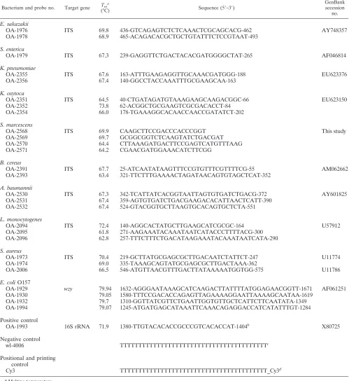
primer pair wl-5593 (5⬘-TCA GCG GCT AAG TTG ATT-3⬘) and wl-5594 (5⬘-ATT TGC TCC CAT GTC TCC-3⬘) was designed using Primer Premier 5.0 (Premier Biosoft International, Palo Alto, CA). The ITS probe specificity was examined by comparing probe sequences with the sequences in GenBank and our sequencing database using BLAST. Thewzyprobes were designed by Oli-goArray 2.0 software and were based on sequences available in GenBank. For each bacterial target, one to four specific capture probes were designed. In addition, one probe, based on the conserved region of 16S rRNA gene, was used as the positive control, one probe containing a 40-poly(T) oligonucle-otide tail was used as the negative control, and one 3⬘-Cy3-labeled probe was used as the positional reference and printing control. Each probe was chem-ically synthesized and 5⬘-amino modified with a space linker of 15 poly(T) oligonucleotides. All of the probes are listed in Table 2.
Serratiasp. ITS sequences.PCR amplicons were cloned into the pGEM-T Easy vector (Promega, MA) and transformed intoE. coliDH5␣. Transformants (white colonies grown on an ampicillin plates containing isopropyl-beta-D -thio-galactopyranoside and 5-bromo-4-chloro-3-indolyl-aˆ-D-galactopyranoside) were selected randomly. Plasmid DNA was isolated using the conventional alkaline lysis method, digested with EcoRI, and visualized on agarose gels to confirm the presence of the corresponding inserts. Sequences were verified using an ABI 3730 automated DNA sequencer. Seven to 16 transformants per strain were examined.
Target DNA amplification and labeling.The primer concentrations were op-timized based on the final intensity of the microarray hybridization signals. Duplex PCR mixtures contained 1⫻PCR buffer (50 mM KCl, 10 mM Tris-HCl, pH 8.3), 2.5 mM MgCl2, 400M deoxynucleoside triphosphates, 0.15M ITS, 0.2M primer for eachwzygene, 2.5 UTaqDNA polymerase, and 50 to 100 ng of DNA template in a final volume of 50l. PCR conditions used consisted of an initial denaturation at 95°C for 5 min, followed by 30 cycles of 95°C for 30 s, 50°C for 30 s, and 72°C for 1 min, with a final extension at 72°C for 5 min. PCR products were purified with the Microcon Centrifugal Filter Devices kit (Millipore Corporation, MA). To label the PCR products, the two reverse primers were used during the PCR, and 0.3l of 25 nM Cy3-dUTP was added. Ten microliters of the purified products generated from the duplex PCR described above was added as the template, and PCR was carried out as described above.
Microarray preparation. Probes were dissolved in 50% dimethylsulfoxide (DMSO) to a final concentration of 1g/l and coated onto aldehyde group-modified glass slides (CapitalBio Corporation, Beijing, China) using a Spot-Array72 (Perkin-Elmer Corporation, CA). Each probe was spotted in triplicate, and coated slides were dried and stored at room temperature in the dark. Each glass slide contained eight individual arrays framed with an 8-sample cover slip that constituted individual reaction chambers (see Fig. S1 in the supplemental material).
Microarray hybridization and data analysis.All labeled PCR products were precipitated using 100% cold ethanol, centrifuged at 13,000⫻gfor 10 min, washed with 75% ethanol, and dried at room temperature. The dried, labeled DNA was diluted in 16l hybridization buffer (25% formamide, 0.1% sodium dodecyl sulfate [SDS], 6⫻SSPE [1⫻SSPE is 0.18 M NaCl, 10 mM NaH2PO4, and 1 mM EDTA {pH 7.7}]), and then hybridized with the prepared microarray at 40°C for 12 h. After hybridization, the slide was washed with solution A (1⫻ SSC [1⫻SSC is 0.15 M NaCl plus 0.015 M sodium citrate], 0.1% SDS) for 3 min, solution B (0.05⫻SSC) for 3 min, and solution C (95% ethanol) for 1.5 min. The microarray then was dried under a gentle air stream and scanned with a laser beam of 532 nm using a GenePix biochip scanner 4100A (Axon Instruments, CA) set to the following parameters: photomultiplier tube gain, 600; pixel size, 5m. The signal-to-noise ratio (SNR) was calculated for each spot using the Bactarray Analyzer software developed in house with the threshold set at 3.0. A signal was considered positive when all probes to a respective target gene generated hy-bridization signals above the SNR threshold.
Nucleotide sequence accession numbers.The ITS sequences ofSerratiaspp. were deposited in GenBank under accession numbers GQ332578 to GQ332604.
RESULTS
Serratiasp. ITS regions.A total of 27 ITS sequences from
Serratiaspp. were obtained, including 11 from fiveS.
marc-escensstrains, 4 from oneS. odoriferastrain, 8 from oneS.
rubidaea strain, and 4 from one S. fonticola strain. ITS
regions were analyzed using tRNA-ScanE software (http: //lowelab.ucsc.edu/tRNAscan-SE/). Two distinct ITS types were identified (see Table S1 in the supplemental material): ITSglu(with the tRNAGlugene) and ITSile⫹ala(with tRNAIle
and tRNAAlagenes). For ITSglu, a 403-bp product was
de-tected in all five S. marcescens strains, 358- and 380-bp products fromS. rubidaea, a 454-bp product fromS.
odor-ifera, and a 463-bp product fromS. fonticola. For ITSile⫹ala,
a 471-bp product was detected fromS. marcescens, a 571-bp product fromS. odorifera, 559- and 672-bp products fromS.
rubidaea, and 556- and 602-bp products from S. fonticola.
[image:2.585.42.284.81.201.2]Alignments of the ITS sequences described above revealed significant interspecies variations (0.608 to 0.794) but low
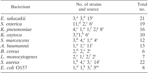
TABLE 1. Target pathogen strains used for microarray analysis
Bacterium No. of strains
and source
Total no.
E. sakazakii 3,a3,b15c 21
S. enterica 11,d2,e6c 19
K. pneumoniae 4,e1,a1,f2,g8c 16
K. oxytoca 3,a1,b6c 10
S. marcescens 3,h4,e1,a4c 12
A. baumannii 1,a1,e11i 13
B. cereus 2,h2,e2c 6
L. monocytogenes 2,e1,j2,f2c 7
S. aureus 1,h4,e3,c14i 22
E. coliO157 1,d1,k3,l3m 8
aATCC.
bCzech Collection of Microorganisms (CCM), Czech Republic.
cEnvironmental isolates from Tianjin Entry-Exit Inspection and Quarantine
Bureau, China.
dInstitute of Medical and Veterinary Science (IMVS), Adelaide, Australia. eNational Center for Medical Culture Collection (CMCC), China. fCenter for Veterinary Culture Collection (CVCC), China. gNational Collection of Type Cultures (NCTC), United Kingdom. hInstitute of Microbiology, Chinese Academy of Sciences (IMCAS). iClinical isolates from General Hospital of Tianjin Medical University, China. jAgricultural Culture Collection of China (ACCC).
kChinese Center for Disease Control and Prevention (CDC). lNational Reference Laboratory forE. coli, Berlin, Germany. mRobert Koch-Institut (RKI), Germany.
on May 16, 2020 by guest
http://jcm.asm.org/
intraspecies polymorphisms (0.985 to 0.997), suggesting that the ITS can be used as a target for species-specific probe designs.
Optimization of duplicate PCR.Primer pair efficiency for 10
target pathogens was determined based on the amplicons of
[image:3.585.43.542.85.629.2]anticipated sizes by testing different primer concentrations (0.10 to 0.50M). Using the optimized ITS andwzy primer concentrations (0.15M ITS primers and 0.2Mwzy prim-ers), target sequences from 10 representative pathogenic strains were amplified (Fig. 1).
TABLE 2. Oligonucleotide probes used
Bacterium and probe no. Target gene Tma
(°C) Sequence (5⬘–3⬘)
GenBank accession
no.
E. sakazakii
OA-1976 ITS 69.8 436-GTCAGAGTCTCTCAAACTCGCAGCACG-462 AY748357
OA-1978 68.9 465-ACAGACACGCTGCTGTATTTCTCCGTAAT-493
S. enterica
OA-1979 ITS 67.3 239-GAGGTTCTGACTACACGATGGGGCTAT-265 AF046814
K. pneumoniae
OA-2355 ITS 67.6 163-ATTTGAAGAGGTTGCAAACGATGGG-188 EU623376
OA-2356 67.4 140-GGCCTACCAAATTTGCGAAGCAA-163
K. oxytoca
OA-2351 ITS 64.5 40-CTGATAGATGTAAAGAAGCAAGACGGC-66 EU623150
OA-2352 73.8 62-ACGGCTGCGAAGTCGCGACACCT-84
OA-2354 66.0 178-TGAAAGGCACAACCAACCGATATCT-202
S. marcescens
OA-2568 ITS 69.9 CAAGCTTCCGACCCACCCGGT This study
OA-2569 69.7 GCGGCGGTCTCAAGTATCTGACGAT
OA-2570 64.4 CTTAAAGATGACTTCCGAGTCATGTTTAAG
OA-2571 64.2 CGAACGATGGAAACATCTTCGG
B. cereus
OA-2391 ITS 67.7 25-ATCAATATAAGTTTCCGTGTTTCGTTTTCG-55 AM062662
OA-2393 63.4 321-TTCTTTGAAAACTAGATAACAGTGTAGCTCAT-352
A. baumannii
OA-2530 ITS 67.3 342-TCATTATCACGGTAATTAGTGTGATCTGACG-372 AY601825
OA-2531 67.4 359-AGTGTGATCTGACGAAGACACATTAACTCATT-390
OA-2532 67.4 524-GTACGGTGCTTAAGTGCACAGTGCTCTA-551
L. monocytogenes
OA-2094 ITS 72.4 140-AGGCACTATGCTTGAAGCATCGCGC-164 U57912
OA-2095 61.8 271-AAGAAATACAAATAATCATACCCTTTTACG-300
OA-2096 62.8 257-TTTCTTTCTGACATAAGAAATACAAATAATCATA-290
S. aureus
OA-1973 ITS 70.4 219-GCTTATGCGAGCGCTTGACAATCTATTCT-247 U11774
OA-1974 69.0 335-TAAAGCAGTATGCGAGCGCTTGACTAAA-362
OA-2006 66.5 546-ATGTTAACGTTTGACTTATAAAAATGGTGG-575 U11786
E. coliO157
OA-1929 wzy 79.94 1632-AGGGAATAAAGCATCAAGACTTATTTTATGGAGAACGGTT-1671 AF061251
OA-1930 79.05 1580-TTTCCGACACCAGAGTTAGAAAAGGAATTAAAAGCAATAA-1619
OA-1932 79.7 1310-GGTTATCGTTCTGAATTGGTGTTGCTCATTCTTCAATATA-1349
OA-1994 79.07 1245-ATGATGAGCATAAATTCAAACAGAGGACCATCATATTTGT-1284
Positive control
OA-1993 16S rRNA 71.9 1380-TTGTACACACCGCCCGTCACACCAT-1404b X80725
Negative control
wl-4006 TTTTTTTTTTTTTTTTTTTTTTTTTTTTTTTTTTTTTTTTc
Positional and printing control
Cy3 TTTTTTTTTTTTTTTTTTTTTTTTTTTTTTTTTTTTTTTT_Cy3d
aMelting temperature.
bThe 16S rRNA gene-based probe. The 1380 to 1404 position inE. coliwas used as the positive control. cThe probe containing the 40 poly(T) oligonucleotide tail was used as the negative control.
dThe 3⬘-Cy3-labeled probe was used as the positional reference and printing control.
on May 16, 2020 by guest
http://jcm.asm.org/
Probe specificity.A total of 185 strains (134 target pathogen strains and 51 closely related bacteria) (see Table S2 in the supplemental material) belonging to 15 different genera and 47 species were used to test the specificity of the designed probes. Probes that cross-hybridized or did not produce signals were removed from the test panel. From 269 hybridization reac-tions, 30 probes (including 27 species-specific probes, 1 posi-tive control probe, 1 negaposi-tive control probe, and 1 positional and printing control probe) were selected from the 79 probes initially screened (Table 2). The microarray procedure de-signed identified all 134 target strains specifically; none of the 51 closely related bacteria tested yielded positive signals, sug-gesting that the designed probes were species specific (Fig. 2A to K).
Microarray sensitivity. The sensitivity was further tested
by hybridization with serially diluted genomic template DNA (0.001 to 10 ng). Representative hybridized results usingE.
sakazakiigenomic template DNA are shown in Fig. S2A to D
in the supplemental material. Based on the positive signals generated, the sensitivity of the assay using genomic DNA was 0.001 ng DNA forA. baumannii; 0.010 ng DNA forS. enterica,
E. sakazakii, and E. coli O157; and 0.100 ng DNA for K.
pneumoniae, K. oxytoca,S. marcescens,B. cereus, L.
mono-cytogenes, and S. aureus. The microarray also was tested
against pure cultures of gram-negative and -positive organ-isms. Genomic DNA was isolated and used to test the sensi-tivity of the microarray.E. sakazakiiATCC 29544 was selected as a representative gram-negative bacterium and was serially diluted to 101to 106 CFU/ml. The positive signals were
ob-tained at 104CFU/ml (see Fig. S2E to H in the supplemental
material).S. aureusCMCC 46112 was selected as a represen-tative gram-positive bacterium, and the probes were sensitive using 104 CFU/ml (see Fig. S2I to L in the supplemental
material). Likewise, the remaining eight strains were identified using 104CFU/ml pure culture (data not shown), suggesting
that this was the minimal dose needed for detection.
Simultaneous detection of multiple pathogens. Since the
detection of pathogens from a single sample would be carried out most efficiently if multiple pathogens could be detected simultaneously, genomic DNA from two pathogens (A.
bau-manniiandL. monocytogenes orE. coli O157 andB. cereus)
were mixed and used as templates to further test the spec-ificity of the microarray assay. The data demonstrated that the probes were able to hybridize the target regions, further demonstrating the specificity of the designed probes to their respective species (data not shown). Mixing genomic DNA from three pathogens (E. sakazakii,S. enterica, andB. cereus [Fig. 2L] or S. marcescens, K. oxytoca, and S. aureus [Fig. 2M]) also demonstrated that the array probes could detect multiple pathogens from a sample containing multiple genomic profiles.
Blinded testing.The specificity and sensitivity of the designed
microarray detection system described was further tested using a double-blind approach. Coded DNA samples from 26 strains (see Table S3 in the supplemental material) were randomly selected and used to hybridize to the microarrays. These results revealed that five isolates were E. sakazakii, four isolates wereS. enterica, three isolates wereK. pneumoniae, two isolates wereK. oxytoca, two isolates wereA. baumannii, two isolates wereS. marcescens, two isolates wereB. cereus, two isolates were L. monocytogenes, three isolates were S.
aureus, and one isolate was E. coli O157. These results
matched the identity according to the Vitek system and serotyping methods in the Tianjin Entry-Exit Inspection and Quarantine Bureau and General Hospital of Tianjin Medi-cal University.
Detection of mock PIF samples.E. sakazakiiATCC 29544,
S. entericaCMCC 50071,K. pneumoniaeATCC 10031,S.
au-reusCMCC 26058, andE. coliO157 IMVS 1332 (8) were used to inoculate PIF and cultured as mentioned in Materials and Methods. Pathogen-free PIF was used as a negative control. DNA was extracted from the respective samples and tested using the microarray probes described above. Hybridization results demonstrated that 25 g PIF contaminated by 3 CFUE.
sakazakii, 3 CFUS. enterica, 2 CFUK. pneumoniae, 6 CFUK.
oxytoca, 9 CFUS. marcescens, 3 CFUA. baumannii, 5 CFUB.
cereus, 3 CFUL. monocytogenes, 7 CFUS. aureus, and 4 CFU
E. coliO157 could be correctly detected (see Fig. S3A to J in
[image:4.585.137.446.70.205.2]the supplemental material).
FIG. 1. PCR amplification of 10 pathogens. Genomic DNA from 10 representative strains was amplified with the two primer pairs wl-5793/ wl-5794 and wl-5593/wl-5594, and amplified products were subjected to agarose gel electrophoresis. Lanes 1 and 12, DL2000 DNA marker; lane 2,E. coliO157 38/99; lane 3,S. entericaCMCC 50071; lane 4,S. aureusCMCC 26058; lane 5,A. baumanniiATCC 19606; lane 6,K. pneumoniae
ATCC 10031; lane 7,K. oxytocaATCC 49334; lane 8,L. monocytogenesCMCC 54001; lane 9,B. cereusAS1.196; lane 10,E. sakazakiiATCC 29544; and lane 11,S. marcescensATCC 13880.
on May 16, 2020 by guest
http://jcm.asm.org/
Detection of PIF samples.A total of 21 batches of PIF from the United States (n ⫽9), Ireland (n⫽ 3), France (n⫽2), Argentina (n⫽2), Holland (n⫽3), and India (n ⫽2) were collected from the Tianjin Entry-Exit Inspection and Quaran-tine Bureau and detected according to inspection and quaran-tine trade standard methods. In the meantime, the PIF samples were detected by the designed microarray. The hybridization profiles showed that two samples were detected asE. sakazakii
and B. cereus, respectively (see Fig. S3K to L in the
supple-mental material), which was affirmed by the Vitek system in the Tianjin Entry-Exit Inspection and Quarantine Bureau and 16S rRNA sequencing in our laboratory. The other 19 samples
were signals of the positive control probes being detected, suggesting that there are other bacteria beyond the 10 patho-gens studied. These results of 100% accuracy indicate that the microarray has the practical ability to detect and differentiate pathogens in PIF samples.
DISCUSSION
DNA microarrays currently are used for the detection of food-borne pathogens, since they are rapid, sensitive, specific, and allow for high-throughput analysis (25). Recently, various reports have demonstrated the efficacy of this approach in the
FIG. 2. Hybridization results. Cy3, which was used as the printing and positional control, is weak or invisible due to fluorescent attenuation. (A)E. sakazakiiATCC 29594; (B)S. entericaCMCC 50071; (C)E. coliO157 IMVS 1332; (D)A. baumanniiATCC 19606; (E)K. pneumoniae
ATCC 10031; (F)B. cereusAS1.196; (G)K. oxytocaATCC 49334; (H)S. aureusCMCC 26058; (I)S. marcescensATCC 13880; (J)L. monocytogenes
CMCC 54001; (K)S. epidermidisCMCC 26069; (L)E. sakazakiiATCC 29544,Salmonellasp. strain M44, andB. cereusAS1.196; (M)S. marcescens
ATCC13880,K. oxytocaATCC49334, andS. aureusCMCC26058.
on May 16, 2020 by guest
http://jcm.asm.org/
detection of waterborne pathogens (21), marine fish pathogens (11), and other food-borne pathogens (7, 10, 19, 28, 30). To our knowledge, this study describes for the first time the ap-plication of DNA microarray technology for the rapid and reliable detection of pathogens associated with PIF contami-nation. The rRNA genes (i.e., 16S, 23S, and 5S) are ideal genetic targets that can be used for bacterial identification, since these sequences are highly conserved between species (14). The primary caveat associated with ribosomal sequences as targets is that their variable regions make the identification of closely related organisms imprecise (23). However, the ITS sequence is not subject to the same selective pressures as the rRNA genes, therefore targeting these sequences overcomes the specificity issues associated with rRNA sequences (1, 13). Sequence and length polymorphisms associated with ITS in-creasingly are being used as targets for bacterial species and subspecies identification (5, 32) and typing (20, 31), as well as being used in evolutionary studies (12, 24). Another advantage of targeting ITS sequences is that they are relatively short. Specific capture probes can be designed for the variable re-gions, and universal primers can be designed for the conserved 16S and 23S rRNA gene sequences, respectively.
Lin et al. reported the detection of food-borne pathogens using microarrays designed to target ITS sequences (19). B.
cereus,E. coli,L. monocytogenes,Pseudomonas aeruginosa,S.
enterica,S. aureus, andVibrio parahaemolyticuswere detected.
In the present study, five additional pathogens,E. sakazakii,K.
pneumoniae,K. oxytoca,S. marcescens, andA. baumanniiwere
added to the panel of pathogens associated with PIF contam-ination.
FiveBacillus species, i.e.,B. anthracis,B. cereus,B.
thurin-giensis,B. mycoides, andB. weihenstephanensis, are closely
re-lated organisms that also posses a high degree of homology at the DNA level, making them difficult to differentiate (26). In this report, theB. cereus-specific probes described correspond to the conserved ITS regions of all five of these species, there-fore all five species could be identified by the probes designed in this study.
Another obstacle associated with microarray-related ap-proaches is that the detection of different DNA sequences is subject to different levels of sensitivity (0.100 to 0.001 ng). One possible reason is that each species possesses different operon copy numbers (rrn) and ITS types. For example,A.baumannii has six RNA rrn operons and one ITS type (ITSile⫹ala, with
tRNAileand tRNAalagenes);E.sakazakiihas sevenrrn
oper-ons, and four of them are ITSglu, the rest are ITSile⫹ala, and the
specific probes were designed based on the ITSglusequences;
K. pneumoniae has eight rrn operons, including three
ITSile⫹ala, four ITSglu, and one ITSnone(with no tRNA genes),
and the specific probes were designed based on the ITSile⫹ala
regions. Therefore, their target sequences possess different copy numbers (six forA.baumannii, four of seven forE.
saka-zakii, and three of eight forK.pneumoniae), and the final ratios
of the amplicons of those three species are 56:32:21. Another possible reason for different levels of sensitivity is different amplification efficiencies. Although they employed the same primers, different templates hold different levels of DNA qual-ity, DNA structure, and GC content, which have profound effects on the final amplicon amount. Our results also showed that the closely related bacteria Salmonellaand E. sakazakii
share the same DNA sensitivity, which is also the same as that
for K. pneumoniae, K. oxytoca, and S. marcescens. For the
sensitivity of the microarray as a whole, the lowest one, 0.100 ng bacterial genomic DNA, should be reported.
Another challenge associated with food pathogen detection is that organisms are surrounded by the biological matrix that comprises food or food products, and the nature of the food product can critically affect sample preparation. In this study, we employed preenrichment along with selective culture for the preparation of specific pathogens. Although this sample preparation method is laborious, it has its advantages. First, the two-step culture method is prone to yield more bacteria, which will increase the amount of genomic DNA available for analysis and enhance hybridization sensitivity. Second, the two-step culture method also will reduce contaminants of food samples. If the DNA obtained for hybridization was contami-nated with food product, it could interfere or inhibit the PCR by reducing hybridization efficiency or producing a false-posi-tive signal (27). In the second step, only 10 ml of the preen-richment was inoculated into 100 ml of selective culture, re-ducing the amount of food sample.
At present, the detection of food-borne pathogens is crucial for the safety of PIF because the consumers are infants and children, who are highly susceptible to outbreaks of life-threat-ening neonatal mlife-threat-eningitis, bacteremia, and diarrhea illness (4). Although our initial efforts focused on only 10 pathogens, efforts now are under way to include most pathogens com-monly associated with PIF contamination. A diagnostic tool with such a high-throughput value undoubtedly will facilitate the identification of pathogenic bacteria, thereby making pre-ventive measures timely and also expediting epidemiologic in-vestigations.
ACKNOWLEDGMENTS
This study was supported by grants from the National High Tech-nology Research and Development Program of China (863 Program) (2006BAK02A14, 2006AA06Z409, and 2006AA020703), the Na-tional 973 Program of China (2009CB522603), the NaNa-tional Key Programs for Infectious Diseases of China (2008ZX10004-002 and 2008ZX10004-009), and the Tianjin Municipal Science and Tech-nology Committee, China (07JCYBJC08500).
REFERENCES
1.Barry, T., G. Colleran, M. Glennon, L. K. Dunican, and F. Gannon.1991. The 16s/23s ribosomal spacer region as a target for DNA probes to identify eubacteria. PCR Methods Appl.1:51–56.
2.Bej, A. K.2003. Molecular based methods for the detection of microbial pathogens in the environment. J. Microbiol. Methods53:139–140. 3.Brown, P. O., and D. Botstein.1999. Exploring the new world of the genome
with DNA microarrays. Nat. Genet.21:33–37.
4.Cahill, S. M., I. K. Wachsmuth, L. Costarrica Mde, and P. K. Ben Embarek.
2008. Powdered infant formula as a source ofSalmonellainfection in infants. Clin. Infect. Dis.46:268–273.
5.Chun, J., I. N. Rivera, and R. R. Colwell.2002. Analysis of 16S–23S rRNA intergenic spacer ofVibrio choleraeandVibrio mimicusfor detection of these species. Methods Mol. Biol.179:171–178.
6.de Boer, E., and R. R. Beumer.1999. Methodology for detection and typing of foodborne microorganisms. Int. J. Food Microbiol.50:119–130. 7.Eom, H. S., B. H. Hwang, D. H. Kim, I. B. Lee, Y. H. Kim, and H. J. Cha.
2007. Multiple detection of food-borne pathogenic bacteria using a novel 16S rDNA-based oligonucleotide signature chip. Biosens. Bioelectron.22:845– 853.
8.Feng, L., W. Wang, J. Tao, H. Guo, G. Krause, L. Beutin, and L. Wang.2004. Identification ofEscherichia coliO114 O-antigen gene cluster and develop-ment of an O114 serogroup-specific PCR assay. J. Clin. Microbiol.42:3799– 3804.
9.Forsythe, S. J.2005.Enterobacter sakazakiiand other bacteria in powdered infant milk formula. Matern. Child Nutr.1:44–50.
on May 16, 2020 by guest
http://jcm.asm.org/
10.Gehring, A. G., D. M. Albin, S. A. Reed, S. I. Tu, and J. D. Brewster.2008. An antibody microarray, in multiwell plate format, for multiplex screening of foodborne pathogenic bacteria and biomolecules. Anal. Bioanal. Chem.391:
497–506.
11.Gonza´lez, S. F., M. J. Krug, M. E. Nielsen, Y. Santos, and D. R. Call.2004. Simultaneous detection of marine fish pathogens by using multiplex PCR and a DNA microarray. J. Clin. Microbiol.42:1414–1419.
12.Gu¨rtler, V.1999. The role of recombination and mutation in 16S–23S rDNA spacer rearrangements. Gene238:241–252.
13.Gu¨rtler, V., and V. A. Stanisich.1996. New approaches to typing and iden-tification of bacteria using the 16S–23S rDNA spacer region. Microbiology
142:3–16.
14.Gutell, R. R., N. Larsen, and C. R. Woese.1994. Lessons from an evolving rRNA: 16S and 23S rRNA structures from a comparative perspective. Mi-crobiol. Rev.58:10–26.
15.Iversen, C., and S. Forsythe.2004. Isolation ofEnterobacter sakazakiiand otherEnterobacteriaceaefrom powdered infant formula milk and related products. Food Microbiol.21:771–777.
16.Iversen, C., M. Lane, and S. J. Forsythe.2004. The growth profile, thermo-tolerance and biofilm formation ofEnterobacter sakazakiigrown in infant formula milk. Lett. Appl. Microbiol.38:378–382.
17.Joosten, H., E. Bidlas, and N. Garofalo.2006.Salmonelladetection in pro-biotic products. Int. J. Food Microbiol.110:104–107.
18.Lai, K. K.2001.Enterobacter sakazakiiinfections among neonates, infants, children, and adults. Case reports and a review of the literature. Medicine (Baltimore)80:113–122.
19.Lin, M. C., A. H. Huang, H. Y. Tsen, H. C. Wong, and T. C. Chang.2005. Use of oligonucleotide array for identification of six foodborne pathogens and
Pseudomonas aeruginosagrown on selective media. J. Food Prot.68:2278– 2286.
20.Maggi, R. G., B. Chomel, B. C. Hegarty, J. Henn, and E. B. Breitschwerdt.
2006. ABartonella vinsonii berkhoffiityping scheme based upon 16S–23S ITS and Pap31 sequences from dog, coyote, gray fox, and human isolates. Mol. Cell Probes20:128–134.
21.Maynard, C., F. Berthiaume, K. Lemarchand, J. Harel, P. Payment, P. Bayardelle, L. Masson, and R. Brousseau.2005. Waterborne pathogen de-tection by use of oligonucleotide-based microarrays. Appl. Environ. Micro-biol.71:8548–8557.
22.Muytjens, H. L., H. Roelofs-Willemse, and G. H. Jaspar.1988. Quality of powdered substitutes for breast milk with regard to members of the family
Enterobacteriaceae. J. Clin. Microbiol.26:743–746.
23.Palys, T., L. K. Nakamura, and F. M. Cohan.1997. Discovery and classifi-cation of ecological diversity in the bacterial world: the role of DNA se-quence data. Int. J. Syst. Bacteriol.47:1145–1156.
24.Pe´rez Luz, S., F. Rodriguez-Valera, R. Lan, and P. R. Reeves.1998. Varia-tion of the ribosomal operon 16S–23S gene spacer region in representatives ofSalmonella entericasubspecies. J. Bacteriol.180:2144–2151.
25.Rasooly, A., and K. E. Herold.2008. Food microbial pathogen detection and analysis using DNA microarray technologies. Foodborne Pathog. Dis.5:531– 550.
26.Rhodehamel, E. J., and S. M. Harmon.1998. Bacillus cereus.InFood and Drug Administration bacteriological analytical manual, 8th ed. (revision A), chapter 14. AOAC International, Gaithersburg, MD.
27.Rossen, L., P. Norskov, K. Holmstrom, and O. F. Rasmussen.1992. Inhibi-tion of PCR by components of food samples, microbial diagnostic assays and DNA-extraction solutions. Int. J. Food Microbiol.17:37–45.
28.Sergeev, N., M. Distler, S. Courtney, S. F. Al-Khaldi, D. Volokhov, V. Chizhikov, and A. Rasooly.2004. Multipathogen oligonucleotide microarray for environmental and biodefense applications. Biosens Bioelectron.20:684– 698.
29.Wang, M., B. Cao, Q. Yu, L. Liu, Q. Gao, L. Wang, and L. Feng.2008. Analysis of the 16S–23S rRNA gene internal transcribed spacer region in
Klebsiellaspecies. J. Clin. Microbiol.46:3555–3563.
30.Wang, X. W., L. Zhang, L. Q. Jin, M. Jin, Z. Q. Shen, S. An, F. H. Chao, and J. W. Li.2007. Development and application of an oligonucleotide microar-ray for the detection of food-borne bacterial pathogens. Appl. Microbiol. Biotechnol.76:225–233.
31.Wojciech, L., Z. Staroniewicz, A. Jakubczak, and M. Ugorski.2004. Typing ofYersinia EnterocoliticaIsolates by ITS profiling, REP- and ERIC-PCR. J. Vet. Med. B Infect. Dis. Vet. Public Health.51:238–244.
32.Xiong, L., F. Kong, Y. Yang, J. Cheng, and G. L. Gilbert.2006. Use of PCR and reverse line blot hybridization macroarray based on 16S–23S rRNA gene internal transcribed spacer sequences for rapid identification of 34 mycobac-teriumspecies. J. Clin. Microbiol.44:3544–3550.