Hepatitis A Virus Genotype C
Qiuxue Huang, Hua Yue, Bin Zhang, Peiting Nie, and Cheng Tang
College of Life Science and Technology, Southwest University for Nationalities, Chengdu, People’s Republic of China
Recently, duck hepatitis A virus genotype C (DHAV-C), a causative agent of duck viral hepatitis, has been responsible for
in-creasing economic losses in the duck industry in China and South Korea. In this study, a real-time PCR assay targeting the 2C
gene for detecting DHAV-C was developed. The assay was confirmed to be specific and sensitive, and the minimum detection
limit was 3.36
ⴛ
10
3copies per reaction, making this assay suitable for rapid diagnosis of DHAV-C infection from clinical
sam-ples. In addition, the dynamics of the viral loads in tissues of specific-pathogen-free (SPF) ducklings infected with DHAV-C were
investigated using this method. The DHAV-C could be detected earliest in the liver within 12 h postinfection. Moreover, high
viral loads were identified in the heart, liver, spleen, lung, kidney, bursa of Fabricius, thymus, pancreas, brain, and small
intes-tine after 24 h postinfection. Taking the data collectively, the study described in this report is the first to have developed a
real-time PCR method for detection of DHAV-C and thus contributes to pathogenicity research.
D
uck viral hepatitis causes a highly contagious disease in
do-mestic ducklings characterized by rapid onset and high
mor-tality (
24
). Traditionally, duck hepatitis virus (DHV) strains have
been classified into three serotypes: type 1 1), type 2
(DHV-2), and type 3 (DHV-3) (
6
,
13
,
21
). DHV-2 and DHV-3 are
clas-sified as members of
Astrovirus
(
3
,
5
,
20
), and DHV-1, belonging
to the
Picornaviridae, is widely epidemic in the duck-producing
areas worldwide (
1
,
9
,
14
,
19
). Recently, a type of DHV newly
found in South Korea (
10
), Taiwan (
23
), and mainland China (
24
)
has been identified also as a novel
Picornaviridae
virus (
10
,
23
,
24
).
However, no serological relationships between the new type of
DHV and the traditional DHV-1 has been found.
To better distinguish different DHVs among the members of
the
Picornaviridae, it was suggested that DHV in the
Picornaviridae
should be renamed duck hepatitis A virus (DHAV) (
4
).
Further-more, based on the genetic structures of DHAV, DHAV was
cat-egorized into three genotypes, genotype A (DHAV-A), genotype B
(DHAV-B), and genotype C (DHAV-C), which respectively
cor-respond to the traditional type of DHV-1, the new genotype
iso-lated from Taiwan, and the new genotype isoiso-lated from South
Korea and mainland China (
4
,
22
). DHAV-C mainly affects
duck-lings within 3 weeks of birth, and its epidemiology, clinical
symp-toms, and pathology are very similar to those of DHAV-A, making
it difficult to distinguish between these two viruses. By
compari-sons of the complete VP1, VP0, and VP3 nucleotide and amino
acid sequences and the partial three-dimensional (3D) nucleotide
sequence, the DHAVs belonging to the same genotype were clearly
distinguished from those of heterologous genotypes (
24
). To
quickly diagnose a DHAV-A infection, both conventional reverse
transcription-PCR (RT-PCR) (
11
) and real-time PCR (
25
)
meth-ods have been developed. Recently, a method based on amplifying
the genomic region in the 5
=
untranscribed region (5
=
-UTR) and
sequence analysis has been developed for the detection and typing
of DHAV (
4
). To further differentiate DHAV strains for disease
diagnosis, a duplex RT-PCR method for simultaneous detection
of DHAV-A and DHAV-C was developed in South Korea (
12
).
Based on the amount of information available on the genome
sequences of DHAV, we analyzed the characteristics of the
struc-tures of DHAV and found that the 2C gene could be used to clearly
differentiate the different DHAV subtypes. In this study, a
real-time PCR assay targeting the 2C gene was developed for detecting
DHAV-C from clinical samples and for determining the dynamic
distribution of DHAV-C in experimentally infected
specific-pathogen-free (SPF) ducklings.
MATERIALS AND METHODS
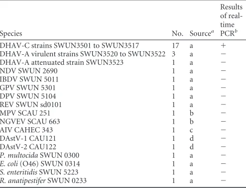
Virus, bacteria, SPF ducklings, and clinical samples.Seventeen clinical
isolates of DHAV-C, 14 virus species, and 4 bacterial species potentially infecting ducklings were used to evaluate the specificity of the real-time PCR assay in this study (Table 1). Fifty specific-pathogen-free (SPF) 3-day-old ducklings were used to determine the virus distribution in tis-sues. The SPF duck eggs were purchased from the experimental animal center of Harbin Veterinary Research Institute of Chinese Academy of Agricultural Science and hatched in the incubator in our laboratory. Thirty-eight clinical liver samples, collected from sick ducklings less than 3 weeks old which were suspected to be infected by the hepatitis virus, were characterized according to the presence of liver swelling with hemorrhagic lesions from April 2007 to July 2011 in China.
Nucleic acid extraction and cDNA synthesis.DNA was extracted
us-ing a DNA extraction kit (TaKaRa Biotechnology, Dalian, China) accord-ing to the manufacturer’s instructions. RNA was extracted usaccord-ing TRIzol reagent (Applied Biosystems Inc., Carlsbad, CA). The general RNA was reverse transcribed into cDNA using a Moloney murine leukemia virus (M-MLV) reverse transcription kit (TIANGEN Biotech, Beijing, China). The cDNA and DNA were stored at⫺20°C for further use.
Development of quantitative real-time PCR.The design of specific
oligonucleotide primers was based on sequence analysis of 20 DHAV-C 2C gene regions published in GenBank. The primer sequences were as follows: forward primer (DHAV-C FP), 5=-TCCAACAGGGTCAAAGC-3=; reverse primer (DHAV-C RP), 5=-AACTACTACAAGTCTGCCACG-3=. The primers amplified a 194-bp fragment of DHAV-C.
Received23 April 2012Returned for modification10 June 2012 Accepted25 July 2012
Published ahead of print1 August 2012
Address correspondence to Cheng Tang, [email protected].
Copyright © 2012, American Society for Microbiology. All Rights Reserved.
doi:10.1128/JCM.01080-12
on May 16, 2020 by guest
http://jcm.asm.org/
The results of conventional RT-PCR amplification with DHAV-C PCR products were verified by electrophoresis using a 2.0% agarose gel and then sequencing. The sequencing result was aligned with other DHAV-C sequences in GenBank. Moreover, the PCR products were de-tected by real-time PCR melting curve analysis using an SYBR PremixEx Taqkit (TaKaRa) according to the manufacturer’s instructions to verify the specificity of the amplification.
The conventional RT-PCR was performed with viral cDNA and the primers mentioned above. The amplicon was purified by the use of a gel extraction kit (Tiangen) according to the manufacturer’s instructions. The purified cDNA fragment was inserted into a clone vector, pMD19-T (TaKaRa), and transformed intoEscherichia coliTOP10 host cells (Tian-gen). The recombinant plasmid was purified using a plasmid extraction reagent (Tiangen). The product was stored at⫺20°C for further analysis. DHAV-C amplification by real-time PCR was performed in a 20-l reaction mixture containing 2.0l of a standard DNA template, 10l of SYBR green I master mix, 0.4l (each) of the primers (10M), and 7.2l of sterilized deionized water. The reactions were carried out in a 7300 Real Time PCR system (ABI). The PCR conditions consisted of one cycle of 3 min at 95°C followed by 30 two-step cycles of 15 s at 95°C and 31 s at 60°C. The standard DNA template was 10-fold serially diluted with sterilized deionized water to generate a standard curve, and each dilution was run in triplicate. The PCR, data acquisition, and analysis were performed using 7300 system software (ABI).
(i) Specificity of the real-time PCR.Specificity of the real-time PCR
was evaluated with the extracts of RNA or DNA templates from duck hepatitis A virus genotype A (DHAV-A) attenuated strain SWUN3523, duck hepatitis A virus genotype A (DHAV-A) virulent strains SWUN3520 to SWUN3522, duck astrovirus 1 1), duck astrovirus 2 (DAstV-2), muscovy parvovirus (MPV), gosling new type viral enteritis virus (NGVEV), avian influenza virus (AIV [H5N1]), Newcastle disease virus (NDV), infection bursal disease virus (IBDV), gosling parvovirus (GPV), duck plaque virus (DPV), reticuloendotheliosis virus (REV),Pasteurella multocida,E.coli(O46),Salmonella enteritidis,Riemerrella anatipestifer, and the allantoic fluid of a normal duck embryo.
(ii) Sensitivity of the real-time PCR.Ten-fold serially diluted
stan-dard DNA templates were assayed by the real-time PCR to determine their sensitivity. Each dilution was also tested with the conventional RT-PCR.
(iii) Repeatability and reproducibility of the real-time PCR.Three
positive samples were used to evaluate the repeatability and
reproducibil-Detection of DHAV-C in experimentally infected ducklings by the
real-time PCR.The animal study was approved by the Ethical and Animal
Welfare Committee from the Universitat Autònoma de Barcelona and followed European Union norms (Council Directive 86/609/EEC). Fifty SPF ducklings were divided into an experimental group (42 ducklings) and a control group (8 ducklings). The experimental group was subcuta-neously infected with 0.2 ml of 4.73⫻104copies of DHAV-C strain SWUN 3504. The controls were injected with the same volume of sodium chloride physiological solution. The experimental and control groups were separated in different SPF incubators, following strict biosafety con-trols. Three SPF ducklings were randomly selected at each time point and were euthanized to collect heart, liver, spleen, lung, kidney, thymus, bursa of Fabricius (BF), pancreas, intestine, and brain as samples at time points 1, 6, 12, 18, 24, 48, and 72 h. All these samples were tested by real-time PCR.
In order to ensure the accuracy of test results, glyceraldehyde-3-phos-phate dehydrogenase (GAPDH), stably expressed in tissues of the DHVA-C-infected duckling tested by our laboratory, was used as the reference gene to normalize the results of viral loads detected by the real-time PCR assay. The primers and conditions of a real-time PCR assay for detecting mRNA expression of GAPDH were as follows: the forward primer (GAPDH FP) (5=-CACAGCCACACACGAAGACA-3=) and reverse primer (GAPDH RP) (5=-CCTTAGCCAGCCCCAGTAGA-3=) were used to amplify a 107-bp fragment of duck GAPDH gene (GenBank accession no.AY436595). The amplification system contained the following com-ponents: 2.0l of standard DNA template, 12.5l of SYBR green I master mix, 0.5l (each) of the primers (10M), and 9.5l of sterilized deion-ized water. The PCR conditions consisted of one cycle of 5 min at 95°C followed by 40 two-step cycles of 15 s at 95°C and 30 s at 60°C.
Data analysis.Quantitative real-time PCR data were analyzed with
7300 system software. Linear regression analysis was used to compare the threshold cycle colonization density data (⌬CT) from the DHAV-C samples (⌬CTsample⫽CTDHAV-C sample⫺CTGAPDH sample) to the data from the calibration sample (⌬CTcalibration ⫽ CTDHAV-C calibration⫺ CTGAPDH calibration). The relative colonization density data were quanti-fied as the⌬⌬CTdetermined by comparisons between the DHAV-C sam-ples and calibration samsam-ples (⌬⌬CT⫽ ⌬CTsample⫺ ⌬CTcalibration), and the relative quantifications of the virus load were assessed by the 2⫺⌬⌬CT method (15).
RESULTS
Specificity of primers.
The conventional RT-PCR performed
with primers (DHAV-C FP and DHAV-C RP) amplified the
expected 194-bp fragment from DHAV-C, as observed by 2.0%
gel electrophoresis. The alignment showed that the fragment
sequence was 100% identical to those of the 2C genes of all the
20 genomes of DHAV-C strains in GenBank. The melting
curves of DHAV-C displayed a single specific peak with a
melt-ing temperature (T
m) value at 81.0
⫾
0.4°C, suggesting a single
product (
Fig. 1
).
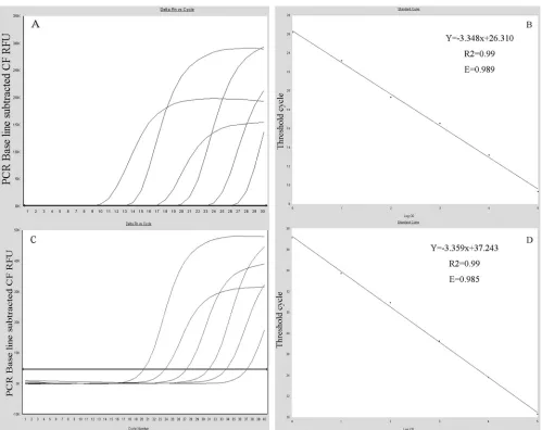
Standard curves of the real-time PCR.
The 10-fold serially
diluted standard plasmid DNAs of DHAV-C and GAPDH
gener-ated two typical standard curves. The standard curves for
⫺
DPV SWUN 5104 1 a ⫺
REV SWUN sd0101 1 a ⫺
MPV SCAU 251 1 b ⫺
NGVEV SCAU 663 1 b ⫺
AIV CAHEC 343 1 c ⫺
DAstV-1 CAU121 1 d ⫺
DAstV-2 CAU122 1 d ⫺
P. multocidaSWUN 0300 1 a ⫺
E. coli(O46) SWUN 0314 1 a ⫺
S. enteritidisSWUN 5223 1 a ⫺
R. anatipestiferSWUN 0233 1 a ⫺
aa, Southwest University for Nationalities, China; b, Sichuan Agricultural University,
China; c, China Animal Health and Epidemiology Center, China; d, China Agricultural University, China.
b⫹
, positive;⫺, negative.
on May 16, 2020 by guest
http://jcm.asm.org/
[image:2.585.41.286.87.274.2]DHAV-C and GAPDH were generated with a range of 3.36
⫻
10
3to 3.36
⫻
10
8gene copies per reaction and 8.32
⫻
10
2to 8.32
⫻
10
7gene copies per reaction (
Fig. 2
), respectively. Both the DHAV-C
and GAPDH assays were linear over a 10
6dilution range of
tem-plate DNA. The
R
2value for both was 0.99, and the reaction
effi-ciencies were 98.9% and 98.5%, respectively. The cDNA copy
numbers of DHAV-C and GAPDH for unknown samples were
quantified using the equations
Y
DHAV-C⫽ ⫺
3.348X
DHAV-C⫹
26.310 and
Y
GAPDH⫽ ⫺
3.359X
GAPDH⫹
37.243, where
Y
is the
threshold cycle and
X
is the log of the starting quantity.
Specificity of the real-time PCR.
All 17 DHV-C strains were
able to detect the DHAV-C RNA, while other non-DHAV-C
vi-ruses and bacteria, including 2 negative-control samples,
pre-sented no amplification by the real-time PCR. The results are
shown in
Table 1
.
Sensitivity of the real-time PCR.
The detection limit of the
real-time PCR developed in this study was about 3.36
⫻
10
3copies
per reaction for DHAV-C cDNA. The conventional RT-PCR was
performed with the same primers as were used for real-time PCR,
and the detection limit was about 3.36
⫻
10
4copies per reaction
for DHAV-C cDNA. Thus, the sensitivity of the real-time PCR was
10 times higher than that of the conventional RT-PCR.
Repeatability and reproducibility test.
The interassay and
in-tra-assay coefficients of variation (CVs) for the detection of the
three positive samples were calculated to be within the ranges of
1.02% to 1.77% and 0.15% to 2.0%, respectively, for viral cDNA
(
Table 2
).
Detection and validation of clinical sample results.
Twenty-two (57.9%) DHAV-C RNAs were identified from a total of 38
clinical samples by real-time PCR detection. Moreover, other,
non-DHAV-C RNAs (16/38 [42.1%]) were further identified in
samples as DHAV-A positive. The results of virus isolation from
15 DHAV-C-positive samples were all confirmed as positive by
the real-time PCR assay.
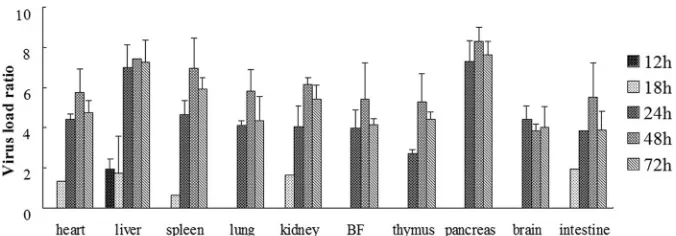
Dynamic and virus titer results in experimentally infected
ducklings.
The DHAV-C was successfully used to artificially
in-fect SPF ducklings in this experiment. Clinical symptoms were
observed at 24 h postinfection, and the SPF ducklings began to die
at 32 h postinfection, with pathological changes typical of duck
hepatitis.
As shown in
Fig. 3
, DHAV-C was first detected in the liver at 12
h postinfection. DHAV-C could be detected in the heart, liver,
spleen, kidney, and intestine tissues at 18 h postinfection. Viral
cDNA was detected in all samples of the infected ducklings from
24 h postinfection. Furthermore, high levels of virus titers were
detected in all samples from 24 to 72 h postinfection. The positive
detection rates of the virus are shown in
Table 3
.
DISCUSSION
The epidemiology, clinical symptoms, and pathological changes
of DHAV-C infection are very similar to those of DHAV-A
infec-tions, making molecular biological methods necessary to
distin-guish DHAV-C infection from other DHV infections (
8
,
16
).
DHAV strains were categorized into three distinct genetic groups
based on the phylogenetic analysis of complete VP1, VP0, and
VP3 and partial 3D sequences (
24
). To screen the molecular
tar-gets for detecting DHAV-C, we analyzed 42 genome sequences of
DHAV, including 20 DHAV-C, 20 DHAV-A, and 2 DHAV-B
se-quences published in GenBank. The analysis of phylogenetic
rela-tionships of the 2C genes of three different genotypes of DHAV
FIG 1Melting curve of the DHAV-C PCR products. The melting curve analysis showed a single specific peak, suggesting there were not nonspecific amplification and primer dimers.
on May 16, 2020 by guest
http://jcm.asm.org/
showed that the three genetic groups of DHAV could be clearly
distinguished based on 2C nucleotide sequences. The result was in
agreement with the analysis of phylogenetic relationships based
on other regions of the DHAV genome (
24
). However, the
muta-tion rate of 2C gene nucleotides was significantly lower than that
of other regions in the DHAV-C genome, making this gene a
po-tential target for molecular detection of DHAV-C. Based on the
bioinformation analysis described above, a real-time PCR assay
was established for detecting DHAV-C in this study. This assay
was sensitive and specific, enabling it to be used a practical
tech-nique for rapid diagnosis and pathogen monitoring of DHAV-C
infection.
In this study, 38 clinical liver samples, which were collected
from April 2007 to July 2011 in China from sick ducklings
sus-pected of duck hepatitis virus infection, were tested by the
real-time PCR assay. Interestingly, all 22 samples collected from
Sep-FIG 2Standard curve and amplification curves. Standard curves (based on plasmid DNA) indicating the linearity and efficiency of the reactions are shown. The results of serial 10-fold dilutions of standard DHAV-C (A) and GAPDH (C) from 3.36⫻103to 3.36⫻108gene copies per reaction and 8.32⫻102to 8.32⫻107
[image:4.585.43.542.63.459.2]gene copies per reaction were determined by real-time PCR, respectively. Linearity of two assays of DHAV-C (B) and GAPDH (D) spanned 6 orders of magnitude. The slope of the curve (logarithmic dilution versus threshold cycle) and the intercept are given in the equation in the figure. In the linear equation, the X represents the log of the starting quantity and the Y represents the threshold cycle. R2, correlation coefficient; E, efficiency.
TABLE 2Repeatability and reproducibility of the assay for selected samples containing DHAV-Ca
Species
Intra-assay result (CT)
SD % CV
Interassay result (CT)
SD % CV
Sample 1 Sample 2 Sample 3 Mean Sample 1 Sample 2 Sample 3 Mean
SWUN3501 13.48 13.51 13.52 13.50 0.021 0.15 13.50 13.73 13.26 13.50 0.26 1.74 SWUN3502 15.50 15.67 15.54 15.57 0.089 0.57 15.57 15.37 15.26 15.40 0.16 1.02 SWUN3503 17.04 16.40 16.56 16.67 0.333 2.0 16.67 16.83 17,25 16.92 0.30 1.77 aAll repeat assays were performed at the same time and under the same conditions. SD, standard deviation; CV, coefficient of variation.
on May 16, 2020 by guest
http://jcm.asm.org/
[image:4.585.40.545.653.716.2]tember 2009 to July 2011 were determined as representing
DHAV-C infection. Nevertheless, the 16 liver samples collected
before September 2009 were negative for DHAV-C and were later
identified as representing DHAV-A infection. No coinfections by
both DHAV-A and DHAV-C were found in this study. The results
presented above suggest that DHAV-C is a recent epidemic DHV
strain in China, which is consistent with reports about the
epide-miology of DHV in other regions of China (
2
,
7
), although the
number of samples investigated in this study was limited. In recent
years, the spread of DHAV-C has represented a serious threat and
has had a significant economic impact on the duck industry in
China. Moreover, the report of the prevalence of DHAV-C in
South Korea showed that DHAV-C has spread in Asia (
12
).
Hence, the development of rapid methods for detection of the
strain from clinical samples is important for pathogenic diagnosis
and disease control.
The dynamic quantitative detection of viral loads in tissues
is useful for understanding the pathogenicity of viruses and
determination of the target tissues collected for clinical virus
detection (
17
,
18
). The results showed that high DHAV-C loads
were detected in various tissues and organs at 24 h
postinfec-tion and suggested that DHAV-C is a pantropic virus and
causes extensive tissue damage in ducklings. In this study, the
DHAV-C was first detected in liver, and persistence in this
organ at high levels suggests that the liver is an ideal target
organ for detecting this virus. In addition, the viral loading in
the brain clearly showed that the DHAV-C could directly
in-vade the central nervous system of ducklings, which also
ex-plained the presence of neurologic signs, including ataxia and
opisthotonos, in the sick ducklings infected by DHAV-C.
In-terestingly, no obviously visible pathological change was
ob-served in immune organs, but presence of the virus could be
determined in the thymus, bursa of Fabricius, and spleen,
which suggests that the immune function of duckling might be
affected by DHAV-C. Collectively, the data in this report
pro-vide a valuable basis for researching pathogenicity and for
pre-vention and control of DHAV.
In conclusion, a real-time PCR assay for detecting DHAV-C
was developed in this study. The assay could be applied as a rapid,
sensitive, and specific molecular tool for diagnosis of DHAV-C
infection and for epidemiological surveys.
ACKNOWLEDGMENTS
We gratefully acknowledge Anchun Chen (Sichuan Agricultural Univer-sity, China) for kindly presentation of muscovy parvovirus and gosling new type viral enteritis virus. We also thank Hanchun Yang and Dabin Zhang (China Agricultural University, China) for kindly presentation of duck astrovirus 1 and duck astrovirus.
This work was supported by funding from the National Hightech R&D Program (863 Program-2012AA101304) and by Veterinary Medi-cine Discipline Program of Southwest University for Nationalities (2011XWD-S0906).
REFERENCES
1. Asplin FD.1965. Duck hepatitis: vaccination against two serological types. Vet. Rec.77:1529 –1530.
2.Fan SC, et al.2009. Isolation and characterization of a new serotype of duck hepatitis virus. Chin. J. Prev. Vet. Med.31:770 –775. (In Chinese.) 3.Fauquet CM, Mayo A, Maniloff J, Desselberger U, Ball LA.2005. Virus
taxonomy: VIIIth report of the International Committee on Taxonomy of Viruses. Elsevier Academic Press, San Diego, CA.
4.Fu Y, et al.2008. Molecular detection and typing of duck hepatitis A virus directly from clinical specimens. Vet. Microbiol.131:247–257.
5.Gough RE, Collins MS, Borland E, Keymer LF. 1984. Astrovirus-like particles associated with hepatitis in ducklings. Vet. Rec.114:279. 6.Haider SA, Calnek BW.1979. In vitro isolation, propagation, and
char-acterization of duck hepatitis virus type III. Avian Dis.23:715–729. 7.He RY, Yu M, Zhang YL, Zhang DY, Cao ZX, Zhang GH. 2010.
Epidemiological investigation and genetic variation in VP1 gene of duck hepatitis virus isolates from in southwestern China in 2007-2009. Chin. J. Anim. Infect. Dis.18:7–15. (In Chinese.)
FIG 3Virus titer of DHAV-C in the internal organs of experimentally infected ducklings. Seven organs were obtained from the experimentally infected ducklings, and viral loads were quantified by the real-time PCR at 1 to 72 h postinfection. One of the spleen samples at 18 h generated the lowest normalizedCT
values (⌬CT), and this was used to calibrate the data. The calibrated quantification ratios assessed by 2⫺⌬⌬CTwere converted into logarithmic values, and the
[image:5.585.123.461.65.183.2]results are shown in histograms. All histograms are arranged according to the time after infection (i.e., 12 to 72 h). The virus titer was not detected at 1 and 6 h postinfection.
TABLE 3The rates of detection of DHAV-C in different organs of experimentally infected SPF ducklings
Organ
No. of positive real-time PCR results/total. no. of results at indicated time (h)
1 6 12 18 24 48 72
Brain 0 0 0 0 3/3 3/3 3/3
Bursa of Fabricius 0 0 0 0 3/3 3/3 3/3
Heart 0 0 0 1/3 3/3 3/3 3/3
Intestine 0 0 0 1/3 3/3 3/3 3/3
Kidney 0 0 0 1/3 3/3 3/3 3/3
Lung 0 0 0 0 3/3 3/3 3/3
Liver 0 0 3/3 2/3 3/3 3/3 3/3
Pancreas 0 0 0 0 3/3 3/3 3/3
Spleen 0 0 0 1/3 3/3 3/3 3/3
Thymus 0 0 0 0 3/3 3/3 3/3
on May 16, 2020 by guest
http://jcm.asm.org/
[image:5.585.41.287.580.723.2]12. Kim MC, Kwon YK, Joh SJ, Kwon JH, Lindberg AM.2008. Differential diagnosis between type-specific duck hepatitis virus type 1 (DHV-1) and recent Korean DHV-1-like isolates using a multiplex polymerase chain reaction. Avian Pathol.37:171–177.
13. Levine PP, Fabricant J.1950. A hitherto-undescribed virus disease of ducks in North America. Cornell Vet.40:71–76.
14. Levine PP, Hofstad MS.1945. Duck disease investigation. Annu. Rep. New York State Vet. Coll.1945:55–56.
15. Liu J, et al.2011. Dynamic distribution and tissue tropism of classical swine fever virus in experimentally infected pigs. Virol. J.8:201. 16. Liu M, et al.2011. Goose haemorrhagic hepatitis caused by a new subtype
duck hepatitis type 1 virus. Vet. Microbiol.152:280 –283.
immune to duck virus hepatitis. Avian Dis.13:834 – 846.
22. Tseng CH, Knowles NJ, Tsai HJ.2007. Molecular analysis of duck hep-atitis virus type 1 indicates that it should be assigned to a new genus. Virus Res.123:190 –203.
23. Tseng CH, Tsai HJ.2007. Molecular characterization of a new serotype of duck hepatitis virus. Virus Res.126:19 –31.
24. Wang L, Pan M, Fu Y, Zhang D.2008. Classification of duck hepatitis virus into three genotypes based on molecular evolutionary analysis. Virus Genes37:52–59.
25. Yang M, Cheng A, Wang M, Xing H.2008. Development and application of a one-step real-time Taqman RT-PCR assay for detection of duck hep-atitis virus type1. J. Virol. Methods153:55– 60.