IJPSR (2014), Vol. 5, Issue 11 (Research Article)
Received on 29 April, 2014; received in revised form, 30 September, 2014; accepted, 15 October, 2014; published 01 November, 2014
FORMULATION AND EVALUATION OF NIFEDIPINE LOADED CHITOSAN MICROSPHERS FOR MUCOADHESIVE DRUG DELIVERY
Preethi Sudheer *1 and Ramakrishna Shabaraya A. 2
Department of Pharmaceutics, KrupanidhiCollege of Pharmacy 1, Bangalore, Karnataka, 560035, India.
Srinivas College of Pharmacy 2, Valachil, Mangalore, Karnataka, India.
ABSTRACT: Nifedipine loaded mcoadhesive micropshres were
prepared by o/w/o emulsification cross linking method using natural polymer chitosan. Microsphres with particle size range b/n 50-300 µm were obtained by this method. The prepared microspheres were evaluated for its drug loading, surface properties, in-vitro release profile. The diffusion and permeation characteristic of the drug is studied by using cellophane membrane and excised porcine intestinal mucosa respectively. The mucoadhesion properties of the microsphers were determined by using modified USP disintegration test apparatus model. In vivo animal studies were studies using rabbit model indicated a controlled blood level profile in comparison with pure nifedipine. IR studies indicated no appreciable drug excipient interaction.
INTRODUCTION: Drug delivery systems (DDS) that can precisely control the release rates or target drugs to a specific body site have had an enormous impact on the healthcare system. Carrier technology offers an intelligent approach for drug delivery by coupling the drug to a carrier particle such as microspheres, nanoparticles, liposomes, etc. which modulates the release and absorption characteristics of the drug. Microspheres constitute an important part of these particulate DDS by virtue of their small size and efficient carrier characteristics. However, the success of these novel DDS is limited due to their short residence time at the site of absorption.
QUICK RESPONSE CODE
DOI:
10.13040/IJPSR.0975-8232.5(11).5019-24
Article can be accessed online on:
www.ijpsr.com
DOI link: http://dx.doi.org/10.13040/IJPSR.0975-8232.5(11).5019-24
It would, therefore, be advantageous to have means for providing an intimate contact of the DDS with the absorbing membranes. It can be achieved by coupling bioadhesion characteristics to microspheres and developing novel delivery systems referred to as “bioadhesive microspheres”. 1
Mucoadhesive polymers are used to immobilize a drug delivery device on a specific site for targeted release and optimal drug delivery due to intimacy and duration of contact. 2
Chitosan is a linear polysaccharide composed of randomly distributed β-(1-4)-linked D-glucosamine and N-acetyl-D-glucosamine units. Commercially, it is produced by the exhaustive deacetylation of chitin (>60%), a structural element in the exoskeleton of crustaceans and insects, which is the second most abundant natural biopolymer after cellulose. Chitosan, the N-deacetylated product of the polysaccharide chitin, is an interesting biopolymer to prepare microspheres owing to its unique polymeric cationic character, good Keywords:
nifedipine, chitosan, microsphres, mucoadhesion, emulsification,
In-vitro release
Correspondence to Author: Preethi Sudheer
Associate Professor,
Department of Pharmaceutics, Krupanidhi College of Pharmacy Bangalore -35, India.
biocompatibility, non-toxicity, biodegradability and its mucoadhesive and absorption-enhancing effect.3
Nifedipine, a calcium channel blocking agent, is frequently used for the treatment of angina pectoris and hypertension. Nifedipine is absorbed rapidly in gastrointestinal tract after an oral administration with a bioavailability of 45% to 75% due to its significant first-pass metabolism. Conventional preparations, which would increase heart rate reflectly and activate the sympathetic nervous system, are not convenient clinically. Nifedipine sustained-release preparation is relatively safe, well in compliance with mild adverse effects owing to its less variation of blood concentration and longer maintenance time.4
MATERIALS AND METHODS:
Chitosan with >85% deacetylation was supplied from CIFL ltd, Cochin. Nifedipine was a gift samples from M/S Micro labs ltd, Bangalore. Other
reagents used were of analytical grade from local suppliers. Porcine intestine was obtained from slaughter house. In vivo animal studies were conducted after the ethical clearance from the institutional ethical clearance committee.
Preparation of microspheres
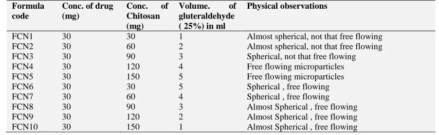
[image:2.612.79.530.354.493.2]The microsphres were prepared by o/w/o double emulsion cross linking method. In this method, initially 1% w/v chitosan solution in 1% v/v acetic acid was prepared by stirring for 4-5 hrs. Nifedipine was dissolved in 1:1 ratio of methanol: acetone mixture, and the resultant solution was added to chitosan solution under mechanical stirring for 2 hrs, followed by gluteraldehyde (25%) was added as rigidising agent. The resultant microparticulate suspension was continuous stirred overnight; the resultant microsphers were filtered by whatman filter paper, washed with petroleum ether and dried. 5
TABLE 1: COMPOSITION OF MICROSPHERS OF NIFEDIPINE Formula
code
Conc. of drug (mg)
Conc. of Chitosan (mg)
Volume. of gluteraldehyde ( 25%) in ml
Physical observations
FCN1 30 30 1 Almost spherical, not that free flowing
FCN2 30 60 2 Almost spherical, not that free flowing
FCN3 30 90 3 Spherical, not that free flowing
FCN4 30 120 4 Free flowing microparticles
FCN5 30 150 5 Free flowing microparticles
FCN6 30 30 5 Spherical , free flowing
FCN7 30 60 4 Spherical , free flowing
FCN8 30 90 3 Almost Spherical , free flowing
FCN9 30 120 2 Almost Spherical , free flowing
FCN10 30 150 1 Almost Spherical , free flowing
Evaluation of microsphers Drug content determination
Microspheres equivalent to 100 mg of the drug was crushed, dissolved in methanol, made up to volume with methanol, and diluted to Beer’s concentration, and absorbance was measured at 235 nm.7
Percentage drug loading
Microspheres equivalent to 100 mg of the drug was taken, washed with 3x 10 ml methanol, the resultant microspheres were dried, was crushed, dissolved in methanol, made up to volume with methanol, and diluted to Beer’s concentration, and measured absorbance at 235 nm.
% drug loading is determined by the following formula= total drug content- free drug content*100 and encapsulation efficiency by total drug content- free drug content / total drug content * 100
Particle size and distribution
The particle size distribution studies were carried out by using optical microscopic method using stage micrometer. The microsphers were taken on a glass slide, added one drop of liquid paraffin and checked under optical microscope under 100X magnification. Particle size of 100 particles was measured.
SEM studies
The surface morphology of the microsphres was studied by using JSM 840 A, scanning electron microscope, Jeol, Japan. The samples were analysed after they haven gold sputtered using 25nm film thickness. 7
In -Vitro release studies
was used as dissolution medium. The volume of dissolution medium was 900 ml and maintained at 37±0.50 C at a rotation speed of 50 rpm. Microsphres equivalent of 100 mg of drug was placed into the dissolution medium. and the release of the drug from microspheres were studied by using collecting the samples at regular time intervals, replacing with fresh buffer, concentration of drug in each sample was measured UV spectrophotometrically at 235 nm.9
In-vitro diffusion studies
In-vitro diffusion studies were carried out by using Franz- diffusion cell using cellophane membrane as model membrane. Microsphres equivalent to 100 mg of the drug was taken on a treated cellophane membrane, which was placed b/n the donor compartment and the receptor compartment of the diffusion cell assembly. The receptor compartment is filled with 25 ml of phosphate as receptor fluid. The microsphres were wetted by 1 ml phosphate buffer pH 7.2. The drug diffusion was studied by collecting receptor fluid at regular time intervals, replacing with fresh buffer solution to maintain sink conditions. The samples were analysed UV spectrophotometrically at 235 nm.9
Ex-vivo permeation studies
The ex-vivo permeation studies carried out by using porcine intestinal mucosa. The fresh intestine was collected, which was cut through lumen. The microsphers equivalent to 100 mg of the drug was taken on mucosal side of the intestine, which was placed b/n donor and receptor compartment; in such a way that mucosal side is facing towards donor compartment. The receptor compartment is filled with 25 ml of phosphate buffer pH 7.2 as receptor fluid. The drug permeation is studied by using collection of receptor fluid at regular time intervals, and analysed for drug content UV spectrophotometrically at 235 nm.10
Extent of mucoadhesion
The mucoadhesion time was determined by using porcine intestinal mucosa. The microspheres were placed on to the mucosal side of the intestine, with minimum pressure; the mucosa was tied to a glass slide, which in turn was tied to USP disintegration test basket rack assembly, the entire assembly was allowed to move up and down in 900 ml of
phosphate buffer. The time required for microsphres to detach from the mucosa is determined.11
Drug excipient interaction studies IR studies
IR studies were carried out for both pure drug and microsphres using KBr pellet technique to see, if any physicochemical interaction b/n pure drug and excipients.
In- vivo studies
The optimized formulations of microsphere using chitosan polymer, which showed the best results in terms of mucoadhesive properties were selected for in -vivo study.
Albino rabbits weighing 1.5–2.5 kg (housed in a temperature (22±1 °C) and relative humidity (55±10%) controlled room) were used in the experiments. The rabbits were maintained in accordance with the principles declared by the Committee for the Purpose of Control and Supervision of Experiments on Animals (CPCSEA, Government of India). They were housed with free access to food and water, except for the final 12 h before experimentation.
Each group consisted of 3 animals with group 1 was treated with best formula of Nifedipine 100 mg of microspheres were given as oral suspension. Group 2 with Nifedipien pure drug suspension 10mg/kg of body weight. After a single administration of above formulations, 2.5 ml of blood samples were collected from the marginal ear vein at, 0.5, 1, 2, 4 and 8 h time points into heparinised collection tubes. The blood was immediately centrifuged at 1000 rpm for 10 min at cooling temperature. The supernatant plasma layer was separated and stored at -200 C until analyzed.
Methanol (40:60 v/v) the pH was adjusted to 4.5 with 10% ortho phosphoric acid. Analysis was run at a flow rate of 1.0 ml/min and the detection wavelength was 235nm for nifedipine.11, 12
RESULTS AND DISCUSSIONS:
In the present study nifedipine loaded micro spheres were prepared by using chitosan polymer by o/w/o emulsification method. Ten formulations of micro spheres were prepared by using various ratios of drug to polymer ranging from 1:1 to 1:5, and also by varying concentration of cross linking agent gluteraldehyde. It was found that increasing concentration of polymer had improved the physical properties, free flowing nature and spherical properties. (Table 1). The drug content of formulation ranged b/n 73.34+/1.23% to 80.60+/-2.12% and drug loading ranged b/n 68.65+/-0.09%-79.56+/-0.87%. The polymer concentration had little effect on drug content and drug loading. And also increased concentration of gluteraldehyde had shown an effect on increased drug loading, may be due to the rigidising properties of gluteraldehyde. The mucoadhesion time ranged b/n 423 sec- 478 sec. (Table 2)
TABLE 2: EVALUATION OF DRUG LOADED MICROSPHERS
S. No. Drug content (%)
Drug loading %
Mucoadhesion Time (sec)
FCN1 73.34 +/-1.23 68. 65+/-0.09 423
FCN2 75.56 +/- 1.44 68.87+/-0.21 445
FCN3 76.12+/- 2.76 69.32 +/0. 65 450
FCN4 73.67 +/- 2.33 69.09+/- 1. 87 476
FCN5 73.14 +/-3.21 70.12+/- 1.22 444
FCN6 80.60+/-2.12 79.56+/- 0.87 475
FCN7 74.25 +/-3.43 71.67+/-2.34 473
FCN8 78.11+/-1.88 76.23+/-3.12 478
FCN9 75.23+/-2.54 70.67+/-2.43 467
FCN10 73.24+/0.65. 70.21+/-0.45 458
The obtained microspheres were characterised by using camera lucida, all the microsphers spherical shape, but the formula FCN6 was selected on the basis of drug content also was more or less spherical particle with size distribution b/n 10- 800 μm. (Fig 1).
SEM was utilized to observe the surface properties of the microsphers. The surface properties of the formula FCN6 studied indicated that the surface of the microsphers was uneven, and slightly porous. This property could have contributed to an extent the mucoadhesive nature of microsphers. (Fig.2)
Particle size distribution data
0 10 20 30 40 50 60
0-200 200-400 400-600 600-800 800-1000
Particle size in micrometer
Fre
qu
e
nc
y
Frequency
[image:4.612.315.565.56.222.2]FIG I: PARTICLE SIZE DISTRIBUTION DATA OF SELECTED FORMULA (FCN6)
FIG 2: SEM PHOTOGRAPH OF (FCN6)
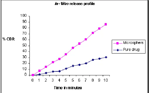
[image:4.612.315.566.250.429.2]In–Vitro release profile of the selected formula (FCN6) was studied against pure drug profile in phosphate buffer pH 7.2, where as microsphers were showing a controlled, but an enhanced drug release pattern in comparison with pure drug. About 80% of the drug was released in a time period of 10hrs. This would be due to from solubilising property of chitosan. (Fig.3)
[image:4.612.314.568.572.731.2]The release data was fitted into various kinetic model, it was seen that, the release profile was fitted to zero order kinetic profile with R2 valueof 0.9923, showed Korsmeyre-Peppas model mechanism with n values of 0.8169.
The pure sample of nifedipine was showing a slow diffusion profile with 30.34 % 10 hrs, whereas drug release from microspheres were showing a release up to 55.55% which may be due to added solubility enhancement property of chitosan polymer. (Fig.4)
In-Vitro diffusion studies
0 10 20 30 40 50 60
0 5 10 15
Time in hrs
p
e
rc
e
n
ta
g
e
d
ru
g
d
if
fu
s
e
d
Microspheres
[image:5.612.315.564.131.296.2]Nifedipine
FIG 4: IN -VITRO DIFFUSION PROFILE FCN6
The ex-vivo permeation studies using porcine intestinal mucosal membrane indicated drug permeation of 33% which was similar to in- vitro diffusion profile, where as the microsphers had shown a controlled release pattern and about 48.67 % drug was permeated in 10 hrs. (Fig. 5)
Extent of mucoadhesion was studied by using modified disintegration test apparatus was ranged
b/n 423-478 sec. This increase in extent of mucoadhesion may be due to uneven/ rough surface and also due to particle size in micrometer range would have contributed to effective surface area of contact.
comparative permeation studies of microsphers
0 10 20 30 40 50 60
0 1 2 3 4 5 6 7 8 9 10
Time in hrs
C
um
ul
a
t
i
v
e
pe
r
c
e
nt
a
ge
pe
r
m
e
a
t
e
d
microsphres
pure drug
FIG 5: COMPARATIVE PERMEATION STUDIES OF FCN6 WITH PURE DRUG
In-vivo studies indicated the mean plasma concentration of nifedipine in rabbits shows that the plasma concentration increased and attained a maximum after which average steady state level of the declined till in the 8 hr study, in case pure sample of nifedipine. In case of microsphres the plasma concentration 2.02µg/ml was observed in 1 hr and controlled pattern was observed with a plasma conc. of 5.46µg/ml in an 8 hr study. The pure drug exhibited a plasma concentration 6.075µg/ml at 1 hr and from then the conc. was declined to 0.45µg/ml in an 8 hr study.
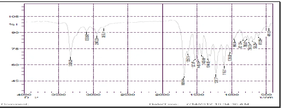
FIG 6: IR SPECTRA OF FCN6
The IR studies showed the principal peaks which was present in the pure drug was also present in
[image:5.612.50.297.214.372.2] [image:5.612.77.540.516.695.2]exicipients. Nifedipine exhibited characteristic peak at the regions of 3329 due to amine group , 3093 due to aromatic group, 2947 due to methyl group, c-c symmetrical n band, 1685 aryl carboxylic acid, C=O stretching vibration, 1633 due to pyridine ring vibration, 1226 due to carbonyl group-C-O or O-H and aryl nitro C-N due stretching vibration.
CONCLUSIONS: Mucoadhesive microsphers of nifedipine using chitosan was prepared using emulsification technique. The microsphres with slightly uneven surface obtained could have contributed to the increased mucoadhesive properties. o/w/o method is found to be method for good drug loading. Drug interaction studies such as IR indicated no appreciable interaction. The in- vitro release profiles indicated controlled, but an enhanced drug release from microsphers. In -vivo studies too indicated a controlled release of the drug in comparison with an oral suspension of drug.
ACKNOWLEDGEMENTS: The authors would
like to acknowledge CIFL Cochin for providing gift sample of chitosan, IISC, Bangalore for carrying SEM Studies.
REFERENCES:
1. Jaspreet Kaur et al, Bioadhesive microspheres as a
controlled drug delivery system Int. J. Pharm 2003; 255: 13–32).
2. Nartaya T et al Mucoadhesive properties of various pectin
on gastrointestinal mucosa: An in vitro evaluation using texture analyzer Eur. J. Pharm. Biopharm.2007; 67:132– 140.
3. Anil K. Anal et al Ionotropic cross-linked chitosan
microspheres for controlled release of ampicillin, Int. J. Pharm 2006; 312: 166–173.
4. Jing Wu et al Pharmacokinetics of nifedipine
sustained-release tablets in healthy Chinese volunteers, Journal of
Chinese Pharmaceutical Sciences 2007; 16:192–196.
5. Bijan KGet al Design, evaluation and optimization of
microcapsules of leflunomide with EudragitRL100 and
EudragitRS. 100 by solvent evaporation technique Int. J.
Pharm 2009; 3 4: 309-313.
6. Anil KA et al Ionotropic cross-linked chitosan
microspheres for controlled release of ampicillin Int. J. Pharm. 2006; 312:166–173.
7. Cunaa M, et al Preparation and in vivo evaluation of muco
adhesive micro particles containing amoxicillin resin complexes for drug delivery to the gastric mucosa Eur. J. Pharm. Biopharm 2001; 51: 199-205.
8. Malay KD and Divya P M. Evaluation of diltiazem
hydrochloride-loaded mucoadhesive microspheres
prepared by emulsification-internal gelation technique Acta Pol. Pharm. Drug Res. 2008; (65) 2: 249-259.
9. M. Nappinnai and Sivaneswari C. Formulation
optimization and characterization of gastroretentive cefpodoxime proxetil mucoadhesive microspheres using 32 factorial design J of pharm r e s 2 0 1 3; (7):3 0 4- 3 0 9.
10. Kaur A and Kaur G, Mucoadhesive buccal patches based
on inter polymer complexes of chitosan–pectin for delivery of carvedilol Saudi Pharm. J 2011; ( 20) 1:21-27.
11. Jing W et al. Pharmacokinetics of nifedipine
sustained-release tablets in healthy chinese volunteers J Chin 192
Pharm Sci 2007, 192(3): 1005-1057.
12. Vivekanand A. et al Validated gradient stability indicating
HPLC method for determining diltiazem hydrochloride and related substances in bulk drug and novel tablet formulation J of Pharm Anal 2012; (2):3:226–237.
13. Ioannis AS, Adrian CW, Vitaliy VK. Chitosan-based
mucoadhesive tablets for oral delivery of ibuprofen Int. J. Pharm 2012; 436:602– 610.
14. Christina Woertz, Maren Preis, Jorg Breitkreutz, Peter
Kleinebudde, Assessment of test methods evaluating mucoadhesive polymers and dosage forms: An overview J. Pharm. Biopharm. 2013; 85(3) 843-853.
15. Laura ME et al Oral drug delivery with polymeric
nanoparticles: The gastrointestinal mucus barriers Adv. Drug Deliv. Rev 2012; 64: 557–570.
16. Vanessa H et al. New developments and opportunities in
oral mucosal drug delivery for local and systemic disease Adv. Drug Deliv. Rev 2012; 64: 16–28.
All © 2014 are reserved by International Journal of Pharmaceutical Sciences and Research. This Journal licensed under a Creative Commons Attribution-NonCommercial-ShareAlike 3.0 Unported License.
This article can be downloaded to ANDROID OS based mobile. Scan QR Code using Code/Bar Scanner from your mobile. (Scanners are
available on Google Playstore)
How to cite this article:
Sudheer Pand Shabaraya AR: Formulation and Evaluation of Nifedipine Loaded Chitosan Microsphers for Mucoadhesive Drug Delivery.