UNCORRECTED
PROOF
A R T I C L E I N F O
Article history: Received 26 August 2016
Received in revised form 22 December 2016
Accepted 25 December 2016 Available online xxx
Keywords: Enterococcus faecium Vancomycin-resistant Comparative genomics Phylogenomic Virulence Multidrug resistant
A B S T R A C T
Enterococcus faeciumis both a commensal of the human intestinal tract and an opportunistic pathogen. The increasing incidence of enterococcal infections is mainly due to the ability of this organism to develop resistance to multiple antibi-otics, including vancomycin. The aim of this study was to perform comparative genome analyses on four vancomycin-re-sistantEnterococcus faecium(VREfm) strains isolated from two fatal cases in a tertiary hospital in Malaysia. Two se-quence types, ST80 and ST203, were identified which belong to the clinically important clonal complex (CC) 17. This is the first report on the emergence of ST80 strains in Malaysia. Three of the studied strains (VREr5, VREr6, VREr7) were each isolated from different body sites of a single patient (patient Y) and had different PFGE patterns. While VREr6 and VREr7 were phenotypically and genotypically similar, the initial isolate, VREr5, was found to be more similar to VRE2 isolated from another patient (patient X), in terms of the genome contents, sequence types and phylogenomic re-lationship. Both the clinical records and genome sequence data suggested that patient Y was infected by multiple strains from different clones and the strain that infected patient Y could have derived from the same clone from patient X. These multidrug resistant strains harbored a number of virulence genes such as theepalocus and pilus-associated genes which could enhance their persistence. Apart from that, a homolog ofE. faecalis beelocus was identified in VREr5 which might be involved in biofilm formation. Overall, our comparative genomic analyses had provided insight into the genetic relat-edness, as well as the virulence potential, of the four clinical strains.
© 2016 Published by Elsevier Ltd. Infection, Genetics and Evolution xxx (2016) xxx-xxx
Contents lists available at ScienceDirect
Infection, Genetics and Evolution
journal homepage: www.elsevier.com
Research paper
Comparative genome analysis of multiple vancomycin-resistant
Enterococcus
faecium
isolated from two fatal cases
Shu Yong Lim
a, Kien-Pong Yap
a, Cindy Shuan Ju Teh
b, Kartini Abdul Jabar
b, Kwai Lin Thong
a,⁎aInstitute of Biological Sciences, Faculty of Science, University of Malaya, Kuala Lumpur, Malaysia bDepartment of Medical Microbiology, Faculty of Medicine, University of Malaya, Kuala Lumpur, Malaysia
1. Introduction
Enterococci are commensals in the gastrointestinal tracts of hu-mans and animals but some members of this genus are also oppor-tunistic nosocomial pathogens which can cause diseases associated with bloodstream and urinary tract (Willems and van Schaik, 2009). Treatment of enterococcal infections is challenging due to the in-trinsic and acquired resistance of enterococci to multiple antibiotics, including the last-line drugs such as vancomycin and daptomycin (Hollenbeck and Rice, 2012).
The first reported cases of vancomycin-resistant enterococci (VRE) occurred in the 1980s in the United Kingdom and France (Leclercq et al., 1988; Uttley et al., 1988). Since then, VRE are in-creasingly reported worldwide, including United States, Europe, and Asia (Bonten et al., 2001; Kuo et al., 2014). Resistance to vancomycin is typically mediated by one of the nine van gene clusters (vanA, vanB,vanC,vanD,vanE,vanG,vanL, vanM,vanN). Among them, vanAand vanB are the predominant resistance genotypes observed (Hollenbeck and Rice, 2012). Horizontal transfer of these genes to other pathogens has been a big concern. In fact, the conjugative trans-fer of enterococcalvanA gene toStaphylococcus aureus strain has been reported (Zhu et al., 2013). This interspecies transfer
⁎Corresponding author.
Email address: [email protected] (K.L. Thong)
of resistance can result in highly resistant pathogens which are diffi-cult to treat with the currently available antibiotics.
A rapid increase in theEnterococcus faeciuminfections has been observed worldwide since late 1980s, coinciding with the acquired vancomycin resistance (Treitman et al., 2005). In the United States, 87% ofE. faecium recovered from nosocomial infections are van-comycin-resistant (Edelsberg et al., 2014). Molecular and compara-tive genomic studies showed that hospital-associated (HA)E. faecium strains are different from community-associated (CA) strains where mobile genetic elements and antimicrobial resistance genes are en-riched in the HA strains (Qin et al., 2012).
Most of the HA strains belong to the clonal complex 17 (CC17) based on multilocus sequence typing (MLST) (Top et al., 2008). Strains from the CC17 pose specific traits that enable them to per-sist in the clinical environment. These include reper-sistance to ampicillin and quinolone, and a pathogenicity island which carries theespgene encoding enterococcal surface protein (Esp) putatively involved in biofilm formation and endocarditis (Heikens et al., 2012; Top et al., 2008). Other genes that contribute to virulence inE. faeciuminclude acmwhich encodes for collagen binding adhesin that contributes to endocarditis (Nallapareddy et al., 2008) andebpfmoperon which
en-codes for pili that are associated with biofilm formation and urinary tract infection (Nallapareddy et al., 2011).
The occurrence of VRE has been reported in several East Asian countries such as China, Taiwan, Japan, and Korea (Cha et al., 2012;
UNCORRECTED
PROOF
Kuo et al., 2014; Matsushima et al., 2012; Zheng et al., 2007). InMalaysia, the first confirmed case of HA VRE was reported in 2006 in Hospital Kuala Lumpur (HKL) in a patient with chronic renal fail-ure (Zubaidah et al., 2006). Other local studies reported a low preva-lence of VRE (1–2%) withE. faeciumbeing the most common species isolated (Ibrahim et al., 2010, 2011). Despite its low prevalence, the growing problems raised by VRE, such as reduced therapeutic options against infections caused by this pathogen, are a major public health concern in Malaysia. Hence, a better understanding on the pathogenic-ity, resistance, and persistence of this organism is important for VRE infection controls.
In this study, we applied pulsed field gel electrophoresis (PFGE) and whole genome sequencing (WGS) to four clinical vancomycin-re-sistantEnterococcus faecium(VREfm) strains isolated from two fatal cases in a tertiary hospital in Kuala Lumpur, Malaysia. We aimed to elucidate the genetic relatedness of these strains through comparative genomic analysis and determine the virulence factors and antimicro-bial resistance determinants harbored by these strains. Our analyses had identified multiple antibiotic resistance genes and virulence gene in the four genomes. Most importantly, our results revealed infection of VREfmstrains from different clones in a single patient, as well as possible spread of the pathogen between the two studied cases.
2. Material and methods
2.1. Clinical data collection and ethical approval
Clinical data including treatments and microbiological results were retrieved from the patient information database and clinical records kept in the hospital. This study had obtained ethical approval from the University of Malaya Research Ethics Committee (UMREC) with eth-ical approval number 20159–1661.
2.2. Bacterial strains
Four VREfmstrains, VRE2, VREr5, VREr6, and VREr7 were from the culture collection of the Biomedical Science Laboratory, Uni-versity of Malaya. These strains were previously isolated from pa-tients admitted to University Malaya Medical Centre (UMMC), Kuala Lumpur, Malaysia between January and June 2011. VRE2 was iso-lated from the blood of patient X and was the first VREfm isolated during that period (an index case as based on retrospective hospital records). VREr5, VREr6, and VREr7 were isolated from the cere-brospinal fluid (CSF), blood, and urine of a patient warded in the In-tensive Care Unit (ICU), patient Y, respectively. These three strains were isolated at one-week interval, with VREr5 being the first isolate, followed by VREr6 and VREr7.
2.3. Pulsed-field gel electrophoresis
Pulsed-field gel electrophoresis (PFGE) was performed as previ-ously described (Turabelidze et al., 2000), with slight modifications. Briefly, the bacteria were first lysed in a combination of lysozyme (100 mg/ml) and mutanolysin (10 kU/ml) at 37 °C for 4 h. Chromo-somal DNA was then prepared in agarose gel block and digested with restriction enzymeSmaI (Promega, Madison, WI, USA) at 25 °C. The restriction fragments were separated by PFGE in 0.5 × TBE buffer for 20 h at 14 °C in a CHEF Mapper system (Bio-Rad, CA, USA) using pulsed times of 3.5–25 s and 1–5 s.XbaI-digested Sal-monella entericaser. Braenderup H9812 was used as the DNA size marker. The PFGE fingerprints were analyzed using BioNumerics version 6.0 software (Applied Maths, Kortrijk, Belgium). The quan-titative differences among the banding patterns were defined by the
Dice coefficient. Cluster analysis was determined based on the un-weighted pair group method with averages (UPGMA), using a posi-tion tolerance 1.5%.
2.4. Antimicrobial susceptibility test
The antibiograms of the four sequenced strains were determined using Kirby-Bauer disc diffusion method (CLSI, 2016). For van-comycin, gentamicin and teicoplanin, minimum inhibitory concen-trations (MICs) were determined using E-test strips (BioMérieux, Marcy-I′Étoile, France). The Clinical and Laboratory Standard Insti-tute (CLSI) guidelines were used to interpret the results (CLSI, 2016).
2.5. Biofilm assay
To study the biofilm forming ability of the studied strains, crys-tal violet assay was performed using the protocol described by Baldassarri et al. (2001) with slight modifications. Biofilm was al-lowed to grow at 37 °C for 48 h and the optical density (OD) of eluted crystal violet stain was measured at 590 nm wavelength. The true OD readings of each strain were acquired after deducting the nega-tive control, which contained only the growth medium. The biofilm forming ability of the studied strains was scored as previously de-scribed (Chelvam et al., 2014; Stepanović et al., 2000). The experi-ment was repeated three times to ensure reproducibility.
2.6. Confocal laser scanning microscopy
Confocal laser scanning microscopy (CLSM) was performed to confirm the biofilm forming ability of the studied strains. Biofilm was grown in an eight-well chamber slide at 37 °C for 48 h. Visualiza-tion of biofilm was performed as previously described (Jurcisek et al., 2011). Confocal images were collected using a Leica TCS SP5 micro-scope. The images were viewed by using the LAS AF Lite software (Leica). The image stacks were acquired using ImageJ software (Fiji, ImageJ, Wayne Rasband National Institutes of Health).
2.7. DNA sequencing, assembly, and annotation
Whole genome sequencing of the four VREfm strains was per-formed on the Illumina Miseq platform, version 2.0 with reads cov-erage ranged from 78 × to 108 ×. Genome assembly was carried out using CLC Genomic Workbench version 5.1 (CLC Bio, Aarhus, Den-mark) and the annotation was done using RAST (Rapid Annotation using Subsystem Technology), Prodigal and Blast2GO (Aziz et al., 2008; Conesa et al., 2005; Hyatt et al., 2010) as previously described (Yap et al., 2014).
2.8. In silico multilocus sequence typing
Multilocus sequence typing (MLST) was performed as described by Homan et al. (2002). The genome sequences were uploaded to the PubMLST website (http://pubmlst.org/efaecium/) in which the seven housekeeping genes (atpA,ddl,gdh,purK,gyd,pstS,adk) were com-pared against the database to predict the respective allele numbers. Se-quence types (STs) were defined based on the allelic profiles of the seven loci in the database.
2.9. Phylogenomic analysis of global strains of E. faecium
UNCORRECTED
PROOF
United Kingdom, Israel, Denmark) with varied sequence types(ST203, 17, 78, 117, 18, 210, 27, 26, 414, 160, 64) were included in our phylogenomic analysis. The names and respective Genbank accession numbers of the sixteen strains are as follows: Aus0085 (CP006620.1), Aus0004 (CP003351.1), TX16 (CP003683.1), E1133 (AHWR00000000.1), E155 (AUWX00000000.1), E1904 (AHX-Q00000000.1), E0120 (AHWI00000000.1), E2560 (AHY-I00000000.1), E1185 (AHWS00000000.1), E161 (JXZA00000000.1), LCT-EF128 (AJUP00000000.1), E1731 (AHXO00000000.1), E6045 (AHYL00000000.1), E1392 (AHWV01000046), E0333 (AH-WL00000000.1), VRE84 (AIVF00000000.1). Genome sequences were submitted to the Reference Sequence Alignment-based Phylo-genic Builder (RealPhy) (Bertels et al., 2014) for the identification of sites that are relevant for the phylogenomic study. E. faecium Aus0085 was chosen as the reference genome as this strain shares sim-ilar features with our Malaysian strains and has the same sequence type (ST203) as two of these strains. Aus0085 is a clinical VRE iso-late with a complete genome and classically used for comparative ge-nomics (Lam et al., 2013). The generated multiple genome sequence alignments were used to construct an approximately-maximum-like-lihood tree using FastTreeMP (Price et al., 2010) as previously de-scribed (Yap et al., 2014).
2.10. Comparative genome analysis
The genome sequences of the four analyzed strains were aligned with the reference strain,E. faecium Aus0085, using Mauve 2.3.1. (Darling et al., 2004) and comparative analysis was performed. The homologous regions between the compared strains and their respec-tive strain-specific regions were obtained by extracting the ortholo-gous file from the Mauve alignment. The nucleotide and amino acid sequences of the predicted homologous regions were aligned and val-idated through BLASTn and BLASTp. A circular genomic map of the comparison was constructed using BLAST Ring Image Generator (BRIG) (Alikhan et al., 2011). Insertion sequence (ISs) elements were analyzed by IS Finder (Siguier et al., 2006). Prophage loci were iden-tified using PHAST (Zhou et al., 2011). The presence of CRISPR was identified using CRISPRfinder (Grissa et al., 2008).
2.11. Identification of putative virulence genes and antibiotic resistance determinants
Putative virulence-associated genes were identified by compar-ing the amino acid sequences of the four studied strains against the sequences of enterococcal virulence factors found in the Virulence Factors of Pathogenic Bacteria Database (VFDB) through BLASTn (Chen et al., 2005). The results were validated through BLAST against the non-redundant database. To identify antibiotic resistance genes, the nucleotide sequences were submitted to the Antibiotic Resistance Gene Database (ARDB) (Liu and Pop, 2009) to generate respec-tive resistance profiles with corresponding resistance genes. The
nucleotide sequences were also submitted to ResFinder 2.1 server (Zankari et al., 2012) to identify acquired antimicrobial resistance genes in the genome of each strain.
2.12. Determination of the plasmid origin of E. faecalis bee homolog
To determine the location of theE. faecalis bee locus identified in VREr5, plasmid DNA was extracted using QIAprep spin miniprep kit (Qiagen, Hilden, Germany) and Polymerase Chain Reaction (PCR) was performed. Novel primers were designed to detect thebee-1 and bee-2 genes whereas primers for detection ofbee-3 gene (P101D12-1, Bee-12) were adopted from Tendolkar et al. (2006).
2.13. Nucleotide accession number
The genome sequences ofE. faeciumstrains VRE2, VREr5, VR-Er6 and VREr7 reported in this study have been deposited in Gen-Bank under accession number LTAA00000000, LTBJ00000000, LT-DQ00000000, and LSZZ00000000, respectively.
3. Results and discussion
3.1. Clinical background
Patient X was admitted into cardiology ward on 24th October 2010. Subsequently after the isolation of first VRE (VRE2) from this patient (an index case), VRE infection cases were frequently reported in the hospital. Patient X was hospitalized for four months and even-tually died on 27th February 2011, approximately one month after pa-tient Y was admitted (21st January 2011). Papa-tient Y was admitted into neuro-ICU ward due to basal ganglia bleed. Microbiological investi-gation revealed that patient Y was infected by multiple pathogens, in-cluding multidrug-resistantAcinetobacter baumannii, methicillin-re-sistant Staphylococcus aureus (MRSA), and Pseudomonas aerugi-nosa. VREr5 was only isolated from patient Y after one month of treatment with vancomycin. Following this, another two strains, VR-Er6 and VREr7, were isolated from the same patient who was given an antibiotic treatment with meropenem, colistin, ceftazidime, and line-zolid. After the linezolid treatment, VRE was no longer detected in pa-tient Y. To note, papa-tient Y had undergone several surgical procedures such as intracranial pressure monitor and external ventricular drain in-sertions during hospitalization. These medical procedures could serve as potential routes of bacterial transmission although this association was unclear. Eventually, patient Y died of sepsis due to infective en-docarditis and pneumonia.
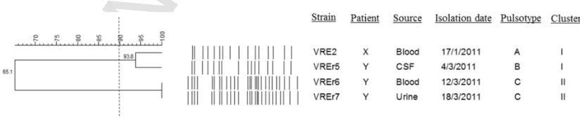
3.2. PFGE revealed genetic difference of strains isolated from a single patient
[image:3.595.90.502.615.700.2]PFGE subtyping of the four clinical strains revealed three pulso-types comprising 16–27 restriction fragments (Fig. 1). Cluster analy-sis at 90% similarity yielded two clusters. VREr6 and VREr7, which
Fig. 1.Dendrogram showing the cluster analysis of four VREfmstrains based on PFGE patterns of theSmaI-digested chromosomal DNA. The dendrogram was constructed using
UNCORRECTED
PROOF
were isolated from the same patient (patient Y), wereindistinguish-able (Cluster II). Interestingly, VREr5, which was also isolated from patient Y, was grouped into a different PFGE cluster (cluster I). In-stead, VREr5 was closely related to VRE2 (with two bands difference) which was isolated from patient X. These observations indicated that VREr5 is likely to be genetically dissimilar with VREr6 and VREr7, despite being isolated from the same host. The close relationship be-tween VRE2 and VREr5 based on the PFGE results suggested that VREr5 could have derived from the same clone as VRE2.
3.3. General genome signatures and multilocus sequence typing (MLST) of four Malaysian VREfmstrains
To understand the macro-restriction differences observed from the PFGE results, WGS of the four Malaysian strains were performed to dissect their genomes. The general genome features of the four VREfm strains are summarized in Table 1. The approximate genome sizes of these strains ranged from 2.8 Mbp to 3.0 Mbp. The guanine-cyto-sine (GC) contents and numbers of predicted protein coding sequences (CDSs) ranged from 37.6% to 37.8% and 2853 to 3057, respectively. Both VRE2 and VREr5 harbored 55 tRNA genes whereas VREr6 and VREr7 harbored 58 tRNA genes.
In silico MLST analysis revealed two sequence types (ST): VRE2 and VREr5 were of ST80 whereas VREr6 and VREr7 were of ST203. Both these sequence types were assigned to the clonal complex 17 (CC17), a specific lineage associated with nosocomial E. faecium strains (Top et al., 2008). ST203 is one of the members of the CC17, apart from ST17, ST18 and ST78. In Australia, ST203 has replaced ST17, the founder group of CC17 (Lam et al., 2013). There were 49 entries on ST203 in the PubMLST database (last accessed on 20th Aug 2016) (Homan et al., 2002) and the data showed that this ST is mainly found in European (49%) and Asia Pacific countries (51%). In Malaysia, ST203 was found in isolates recovered from both in-fected and colonized people (Getachew et al., 2013). In contrast, there were only 13 entries of ST80 in the MLST database which are mainly represented by strains originated from European countries, except for two entries with Asian strains. To the best of our knowledge, this is the first report of ST80 in Malaysia. Strains of CC17 have spread worldwide, including in Malaysia (Getachew et al., 2013; Weng et al., 2013). Our report on ST80 provides new insight into the dissemina-tion of this high-risk clonal complex in Malaysia.
3.4. Comparative genomics revealed variations in genes associated with fitness and adaptive advantage
[image:4.595.345.521.578.685.2]The genomes of the Malaysian strains were compared with that of E. faeciumAus0085 (GenBank accession number CP006620.1), one of the 13 completeE. faeciumgenomes known up to date. Aus0085 was chosen as a reference strain because it shares similar features with our four VREfm strains. This clinical VRE strain was from similar geographical region, isolated from blood sample of a bacteremia pa-tient (Lam et al., 2013). Moreover, this strain also has the same ST
Table 1
General genome features, sequence types, and clonal complex of the four VREfmstrains.
VRE2 VREr5 VREr6 VREr7
Genome size (bp) 2,862,609 2,898,367 3,014,993 3,021,201
GC% 37.85 37.8 37.61 37.6
CDS 2853 2906 3049 3057
tRNA 55 55 58 58
rRNA 3 3 3 3
Sequence type 80 80 203 203
Clonal complex 17 17 17 17
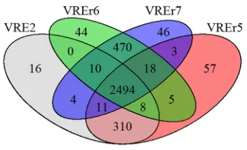
as two of our strains (VREr6, VREr7). Our comparative analyses showed that 2148 open reading frames (ORFs) were shared among all the genomes, which accounted for approximately 71.0% of the total ORFs present in each of the studied strains. This percentage is com-parable to that observed by Lam et al. (2013) who compared the con-served ORFs in Aus0085 with other clinical isolates, demonstrating that the core genome ofE. faeciumis stable. This core genome pro-vides evidence of the conservation of genes amongE. faeciumstrains. When the four local strains were compared among themselves, the number of core genes increased to 2494. There are 839 dispensable or accessory ORFs whereas the number of strain-specific ORFs ranged from 16 in VRE2 to 57 in VREr5 (Fig. 2). Most of these strain-spe-cific ORFs encode for hypothetical proteins and mobile element pro-teins, which might play a crucial role in determining distinct virulence features of each strain.
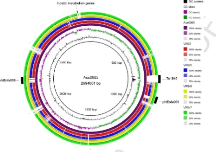
At least four chromosomal regions spanning approximately 169 kbp in Aus0085 showed low or no homology (< 70%) with the four Malaysian strains (Fig. 3). Most of these regions are genomic is-lands of Aus0085, suggesting that genomic isis-lands can contribute to the genome diversity ofE. faecium. Most notably, the four studied strains differed from the reference strain by the absence ofvanB -con-taining Tn1549transposon (Fig. 3). Instead, our VREfm strains har-boredvanAgene which is on the Tn1546transposon. Differences be-tween these twovangenotypes will be discussed in the antibiotic sec-tion. Furthermore, our results showed that VREr6 and VREr7 shared higher similarity with the reference genome compared to VRE2 and VREr5 (Fig. 3). This observation showed that VREr6 and VREr7 have substantial variations with VRE2 and VREr5. This was further sup-ported by the identification of different genes associated with fitness and adaptive advantage found only in both VRE2 and VREr5 and in both VREr6 and VREr7.
Both VRE2 and VREr5 harbored a cluster of proteins involved in inositol metabolism. Inositol is commonly found in soil and can be used as a sole carbon source in various microorganisms such as Rhizo-bium leguminosarum(Fry et al., 2001) andBacillus subtilis(Yoshida et al., 2008). Van Schaik et al. (2010) reported a 7 kb gene cluster encoding a complete inositol metabolism pathway in theesp patho-genicity island (PAI) of another infectiousE. faeciumstrain E1679 and demonstrated the capability of this strain to use inositol as a car-bon source. This 7 kb gene cluster shared identical amino acid sim-ilarity with the gene cluster found in both VRE2 and VREr5. Simi-lar to E1679, this gene cluster was also found integrated into theesp PAI of both VRE2 and VREr5. In contrast, homologs of this 7 kb gene cluster were not detected in the genomes of VREr6 and VR-Er7. The presence of transposases and mobile element proteins in the vicinity of this gene cluster suggested its horizontal origin. Inositol is maintained at a low concentration in blood and urine of healthy peo-ple but it is increased in patients with diabetes and diabetic-associated
Fig. 2.Venn diagram showing the distribution and number of core, dispensable and strain-specific genes of the Malaysian VREfmstrains. Each circle is labelled with the
[image:4.595.37.289.648.731.2]UNCORRECTED
[image:5.595.94.528.66.373.2]PROOF
Fig. 3.Circular genomic map and genome comparison of Aus0085, VRE2, VREr5, VREr6, and VREr7. The inner ring shows coordinate in scale and the total genome size of the reference sequence, Aus0085. The black histogram bar represents GC content whereas the purple-green histogram bar represents GC skew. Colored arches representing orthologous regions of each genome in respect to Aus0085 (purple arch) and are shown in the following order (inside to outside): Aus0085, VRE2, VREr5, VREr6, VREr7. The outermost arch (black) represents the location of Tn1546, phiEnfa005, phiEnfa006, and inositol catabolism genes relative to Aus0085. (For interpretation of the references to color in this figure legend, the reader is referred to the web version of this article.)
renal diseases (Hong et al., 2012). The ability to metabolize this sugar and use it as a carbon source may provide an additional growth advan-tage for these strains to outcompete other bacteria in diabetic patients, such as in patient Y.
It is important to note that the genome sizes of both VREr6 and VREr7 were approximately 140 kb larger than that of VRE2 and VR-Er5. This size variation was probably contributed by accessory genes which code for phage-related and plasmid-associated proteins. Among the plasmid-associated proteins, an atypical nonlantibiotic bacteriocin, Lactococcin 972 (Lcn972), was identified in VREr6 and VREr7 and shared 100% amino acid sequence identity with that of Aus0085. Bac-teriocins are peptides produced by an organism to inhibit the growth of its closely related species (Héchard and Sahl, 2002). Instead of tar-geting the cytoplasmic membrane and forming pores, Lcn972 inhibits septum formation which leads to deformation and eventually lysis of cells (Martinez et al., 2000). Martinez et al. (2008) reported that this bacteriocin disrupts cell wall biosynthesis by interacting with cell wall precursor lipid II, which is the primary docking site for lantibiotics prior to pore formation. Lcn972 is the first nonlantibiotic that specif-ically interacts with lipid II. Besides, Lcn972 also plays a role in the induction of prophages, although it is limited to specific prophage/host system (Madera et al., 2009). With its role in killing closely-related strains, the presence of Lcn972 in both VREr6 and VREr7 may in-crease the competitiveness of these two strains in colonizing and sub-sequently infecting the host.
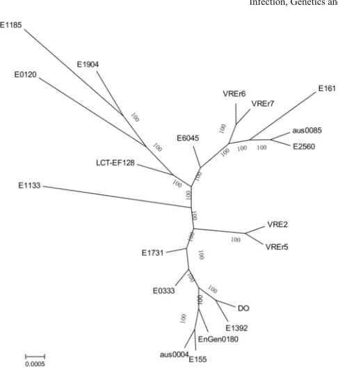
3.5. Phylogenomic analysis reveals shared common ancestry of clinical E. faecium and two distinct lineages of the local VREfm
strains
To better understand the relationship among the Malaysian strains and other global strains, a maximum-likelihood tree was constructed based on the core genome alignments of 20E. faeciumstrains using Aus0085 as the reference (Fig. 4). All these strains, except E0120, are from the CC17. Our results showed that VREr5 was distantly re-lated to VREr6 and VREr7, although they were all isore-lated from the same host. Instead, VREr5 was more closely related to VRE2 isolated from patient X. This observation is in concordance with the PFGE re-sults. In addition, VREr6 and VREr7 were closely related to strain E161 isolated from China. Notably, these two strains were also more closely related to the reference Aus0085 compared to VRE2 and VR-Er5, which concurred with the genomic variations discussed in the previous section (Fig. 3). The close relationship observed among our strains and other strains from the CC17 suggested a shared common ancestor among these clinical strains.
UNCORRECTED
[image:6.595.42.285.49.317.2]PROOF
Fig. 4.Phylogenomic tree inferred from approximately-maximum-likelihood method from aligned core genomes. Multiple genomes alignments are generated from 20 global E. faeciumstrains using Aus0085 as a reference. The unrooted phylogenomic tree is in-ferred via approximately-maximum likelihood method using FastTreeMP (Price et al., 2010). Bootstrap support values are shown in each node.
ferent subgroups using Bayesian Analysis of Population Structure (BAPS) model, with ST78 in BAPS 2-1 and ST17 and ST18 in BAPS group 3-3 (Willems et al., 2012). Due to high recombination rate of E. faecium, the inference that CC17 strains originated from a sin-gle founder using eBURST algorithm is questionable (Willems et al., 2012). BAPS is an alternative approach to study population structure that accounts for recombination events (Tang et al., 2009). Our re-sults agreed with BAPS analysis which indicated that ST17, ST18, and ST78, which were previously believed to have a same evolutionary history using the eBURST algorithm, were from two evolutionary tra-jectories which later converged into the same clonal complex through acquisition of genes that characterized CC17 (Willems et al., 2012).
Based on the phylogenomic analysis, together with the differences in PFGE profiles, sequence types and genomic contents, we showed that patient Y was most probably infected by VREfmstrains from more than one lineages. This was supported by retrospective analysis of clinical records which showed that patient Y underwent nasogastric tube exchange between the isolation period of VREr5 and VREr6. This medical procedure might have introduced new VREfm strains to the patient through contaminated catheters or hands of healthcare workers, hence VREr5, the initial strain isolated from patient Y, was different from the subsequent strains, VREr6 and VREr7. Moreover, since patient Y was admitted when patient X was still in the hospital, the genetic similarity between VRE2 and VREr5 suggested that VRE2 could have persisted and spread in the hospital.
3.6. Mobile genetic elements contribute to genomic plasticity
Several studies have highlighted the roles of mobile genetic el-ements in defining genome diversity of E. faecium strains (Qin et al., 2012; van Schaik et al., 2010). Insertion sequence (IS) elements, prophages, as well as Clustered Regularly Interspaced Short Palin
dromic Repeat (CRISPR) are among the major factors which could contribute to genome dynamics.
A total of 43 to 55 IS elements were identified in our strains. Ma-jority of them were members from the IS3(IS1485, ISEfa8, ISEfa10, ISEnfa3) and ISL3(IS1251, ISEfa11, IS1476, ISEfa5) families. IS16, which has been suggested as the molecular marker for hospital-asso-ciated enterococcus, was also observed (Werner et al., 2011). IS el-ements can contribute to genome plasticity of bacteria by triggering gene rearrangement, as well as gene inactivation that affects the inte-gration of phages and plasmids (Huh et al., 2004; Ooka et al., 2009). The different types of IS elements identified in the four sequenced strains may have different effects on their genomes, not only in the overall genome structure but also virulence and host adaptation.
Prophages ranging from 39.0 kb to 47.8 kb in size were predicted in the four genomes. Interestingly, high sequence similarity was ob-served in prophages from strains of the same sequence type. This included two regions in VREr6 and VREr7 which shared 93% to 95% sequence identity with phages phiEnfa005 and phiEnfa006 of Aus0085 (Fig. 3). The ORFs in all the predicted prophage regions mainly contained phage-specific proteins and hypothetical proteins (83.3% to 98.0%). VREr7 shared a similar prophage region with that of VRE2 and VREr5 except for an additional 10.8 kb region contain-ing genes encodcontain-ing glycopeptide resistance proteins, cadmium trans-porter, and RelB/RelE toxin-antitoxin system. This region is flanked by two transposases, suggesting that these additional genes were being acquired later by the prophage, indicating that prophages could play an important role in genome diversity of these four strains.
CRISPRs are repetitive sequences that, together with CRISPR-as-sociated (cas) genes, protect bacteria against integration of exogenous DNA such as phages and plasmids into their genomes (Palmer and Gilmore, 2010; Sorek et al., 2008). Qin et al. (2012) reported that clinicalE. faeciumlacks CRISPR loci and this probably explains the higher rate of exogenous DNA in their genomes compared to those of non-clinicalE. faeciumisolates. CRISPR-like regions (designated as questionable CRISPR by CRISPRs Finder) were identified in all four Malaysian strains. However, all these CRISPR-like regions ei-ther lacked anycasgenes or are located within a coding region. Simi-lar results were reported for strains E1071, E1679, and E0371 by van Schaik et al. (2010), who suggested that these partial CRISPR-like loci might not have any functional significance in these strains. The diver-sity in prophage sequences and abundance of uncharacterized mobile element proteins found in the genomes of our strains might in part due to these possibly non-functional CRISPR regions.
Our molecular subtyping using PFGE, MLST and phylogenomic tree suggested that VREr6 and VREr7 probably came from the same clone. However, the genomic variations contributed by mobile ge-netic elements allowed us to discriminate these strains. This highlights the discriminatory power of WGS in detecting fine differences of the same clone and hence provides a better resolution in distinguishing strains with high genetic similarity.
3.7. CDS associated with surface polysaccharides, microbial surface components recognizing adhesive matrix molecules (MSCRAMM), and putative virulence factors
The pathogenicity of VRE is contributed by several important vir-ulence factors which are mainly involved in adherence, biofilm for-mation, invasion and antiphagocytic activity. The virulence associated genes found in the genomes of the four analyzed strains are summa-rized in Table 2.
UNCORRECTED
[image:7.595.37.290.85.279.2]PROOF
Table 2
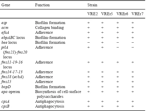
Selected virulence-associated genes identified in the four Malaysian VREfmstrains.
Gene Function Strain
VRE2 VREr5 VREr6 VREr7
esp Biofilm formation + + + +
acm Collagen binding + + + +
efaA Adherence + + + +
ebpABClocus Biofilm-formation + + + +
beelocus Biofilm formation − + − −
pilA (fms21)-fms20 locus
Adherence + + − −
fms11-19-16 locus
Adherence + + + +
fms14-17-13 Adherence + + + +
fms18(ecbA) Adherence + + + +
fms15 Adherence + + + +
bopD Biofilm formation + + + +
epaoperon Biosynthesis of cell-surface polysaccharides
+ + + +
cpsA Antiphagocytosis + + + +
cpsB Antiphagocytosis + + + +
“+” indicates the presence of virulence genes whereas “-” indicates the absence of virulence genes.
Theepagene cluster encodes for proteins involved in the biosynthe-sis of an antigenic cell wall polysaccharide composed of rhamnose, glucose, galactose, N-acetylgalactosamine and N-acetylglucosamine (Teng et al., 2009). Disruption of theepalocus showed attenuation in biofilm formation, phagocytic resistance and tissue invasion inE. fae-calis(Teng et al., 2009; Zeng et al., 2004). Theepagene cluster of E. faecalisconsists of 18 genes (epaAtoepaR) but only 15 of them (epaA-H,epaL-M,epaO-R) are found inE. faecium(Qin et al., 2012). Besides, these 15 genes are ordered differently as those found inE. faecalis(Qin et al., 2012). All the 15epagenes were present in our four VREfm strains, sharing same organization and high amino acid identities (88–100%) with that of the reference strain Aus0085. Qin et al. (2012) had reported the conservation of this gene cluster between TX16 and 21 E. faecium draft genomes. Our comparative genome analyses onepa gene cluster of TX16 further confirmed that these genes are part of the core genome ofE. faecium. The virulence nature as well as the conservation of theseepagenes inE. faeciumsuggests that these genes can serve as a potential target for new drug develop-ment against infections caused by this pathogen.
Surface expressed proteins such as pili and MSCRAMM are im-portant virulence factors involved in adhesion, biofilm formation, and invasion of Gram-positive bacteria. A total of 15 genes encoding cell-wall anchored proteins with MSCRAMM features were previ-ously identified in E. faecium TX16 (Nallapareddy et al., 2003; Sillanpää et al., 2009, 2008). Eleven of these genes form four gene clusters while the rest are present as a single gene. Three gene clusters, ebpABC,fms11-fms19-fms16, andfms14-fms17-fms13, were found in the genomes of all four local strains. Among these, theebpABC clus-ter, which encodes for biofilm-associated pili, was shown to be impor-tant in urinary tract infection in an animal model (Nallapareddy et al., 2011). Another gene cluster,fms21-fms20, was detected only in VRE2 and VREr5 but not in VREr6 and VREr7. Thefms21-fms20cluster is carried on a transferable plasmid (Kim et al., 2010), suggesting that VRE2 and VREr5 acquired these genes horizontally. All the four an-alyzed strains harboredacm, encoding the collagen binding protein Acm, which plays a significant role in endocarditis (Nallapareddy et al., 2008). Another collagen-binding protein, Fms18 (fms18) was also found in our strains. In contrast, the scm gene which encodes the second collagen-binding protein inE. faecium, was absent in all our strains.
Our search for enterococcal virulence factors using the virulence factor database (VFDB) also detected homologs of endocarditis spe-cific antigen EfaA (63% amino acid identity) and a sugar transcrip-tional regulator BopD (87% amino acid identity) of E. faecalisin the genomes of the four studied strains. Two capsule-associate genes (cpsA,cpsB) were also found in all the studied genomes, which may protect the strains from host phagocytic activities. Additionally, the enterococcal surface protein (Esp), which is the hallmark in clinical isolates, was also identified in these strains. Other major virulence fac-tors listed in the VFDB such as gelatinase and hyaluronidase were not detected in any of the four studied strains.
3.8. Homologs of E. faecalis bee locus revealed possible association with biofilm formation in VREr5
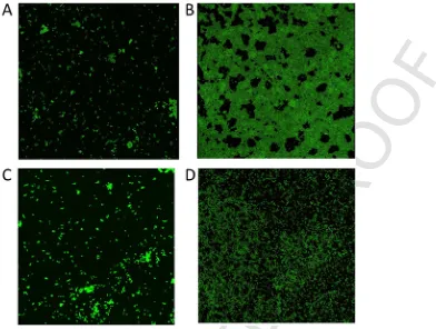
Biofilm formation is an important virulence factor of enterococci as these organisms are frequently isolated from biofilm-mediated in-fections such as endocarditis and those associated with indwelling medical devices (Donlan et al., 2002). Furthermore, there is a poten-tial transfer of enterococcal antibiotic resistance genes to other more pathogenic bacteria such asStaphylococcus aureusin mixed-species biofilm as the horizontal transfer rate of antimicrobial resistance genes is high in this type of biofilm (Donlan et al., 2002). Hence, crystal vi-olet assay was performed to investigate the biofilm forming ability of the four strains.
Based on the interpretation scheme adopted from Stepanović et al. (2000), our results showed that VREr5 was a strong biofilm producer whereas the rest of the strains were non-biofilm producers (Table 3). To confirm these results, CLSM was performed. The acquired CLSM images showed that VREr5 formed dense biofilm (Fig. 5). In con-trast, VRE2 and VREr6 can only form small aggregates. VREr7, on the other hand, produced a thin monolayer on the chamber slide. The CLSM images confirmed the biofilm forming ability of VREr5. How-ever, VREr7, which was classified as non-biofilm producer based on the crystal violet assay, showed potential biofilm forming ability as observed from the CLSM image. One of the possible explanations for these observations is the different abiotic surfaces used to grow biofilm in the crystal violet assay and for CLSM imaging. The effect of different abiotic substrates on bacterial biofilm formation had been demonstrated in other study (Tendolkar et al., 2004). Since VREr5 was the only strong biofilm former, we examined the genome of VR-Er5 and found five genes which resembled thebeelocus ofE. faecalis. The cut-off OD (ODc) was defined as three standard deviations above of the mean OD of the negative control. The biofilm forming ability of each strain was scored as follow: OD ≤ ODc = non-biofilm producer, ODc < OD ≤ (2 × ODc) = weak-biofilm producer, (2 × ODc) < OD ≤ (4 × ODc) = moderate-biofilm producer, OD > (4 × ODc) = strong-biofilm producer (Chelvam et al., 2014; Stepanović et al., 2000).
Thebeelocus (biofilmenhancer inenterococcus) is a cluster of biofilm-associated genes composed of three genes (bee-1, bee-2, bee-3) encoding putative cell wall-anchor proteins and two genes (str1,str2) encoding for sortases (Tendolkar et al., 2006). Insertion of
Table 3
Average results of three replicates of the crystal violet assay to determine biofilm form-ing potential.
Strain OD590 Interpretationa
VRE2 0.016 Non-biofilm producer
VREr5 1.780 Strong-biofilm producer VREr6 0.049 Non-biofilm producer VREr7 0.083 Non-biofilm producer
UNCORRECTED
[image:8.595.137.529.62.358.2]PROOF
Fig. 5.CLSM images of the four Malaysian VREfmstrains grown in TSB. Each image represents the “flatten” three-dimensional (3D) Z-projection of stack images of A) VRE2,
B) VREr5, C) VREr6, and D) VREr7. The non-biofilm formers (VRE2, VREr6) were either scattered around or formed small aggregates on the glass slide. VREr7 formed thin monolayer whereas VREr5 formed dense biofilm.
Tn917withinbee-2has led to a 70% reduction of biofilm formation in E. faecalisstrain E99 (Tendolkar et al., 2006). Four of the five unique genes of VREr5 showed high amino acid identity (99%) with Bee-2, Bee-3 and the two sortase proteins of thebeelocus whereas one of the unique genes showed only 39% amino acid identity with Bee-1. Despite its low amino acid similarity, the putative Bee-1 homolog of VREr5 shared similar structures of the Bee-1 protein. These included a region from residues 705 to 810 that showed low degree of sim-ilarity (E = 1.08e-03) to collagen binding B domain of Staphylococ-cus aureus, and a region from 338 to 469 that encodes for the Von Willebrand factor type A (VWA) domain. The VWA domain is usu-ally associated with pilus and can bind to a variety of ligands, includ-ing collagen, laminin and human epithelial cells (Konto-Ghiorghi et al., 2009; Whittaker and Hynes, 2002). Given the high similarity of the unique gene cluster of VREr5 to that ofbeelocus and being the only strong biofilm former, an association of thebeelocus homolog to biofilm formation cannot be ruled out. However, further analyses are needed to confirm this association.
The E. faecalis beelocus has been reported to be located on a large conjugative plasmid (Coburn et al., 2010). To determine the lo-cation of thebeehomolog in the VREr5, plasmid DNA was extracted and PCR using the primers targetingbee-1,bee-2 andbee-3 was per-formed. Our PCR results showed that all these three genes were suc-cessfully amplified (Data not shown), suggesting that thebeelocus homolog was most probably located on a plasmid.
3.9. Association between antibiotic resistance phenotypes and genotypes
Enterococci, particularlyE. faecium, are resistant to multiple an-tibiotics commonly use in treatment of enterococcal infections. This
resistance can be intrinsic or acquired through mutation of the in-trinsic genes or acquisition of mobile elements carrying the resis-tant determinants. The antibiotic susceptibility profile of the four se-quenced strains is shown in Table 4. All these strains were resistant to vancomycin, ampicillin, kanamycin, streptomycin, erythromycin, clindamycin and tetracycline. VRE2 was susceptible to gentamicin, whereas the other three strains were resistant to gentamicin. VRE2 and VREr5 showed intermediate resistance to teicoplanin whereas VREr6 and VREr7 were resistant to it. None of the sequenced strains was re-sistant to linezolid.
Based on the microbiological data from hospital records, no VRE strain was isolated from patient X and patient Y at the time of admis-sion. Within three months, VRE2 was isolated from patient X. For pa-tient Y, VREr5 was isolated after one month of admission and van-comycin treatment. These observations suggested that patient X and patient Y could have acquired VRE strains due to their prolonged hos-pitalization, or that vancomycin resistance of the colonized VRE was induced after vancomycin was prescribed.
UNCORRECTED
[image:9.794.72.736.69.340.2]PROOF
Table 4
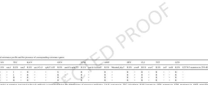
Antimicrobial resistance profile and the presence of corresponding resistance genes.
VAN TEC KAN GEN STM AMP ERY CLI TET LZD
R/I/S vanA R/I/S vanZ R/I/S aac(6′)-Ii aph(3′)-III R/I/S aac(6′)-aph(2″) R/I/S ant(6)-Ia/aadE R/I/S Mutatedpbp5 R/I/S ermB R/I/S msrC R/I/S tetS tetM R/I/S G2576T mutation in 23S rRNA
VRE2 R + I + R + + S − R + R + R + R + R + − R −
VREr5 R + I + R + + R + R + R + R + R + R + − R −
VREr6 R + R + R + + R + R + R + R + R + R − + R −
VREr7 R + R + R + + R + R + R + R + R + R − + R −
UNCORRECTED
PROOF
VanA phenotype (vancomycin MIC > 256 μg/ml, teicoplaninMIC = 16 μg/ml) whereas VREr6 and VREr7 displayed VanB phe-notype-vanA genotype, which were highly resistant to vancomycin (MIC > 256 μg/ml) but susceptible (MIC = 8 μg/ml) to teicoplanin. The occurrence of VanB phenotype-vanAgenotype isolates highlights the importance of MIC determination forvanAVRE in establishing the resistance phenotype and choice of treatment methods against this pathogen. It has been reported that point mutations ofvanSor disrup-tion ofvanYorvanZlead to impaired teicoplanin resistance invanA genotype VRE strains (Gu et al., 2009; Hashimoto et al., 2000). How-ever, no mutations or disruptions of these regions were observed in VREr6 and VREr7. Further analysis is needed to identify the cause of this impairment.
ThevanA-associated Tn1546transposon is polymorphic (Gagnon et al., 2011; Huh et al., 2004; Willems et al., 1999). Most of the known structural variations are caused by IS elements, which usually lead to complete or partial loss of thevanAcluster elements (Huh et al., 2004). Our detailed structural analysis revealed that the Tn1546-like transposons of the four studied strains were similar. The left end of the transposon, encoding a transposase (orf1) was truncated, with one IS1216Vinsertion directly beforeorf2. Moreover, an IS1251was found inserted in the intergenic region ofvanSandvanH. The struc-ture observed resembled type F reported by Willems et al. (1999). In-sertion of IS elements may change the level of vancomycin resistance by disrupting thevanelements, particularly through partial or com-plete deletion ofvanRandvanS(Gagnon et al., 2011). However, we could not verify the effects of IS insertions as all our strains had the same MIC (≥ 256 μg/ml).
Aminoglycoside resistance is mediated by three types of aminogly-coside modifying enzymes: N-Acetyltransferases (AAC), O-Adenyl-transferases (ANT), and O-phosphoO-Adenyl-transferases (APH) (Hollenbeck and Rice, 2012). Three genes of these enzymes,ant(6)-Ia,aac(6′)-Ii andaph(3′)-IIIwere found in all four VREfmstrains, which could con-fer resistance to streptomycin, tobramycin, and kanamycin, respec-tively. Theant(6)-Ia(also known asaadE) gene is often found in a cluster ofant(6)-sat4-aphA, which confers resistance to streptomycin, streptothricin, and kanamycin. This resistance gene cluster was iden-tified in all four studied strains, sharing 100% nucleotide identity with the published sequence reported by Werner et al. (2001) (Gen-Bank accession no. AF330699). Additionally, a bifunctional gene, aac(6′)-aph(2″), which confers high-level acquired resistance to gen-tamicin was identified in the Malaysian strains, except for VRE2. The absence of this resistance gene might explain the susceptibility of VRE2 towards gentamicin (M.I.C = 4 μg/ml). The rest of the three strains showed high-level resistance to gentamicin (M.I.C > 256 μg/ ml). Gentamicin is one of the recommended aminoglycosides used in combination with a cell wall active agent in the synergistic ther-apy of severe enterococcal infections (Hollenbeck and Rice, 2012). High-level resistance to gentamicin eliminates the synergistic killing effect, further challenging the development of new treatment methods against enterococcal diseases.
E. faeciumis intrinsically resistant to β-lactam drugs through the expression of low-affinity penicillin-binding protein 5 (PBP5). In-creased resistance to β-lactam drugs can be developed through the ac-quisition of β-lactamase orpbp5 mutation. Four β-lactamases were identified in each of the studied strains. These included two from met-allo-β-lactamase superfamily, one putative class C β-lactamase and one from unknown-class β-lactamase. However, high- level β-lactam resistance in E. faecium is commonly contributed by mutations in PBP5. In our study, three types of point mutations were observed in thepbp5 genes of the four sequenced strains: insertion of a ser-ine residue at position 466, after a serser-ine residue; replacement of
methionine at position 485 to alanine and replacement of glutamic acid at position 629 by valine. All these mutations are associated with increased MICs of β-lactam drugs (Rice et al., 2004).
The four strains were also resistant to erythromycin, which was conferred by theermBgene. Additionally, all the studied strains car-ried themsrC gene, a homolog of staphylococcal msrA, which en-codes an ABC efflux pump for macrolides and streptogramin B (Singh et al., 2001). Two types of tetracycline resistance genes were ob-served among the four studied strains. VRE2 and VREr5 harbored thetetSgene whereas VREr6 and VREr7 carried thetetMgene. Both these genes confer tetracycline resistance through ribosomal protec-tion and are mostly located on conjugative transposon or self-trans-ferrable plasmids (Charpentier et al., 1993; Martin et al., 1986).
Overall, our data showed high similarity in antibiotic resistance profile in the three strains isolated from patient Y, demonstrating that no additional resistance determinants were acquired or induced dur-ing the isolation period. There was a concordance in the observed an-tibiotic resistance phenotypes and the presence of the respective resis-tance genes or mutations
4. Conclusions
To the best of our knowledge, this is the first comparative genomic analysis on the Malaysian VREfm. Four clinical VREfm strains from two fatal cases were shown to be different based on the PFGE analy-sis, particularly three strains from a single patient (patient Y). Detailed genome analyses revealed that VREr5, the initial isolate from patient Y, was genetically more similar to that of VRE2 isolated from an in-dex case (patient X). Molecular analyses including MLST and phy-logenomics further indicated that VREr5 was different from the other two strains (VREr6, VREr7) isolated from patient Y. Coupled with clinical data, these results revealed that patient Y was infected with multiple strains of VREfmfrom different clones. Additionally, the first isolated strain from patient Y was probably of the same clone as the strain from the index case, patient X. Our study also detected a number of antibiotic resistance- and virulence genes in the four VRE genomes which could contribute to their persistence and pathogenicity. Further-more, there is a possible association of the homolog ofE. faecalis bee locus with the potential to form biofilm. This is the first report on the emergence of ST80 VRE strains in Malaysia.
Overall, our molecular analyses have shed light on the genetic re-latedness of the VREfmstrains isolated from the two clinical cases, as well as the virulence nature of these strains. Future study to include a bigger sample size of the local strains should be considered to provide a better overview on the genome diversity of VREfmin Malaysia.
Uncited reference
Derbise et al., 1997
Acknowledgements
UNCORRECTED
PROOF
References
Alikhan, N.-F., Petty, N.K., Ben Zakour, N.L., Beatson, S.A., 2011. BLAST ring image generator (BRIG): simple prokaryote genome comparisons. BMC Ge-nomics 12, 402.
Arthur, M., Courvalinn, P., 1993. Genetics and mechanisms of glycopeptide resistance in enterococci. Antimicrob. Agents Chemother. 37, 1563–1571.
Aziz, R.K., Bartels, D., Best, A.A., DeJongh, M., Disz, T., Edwards, R.A., Formsma, K., Gerdes, S., Glass, E.M., Kubal, M., Meyer, F., Olsen, G.J., Olson, R., Oster-man, A.L., Overbeek, R.A., McNeil, L.K., Paarmann, D., Paczian, T., Parrello, B., Pusch, G.D., Reich, C., Stevens, R., Vassieva, O., Vonstein, V., Wilke, A., Zag-nitko, O., 2008. The RAST server: rapid annotations using subsystems technology. BMC Genomics 9, 75.
Baldassarri, L., Cecchini, R., Bertuccini, L., Ammendolia, M.G., Iosi, F., Arciola, C.R., Montanaro, L., Di Rosa, R., Gherardi, G., Dicuonzo, G., Orefici, G., Creti, R., 2001. Enterococcus spp. produces slime and survives in rat peritoneal macrophages. Med. Microbiol. Immunol. 190, 113–120.
Bertels, F., Silander, O.K., Pachkov, M., Rainey, P.B., van Nimwegen, E., 2014. Auto-mated reconstruction of whole genome phylogenies from short sequence reads. Mol. Biol. Evol. 31, 1077–1088.
Bonten, M.J.M., Willems, R., Weinstein, R.A., 2001. Vancomycin-resistant entero-cocci: why are they here, and where do they come from?. Lancet Infect. Dis. 1, 314–325.
Cha, J.O., Jung, Y.H., Lee, H.R., Yoo, J.I., Lee, Y.S., 2012. Comparison of genetic epidemiology of vancomycin-resistant Enterococcus faecium isolates from humans and poultry. J. Med. Microbiol. 61, 1121–1128.
Charpentier, E., Gerbaud, G., Courvalin, P., 1993. Characterization of a new class of tetracycline-resistance gene tet(S) in Listeria monocytogenes BM4210. Gene 131, 27–34.
Chelvam, K.K., Chai, L.C., Thong, K.L., 2014. Variations in motility and biofilm for-mation of Salmonella enterica serovar Typhi. Gut Pathog. 6, 2.
Chen, L., Yang, J., Yu, J., Yao, Z., Sun, L., Shen, Y., Jin, Q., 2005. VFDB: a reference database for bacterial virulence factors. Nucleic Acids Res. 33, 325–328. CLSI, 2016. Performance standards for antimicrobial susceptibility testing. In: 26th
In-formational Supplement, Document M100-S26. Clin. Lab. Stand. Inst, Wayne, PA. Coburn, P.S., Baghdayan, A.S., Craig, N., Burroughs, A., Tendolkar, P., Miller, K.,
Najar, F.Z., Roe, B.A., Shankar, N., 2010. A novel conjugative plasmid from Ente-rococcus faecalis E99 enhances resistance to ultraviolet radiation.
Plas-mid 64, 18–25.
Conesa, A., Gotz, S., Garcia-Gomez, J.M., Terol, J., Talon, M., Robles, M., 2005. Blast2GO: a universal tool for annotation, visualization and analysis in functional genomics research. Bioinformatics 21, 3674–3676.
Darling, A.C.E., Mau, B., Blattner, F.R., Perna, N.T., 2004. Mauve: multiple alignment of conserved genomic sequence with rearrangements. Genome
Res. 14, 1394–1403. .
Donlan, R.M., Costerton, J.W., Donlan, R.M., Costerton, J.W., 2002. Biofilms: sur-vival mechanisms of clinically relevant microorganisms. Clin. Microbiol. Rev. 15, 167–193.
Edelsberg, J., Weycker, D., Barron, R., Li, X., Wu, H., Oster, G., Badre, S., Lange-berg, W.J., Weber, D.J., 2014. Prevalence of antibiotic resistance in US hospitals. Diagn. Microbiol. Infect. Dis. 78, 255–262.
Fry, J., Wood, M., Poole, P.S., 2001. Investigation of myo-inositol catabolism in Rhi-zobium leguminosarum bv. viciae and its effect on nodulation competitiveness. Mol. Plant-Microbe Interact. 14, 1016–1025.
Gagnon, S., Lévesque, S., Lefebvre, B., Bourgault, A.M., Labbé, A.C., Roger, M., 2011. vanA-Containing Enterococcus faecium susceptible to vancomycin and te-icoplanin because of major nucleotide deletions in Tn1546. J. Antimicrob. Chemother. 66, 2758–2762.
Getachew, Y., Hassan, L., Zakaria, Z., Saleha, A.A., 2013. Genetic variability of van-comycin-resistant Enterococcus faecium and Enterococcus faecalis isolates from humans, chickens, and pigs in Malaysia. Appl. Environ.
Micro-biol. 79, 4528–4533.
Grissa, I., Vergnaud, G., Pourcel, C., 2008. CRISPRFinder: a website to compare clus-tered regularly interspaced short palindromic repeats. Nucleic Acids
Res. 36, 52–57.
Gu, L., Cao, B., Liu, Y., Guo, P., Song, S., Li, R., Dai, H., Wang, C., 2009. A new Tn1546 type of VanB phenotype-vanA genotype vancomycin-resistant Enterococ-cus faecium isolates in mainland China. Diagn. Microbiol. Infect. Dis. 63, 70–75. Hashimoto, Y., Tanimoto, K., Ozawa, Y., Murata, T., Ike, Y., 2000. Amino acid sub-stitutions in the VanS sensor of the VanA-type vancomycin-resistant enterococcus strains result in high-level vancomycin resistance and low-level teicoplanin resis-tance. FEMS Microbiol. Lett. 185, 247–254.
Héchard, Y., Sahl, H.G., 2002. Mode of action of modified and unmodified bacteri-ocins from gram-positive bacteria. Biochimie 84, 545–557.
Heikens, E., Singh, K.V., Jacques-Pakaz, K.D., Luit-asbroek, M.V., Oostdijk, E.A., Bonten, M.J.M., Murray, B.E., Willems, R.J.L., 2012. Contribution of the entero-coccal surface protein Esp to pathogenesis of Enterococcus faecium endocarditis. Microbes Infect. 13, 1185–1190.
Hollenbeck, B.L., Rice, L.B., 2012. Intrinsic and acquired resistance mechanisms in enterococcus. Virulence 3, 421–433.
Homan, W.L., Tribe, D., Poznanski, S., Li, M., Hogg, G., Spalburg, E., Embden, J.D.a.V., Willems, J.L., Willems, R.J.L., 2002. Multilocus sequence typing scheme for Enterococcus faecium. J. Clin. Microbiol. 40, 1963–1971.
Hong, J.H., Jang, H.W., Kang, Y.E., Lee, J.H., Kim, K.S., Kim, H.J., Park, K.R., Ku, B.J., 2012. Urinary chiro- and myo-inositol levels as a biological marker for type 2 diabetes mellitus. Dis. Markers 33, 193–199.
Huh, J.Y., Lee, W.G., Lee, K., Shin, W.S., 2004. Distribution of insertion sequences associated with Tn1546-like elements among Enterococcus faecium isolates from patients in Korea. J. Clin. Microbiol. 42, 1897–1902.
Hyatt, D., Chen, G.-L., Locascio, P.F., Land, M.L., Larimer, F.W., Hauser, L.J., 2010. Prodigal: prokaryotic gene recognition and translation initiation site identification. BMC Bioinf. 11, 119.
Ibrahim, R., Mohamad, M., Rahman, M., 2010. Enterococci: emerging drug resistant bacteria in hospital acquired infections at Hospital Kuala Lumpur, Malaysia. Int. J. Microbiol. 9, 1–7.
Ibrahim, R.B., Mohamad, M., Rahman, M.M., 2011. Vancomycin resistant enterococci and detection of responsible genes. Pak. J. Med. Sci. 27, 784–788.
Jurcisek, J.A., Dickson, A.C., Bruggeman, M.E., Bakaletz, L.O., 2011. In vitro biofilm formation in an 8-well chamber slide. J. Vis. Exp. 9–10.
Kim, D.S., Singh, K.V., Nallapareddy, S.R., Qin, X., Panesso, D., Arias, C.A., Murray, B.E., 2010. The fms21 (pilA)-fms20 locus encoding one of four distinct pili of En-terococcus faecium is harboured on a large transferable plasmid associated with gut colonization and virulence. J. Med. Microbiol. 59, 505–507.
Konto-Ghiorghi, Y., Mairey, E., Mallet, A., Duménil, G., Caliot, E., Trieu-Cuot, P., Dramsi, S., 2009. Dual role for pilus in adherence to epithelial cells and biofilm formation in Streptococcus agalactiae. PLoS Pathog. 5.
Kuo, A.J., Su, L.H., Shu, J.C., Wang, J.T., Wang, J.H., Fung, C.P., Chia, J.H., Lu, J.J., Wu, T.L., 2014. National surveillance on vancomycin-resistant Enterococcus fae-cium in Taiwan: emergence and widespread of ST414 and a Tn1546-like element with simultaneous insertion of IS1251-like and IS1678. PLoS One 9, 1–13. Lam, M.M.C., Seemann, T., Tobias, N.J., Chen, H., Haring, V., Moore, R.J., Ballard,
S., Grayson, L.M., Johnson, P.D.R., Howden, B.P., Stinear, T.P., 2013. Compara-tive analysis of the complete genome of an epidemic hospital sequence type 203 clone of vancomycin-resistant Enterococcus faecium. BMC Genomics 14, 595. Leclercq, R., Derlot, E., Duval, J., Courvalin, P., 1988. Plasmid-mediated resistance to
vancomycin and teicoplanin in Enterococcus faecium. N. Engl. J. Med. 319, 157–161.
Liu, B., Pop, M., 2009. ARDB - antibiotic resistance genes database. Nucleic Acids Res. 37, 443–447.
Madera, C., Garcia, P., Rodriguez, A., Suarez, J.E., Martinez, B., 2009. Prophage in-duction in Lactococcus lactis by the bacteriocin Lactococcin 972. Int. J. Food Mi-crobiol. 129, 99–102.
Martin, P., Trieu-cuot, P., Courvalin, P., 1986. Nucleotide sequence of the tetM tetra-cycline resistance determinant of the streptococcal conjugative shuttle transposon Tn1546. Nucleic Acids Res. 14, 7047–7058.
Martinez, B., Bottiger, T., Schneider, T., Rodriguez, A., Sahl, H.G., Wiedemann, I., 2008. Specific interaction of the unmodified bacteriocin lactococcin 972 with the cell wall precursor lipid II. Appl. Environ. Microbiol. 74, 4666–4670.
Martinez, B., Rodriguez, A., Suarez, J.E., 2000. Lactococcin 972, a bacteriocin that in-hibits septum formation in lactococci. Microbiology 146, 949–955.
Matsushima, A., Takakura, S., Yamamoto, M., Matsumura, Y., Shirano, M., Nagao, M., Ito, Y., Iinuma, Y., Shimizu, T., Fujita, N., Ichiyama, S., 2012. Regional spread and control of vancomycin-resistant Enterococcus faecium and Enterococ-cus faecalis in Kyoto, Japan. Eur. J. Clin. Microbiol. Infect. Dis. 31, 1095–1100. Mohamed, N.A., Hussin, H., Chang, K.M., Hashim, R., Ahmad, N., 2015.
Van-comycin resistant enterococcus (VRE): prevalence and characteristics in a tertiary hospital in Malaysia. Brunei Int. Med. J. 11, 241–246.
Molton, J.S., Tambyah, P.A., Ang, B.S.P., Ling, M.L., Fisher, D.A., 2013. The global spread of healthcare-associated multidrug-resistant bacteria: a perspective from Asia. Clin. Infect. Dis. 56, 1310–1318.
Nallapareddy, S.R., Singh, K.V., Sillanpaa, J., Zhao, M., Murray, B.E., 2011. Relative contributions of Ebp Pili and the collagen adhesin ace to host extracellular matrix protein adherence and experimental urinary tract infection by Enterococcus fae-calis OG1RF. Infect. Immun. 79, 2901–2910.
Nallapareddy, S.R., Singh, K.V., Murray, B.E., 2008. Contribution of the collagen ad-hesin Acm to pathogenesis of Enterococcus faecium in experimental endocarditis. Infect. Immun. 76, 4120–4128.
Nallapareddy, S.R., Weinstock, G.M., Murray, B.E., 2003. Clinical isolates of Entero-coccus faecium exhibit strain-specific collagen binding mediated by Acm, a new member of the MSCRAMM family. Mol. Microbiol. 47, 1733–1747.
UNCORRECTED
PROOF
(IS) elements on bacterial genome diversification through analysis of small-sizestructural polymorphisms in Escherichia coli O157 genomes. Genome Res. 19, 1809–1816.
Palmer, K.L., Gilmore, M.S., 2010. Multidrug-resistant enterococci lack CRISPR-cas. MBio 1, 1–10.
Price, M.N., Dehal, P.S., Arkin, A.P., 2010. FastTree 2 - approximately maxi-mum-likelihood trees for large alignments. PLoS One 5.
Qin, X., Galloway-Peña, J.R., Sillanpaa, J., Roh, J.H., Nallapareddy, S.R., Chowdhury, S., Bourgogne, A., Choudhury, T., Muzny, D.M., Buhay, C.J., Ding, Y., Dugan-Rocha, S., Liu, W., Kovar, C., Sodergren, E., Highlander, S., Petrosino, J.F., Worley, K.C., Gibbs, R.A., Weinstock, G.M., Murray, B.E., 2012. Complete genome sequence of Enterococcus faecium strain TX16 and comparative genomic analysis of Enterococcus faecium genomes. BMC Microbiol. 12, 135.
Rice, L.B., Bellais, S., Carias, L.L., Hutton-Thomas, R., Bonomo, R.A., Caspers, P., Page, M.G.P., Gutmann, L., 2004. Impact of specific pbp5 mutations on expres-sion of beta-lactam resistance in Enterococcus faecium. Antimicrob. Agents Chemother. 48, 3028–3032.
Siguier, P., Perochon, J., Lestrade, L., Mahillon, J., Chandler, M., 2006. ISfinder: the reference centre for bacterial insertion sequences. Nucleic Acids
Res. 34, D32–D36.
Sillanpää, J., Nallapareddy, S.R., Prakash, V.P., Qin, X., Hook, M., Weinstock, G.M., Murray, B.E., 2008. Identification and phenotypic characterization of a second col-lagen adhesin, Scm, and genome-based identification and analysis of 13 other pre-dicted MSCRAMMs, including four distinct pilus loci, in Enterococcus faecium. Microbiology 154, 3199–3211.
Sillanpää, J., Prakash, V.P., Nallapareddy, S.R., Murray, B.E., 2009. Distribution of genes encoding MSCRAMMs and pili in clinical and natural populations of Ente-rococcus faecium. J. Clin. Microbiol. 47, 896–901.
Singh, K.V., Malathum, K., Murray, B.E., 2001. Disruption of an Enterococcus fae-cium species-specific gene, a homologue of acquired macrolide resistance genes of staphylococci, is associated with an increase in macrolide susceptibility. Antimi-crob. Agents Chemother. 45, 263–266.
Sorek, R., Kunin, V., Hugenholtz, P., 2008. CRISPR—a widespread system that pro-vides acquired resistance against phages in bacteria and archaea. Nat. Rev. Micro-biol. 6, 181–186.
Stepanović, S., Vuković, D., Dakić, I., Savić, B., Švabić-Vlahović, M., 2000. A modi-fied microtiter-plate test for quantification of staphylococcal biofilm formation. J. Microbiol. Methods 40, 175–179.
Tang, J., Hanage, W.P., Fraser, C., Corander, J., 2009. Identifying currents in the gene pool for bacterial populations using an integrative approach. PLoS Comput. Biol. 5.
Tendolkar, P.M., Baghdayan, A.S., Michael, S., 2004. Enterococcal surface protein, Esp, enhances biofilm formation by Enterococcus faecalis. Infect. Im-mun. 72, 6032–6039.
Tendolkar, P.M., Baghdayan, A.S., Shankar, N., 2006. Putative surface proteins en-coded within a novel transferable locus confer a high-biofilm phenotype to Entero-coccus faecalis. J. Bacteriol. 188, 2063–2072.
Teng, F., Singh, K.V., Bourgogne, A., Zeng, J., Murray, B.E., 2009. Further characteri-zation of the epa gene cluster and Epa polysaccharides of Enterococcus faecalis. Infect. Immun. 77, 3759–3767.
Top, J., Willems, R., Bonten, M., 2008. Emergence of CC17 Enterococcus faecium: from commensal to hospital-adapted pathogen. FEMS Immunol. Med. Micro-biol. 52, 297–308.
Treitman, A.N., Yarnold, P.R., Warren, J., Noskin, G.A., 2005. Emerging incidence of Enterococcus faecium among hospital isolates (1993 to 2002). J. Clin. Micro-biol. 43, 462–463.
Turabelidze, D., Kotetishvili, M., Jr, J.G.M., Sulakvelidze, A., Kreger, A., Morris, J.G., 2000. Improved pulsed-field gel electrophoresis for typing vancomycin-resis-tant enterococci. J. Clin. Microbiol. 38, 4242–4245.
Uttley, A., Collins, C., Naidoo, J., George, R., 1988. Vancomycin-resistant entero-cocci. Lancet 331, 57–58.
van Schaik, W., Top, J., Riley, D.R., Boekhorst, J., Vrijenhoek, J.E.P., Schapendonk, C.M.E., Hendrickx, A.P., Nijman, I.J., Bonten, M.J.M., Tettelin, H., Willems, R.J.L., 2010. Pyrosequencing-based comparative genome analysis of the nosoco-mial pathogen Enterococcus faecium and identification of a large transferable pathogenicity island. BMC Genomics 11, 239.
Weng, P.L., Ramli, R., Shamsudin, M.N., Cheah, Y., Hamat, R.A., 2013. High genetic diversity of Enterococcus faecium and Enterococcus faecalis clinical isolates by pulsed-field gel electrophoresis and multilocus sequence typing from a hospital in Malaysia. Biomed. Res. Int. 2013, 938937.
Werner, G., Fleige, C., Geringer, U., van Schaik, W., Klare, I., Witte, W., 2011. IS ele-ment IS16 as a molecular screening tool to identify hospital-associated strains of Enterococcus faecium. BMC Infect. Dis. 11, 80.
Werner, G., Hildebrandt, B., Witte, W., 2001. Aminoglycoside-streptothricin resis-tance gene cluster aadE-sat4-aphA-3 disseminated among multiresistant isolates of Enterococcus faecium. Antimicrob. Agents Chemother. 45, 3267–3269. Whittaker, C.A., Hynes, R.O., 2002. Distribution and evolution of von
Willebrand/in-tegrin A domain: widely dispersed domains with roles in cell adhesion and else-where. Mol. Biol. Cell 13, 3369–3387.
Willems, R.J., van Schaik, W., 2009. Transition of Enterococcus faecium from com-mensal organism to nosocomial pathogen. Future Microbiol 4, 1125–1135. Willems, R.J.L., Top, J., van den Braak, N., van Belkum, A., Mevius, D.J., Hendriks,
G., Van Santen-verheuvel, M., Embden, J.D.A.V., 1999. Molecular diversity and evolutionary relationships of Tn1546-like elements in enterococci from humans and animals. Society 43, 483–491.
Willems, R.J.L., Top, J., van Schaik, W., Leavis, H., Bonten, M., Sirén, J., Hanage, W.P., Corander, J., 2012. Restricted gene flow among hospital subpopulations of Enterococcus faecium. MBio 3, 1–10.
Yap, K., Gan, H.M., Shuan, C., Teh, J., Chai, L.C., Thong, K.L., 2014. Comparative genomics of closely related Salmonella enterica serovar Typhi strains reveals genome dynamics and the acquisition of novel pathogenic elements. BMC Ge-nomics 15, 1–20.
Yoo, S.J., Sung, H., Cho, Y.-U., Kim, M.-N., Pai, C.H., Kim, Y.S., 2006. Role of hori-zontal transfer of the transposon Tn1546 in the nosocomial spread of vanA van-comycin-resistant enterococci at a tertiary care hospital in Korea. Infect. Control Hosp. Epidemiol. 27, 1081–1087.
Yoshida, K.I., Yamaguchi, M., Morinaga, T., Kinehara, M., Ikeuchi, M., Ashida, H., Fujita, Y., 2008. myo-Inositol catabolism in Bacillus subtilis. J. Biol. Chem. 283, 10415–10424.
Zankari, E., Hasman, H., Cosentino, S., Vestergaard, M., Rasmussen, S., Lund, O., Aarestrup, F.M., Larsen, M.V., 2012. Identification of acquired antimicrobial re-sistance genes. J. Antimicrob. Chemother. 67, 2640–2644.
Zeng, J., Teng, F., Weinstock, G.M., Murray, B.E., 2004. Translocation of Enterococ-cus faecalis strains across a monolayer of polarized human enterocyte-like T84 cells. J. Clin. Microbiol. 42, 1149–1154.
Zheng, B., Tomita, H., Xiao, Y.H., Wang, S., Li, Y., Ike, Y., 2007. Molecular charac-terization of vancomycin-resistant Enterococcus faecium isolates from mainland China. J. Clin. Microbiol. 45, 2813–2818.
Zhou, Y., Liang, Y., Lynch, K.H., Dennis, J.J., Wishart, D.S., 2011. PHAST: a fast phage search tool. Nucleic Acids Res. 39, 1–6.
Zhu, W., Clark, N., Patel, J.B., 2013. pSK41-like plasmid is necessary for inc18-like vanA plasmid transfer from Enterococcus faecalis to Staphylococcus aureus in vitro. Antimicrob. Agents Chemother. 57, 212–219.
Zubaidah, A.W., Ariza, A., Azmi, S., 2006. Hospital-acquired vancomycin-resistant enterococci: now appearing in Kuala Lumpur Hospital. Med. J.