5
X
October 2017
164
©IJRASET (UGC Approved Journal): All Rights are Reserved
Performance Analysis of Adaptive NEBF Filter
Using Cellular Automata for Retinal Vessel
Segmentation
Naina Singh1, Aarti2 1,
2 Computer Engineering & Technology Department, Amritsar College of Engineering and Technology
Abstract: Explicit faultless vessel detection in eye retina image is vital but unfortunately time consuming and tedious task. Detecting the exudates in the retina image where number of other abnormalities may exist which makes it more strenuous in nature. The main aim of segmenting the retina image is that to have a clear illustration of the image so that it can be analysed easily. An enhanced version of NEBF filter utilized to inpaint the exudate in the eye in a specific pattern by detecting nearby false positives which has to be deduced in number to get a better improved detection during segmentation of vessels. The Enhanced Novel Exudate Method is utilized for Inpainting for Retinal Vessel Segmentation in Cellular Domain using Cellular Automation.
Keywords: Exudate, Retina image, Cellular Domain, ANEBF Filter, Vessel Segmentation.
I. INTRODUCTION A. Image Segmentation
Image segmentation is the division of an image into meaningful structures. It is often an essential step in image analysis, object representation, visualization, and many other image processing tasks. Image segmentation is definitely the procedure connected with delegating the indicate to each pixel inside a picture along with exactly the same content label share specified visible traits. It is definitely the procedure connected with partitioning an electronic digital image in to several segments.
In computer prescient vision, image segmentation is usually particles partitioning an electronic digital photo into various segments also called super-pixels. The intention of segmentation should be to make simpler and to alter the manifestation associated with a photo into one thing that's more special and much easier in order to analyse. Image segmentation is commonly employed to seek out things and restrictions within images. Image segmentation is usually particles determining a brand to each pixel in the photo techniques pixels with the exact same brand talk about specified characteristics.
B. Retinal Vessel Segmentation
Retinal vessel segmentation algorithms usually are an elementary piece of programmed retinal disease assessment systems. By means of examining and revealing of vasculature components within retinal illustrations, we could very early diagnose a type 2 diabetes within sophisticated levels in comparison of the declares of retinal bloodstream vessels. Segmentation of veins within retinal illustrations will allow very early diagnosis of disease, using this method gives many benefits. A vascular system usually mapped by hand which is the time-consuming method that needs each coaching and skill. Automating the task will allow consistency, above all, saving plenty of time of which an expert professional or even medical doctor would commonly apply regarding information screening.
II. PROPOSEDTECHNIQUE
A. Adaptive NEBF Filter
ANEBF stands for adaptive Neighborhood Estimator Before Filling filter which is an enhanced version of NEBF filter utilized to inpaint the exudate in the eye in a specific pattern by detecting nearby false positives which has to be deduced in number to get a better improved detection during segmentation of vessels
B. Cellular Automation
165
©IJRASET (UGC Approved Journal): All Rights are Reserved
homogeneous houses, cellphone houses, tessellation houses, and also iterative arrays. A cellular automaton is made common grid of skin cells, each inside one among your specific quantity of states, just like on plus off. The grid might be in different specific quantity of dimensions. For each and every cell, some skin cells identified as the local community is defined family of which can help determine the modern point out for each cell with regards to the latest point out in the cell as well as the suggests of cellular structure in their local community.
III.RELATEDWORK
Annunziata et al. [1] has presented precise vessel detection within retinal images is a vital and tedious task. Diagnosis of retinal images is actually more challenging in pathological images with all the latest presence of exudates along with other abnormalities. Retinal vascular enhancement is attained through a multiple-scale Hessian approach whereas Wang et al [2] has presented the choroid which is actually a vascular layer holding the main job of supplying the right amount of oxygen and nourishment to the eye retina. Any kind of change in thickness or thinness in the size of choroid happen to be hypothesized in order to connect with a large variety of retinal diseases while in the pathophysiology. Further Sundaresh et al [3] has presented the lamina cribrosa (LC) is a kind of tissue the connects in the eye within the posterior area with an intricate mesh-like extremely small structure with the help of which all of the retinal ganglion cell axons pass. Gou et al [4] has presented the extraction of retinal exudates in any form is significant for diagnosis of eye diseases. Currently, the usual extracting of the vessels which are exudates in the retinal images with very low contrast and various widths is a bottleneck. Jingyun et al [5] has presented that the branch retinal artery occlusion which is definitely an ocular emergency which can result in partial blindness or complete blindness. Calculative data derived from BRAO region in the eye is important to measure the amount of danger the eye will have to face in case of adverse effect on eye. Shuxia et al [6] has presented the Choroid neo vascularization (CNV) is the outcome of the abnormality that states that a blood stream in the eye is passing through brunch membrane of eye which is undesirable and hazadourous for the health of eye Alauddin et al [7] has presented that this retinal vascular multilevel pattern is unique so that you can every which usually can be used for human being identity throughout biometric authentication. Within this research, this creator have suggested your new biometric verification method working and recognizing all these aspect points. The main veins characterized by way of breadth and time-span will be determined from the segmented vascular circle Priyadarshini et al [8] has presented that an automated circulation system segmentation program protocol for that retinal photograph beneath pathological ailments for Person suffering from diabetes Retinopathy using printed filters in addition to administered distinction techniques. Zhitao et al [9] has presented that the distinction constrained flexible histogram equalization is usually utilized to improve distinction with the picture having anisotropic diffusion equation. The highly advancement result is computed through the superposition within a dozen directions. a non-vessel is taken away in accordance with the bimodality regarding histogram of the graphic after advancement and also smoothing. Yuansheng et al [10] has presented the vessel segmentation with digital retinal photos employs a huge role around diagnosis of illnesses including people with diabetes along with retinopathy with prematurity because of these types of illnesses effect a retina. Some sort of consecutive convolution cellular levels and also pooling cellular levels keep to the input data, so your multilevel can certainly find out the fuzy features so that you can segment retinal vessel. Rossant et al [11] has presented the approach which is committed by using substantial improvements resulting from vasculitis, however it is as well genuine for vessel segmentation by using average need of alteration. That uses a presegmentation measure which is essential for any robustness in addition to accuracy and reliability of your results. Khaliq et al [12] has presented that the Diabetes retinopathy (DR) is definitely the foremost ophthalmic illness owing to variant in blood vessels framework which could induce blndness. The actual retinal problematic vein morphology distinguishes a accelerating stages of development of varied sight debilitating illnesses and thus clears a strategy to help characterize it has the seriousness. Qiaoliang et al [13] has presented a broad and strong nerve organs system with formidable induction potential is actually consist of design of the particular transformation. Chengzhang et al [14] has presented a method for segmentation of retina eye image with the help of a technique known as extreme learning machine which uses 3-D element view of every pixel of an eye image differencing with the help of neighboring pixels and functions implied on those pixels
IV.GAPSINLITERATURE
Number of issues has been discovered by through study of literature survey. These issues have not been yet explored. Mostly the existing exudate segmentation techniques suffer from the following issues in prior studies which are:
166
©IJRASET (UGC Approved Journal): All Rights are Reserved
B. Poor computational speed can also be considered as major gap found in most of the retinal images.
C. Transform based methods which can be used to improve the speed of segmentation which was ignored in this.
V. PROPOSEDMETHODOLOGY
A. Adaptive NEBF Filter
Step 1- Firstly Initialize the image
Step 2- Then, Input the retina eye image which is to be segmented Step 3- Convert the imputed retina eye image into cellular domain
Step 4- Next step will involve applying an Adaptive Neighbourhood Estimator before Filling (ANEBF) Filter on image Step 5- Now Apply vessel segmentation to that image
Step 6- Then exudate the segmented image to have clear view of retina veins in the eye image Step 7 –Then, Apply morphological filter to exudate image of retina eye
Step 8 - Evaluate the parameters on final output retina image
VI.EXPERIMENTALSETUPANDRESULTS
A. Data Set Used
Chase Data Set: The human being retina offers the possibilities to disclose information pertaining to retinal, ophthalmic, and in some cases systemic ailments such as type 2 diabetes, high blood pressure, as well as arteriosclerosis. The purpose is definitely assessed within the openly accessible DRIVE as well as STARE listings, frequently used for this specific purpose with a whole new public retinal vessel reference dataset CHASE_DB1 the subset with retinal pictures with multiethnic children from the Child Heart and Health Study in England (CHASE) dataset.
START
CONVERT IMAGE INTO CELLULAR DOMAIN
APPLY ADAPTIVE NEBF FILTER
APPLY SEGMENTATION
APPLY MORPHILOGICAL FILTER INPUT IMAGE
EXUDATE SEGMENTATED IMAGE
END
167
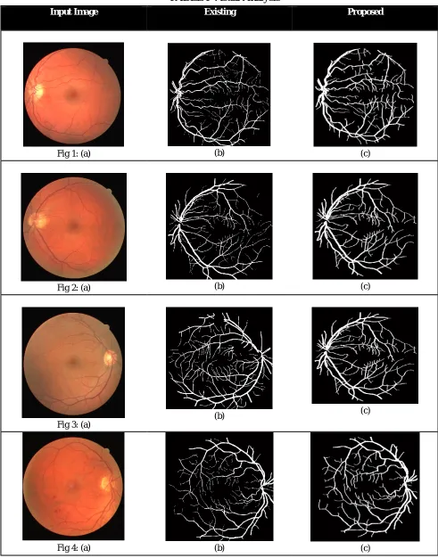
[image:5.612.60.551.114.740.2]©IJRASET (UGC Approved Journal): All Rights are Reserved B. Visual analysis
TABLE I Visual Analysis
Input Image Existing Proposed
Fig 1: (a) (b) (c)
Fig 2: (a) (b) (c)
Fig 3: (a) (b)
(c)
168
©IJRASET (UGC Approved Journal): All Rights are Reserved
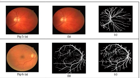
Fig 5: (a) (b) (c)
Fig 6: (a)
(b) (c)
In all above figures, fig1(a)-fig6(a) shows the input retinal image, fig1(b)-fig6(b) shows the resultant existing retinal image, fig1(c)-fig6(c) represents the resultant proposed retinal vessel image. Following parameters has been used namely:
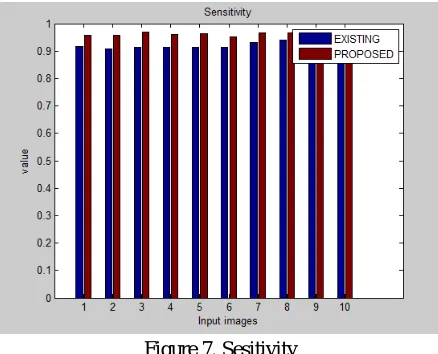
1) Sensitivity: Sensitivity is popularly known as true positive rate or probability of diagnosis in certain domains measures this ratio regarding positives which are accurately recognized while in the image. It is statistical measures of the performance of a binary classification test
[image:6.612.64.555.85.358.2]Where Se (Sensitivity) TP (true positives), FP (false positives), FN (false negatives), and TN (true negatives).
TABLE II
Sensitivity
S.no Existing Proposed
Image 1 0.91618 0.95739
Image 2 0.90659 0.95751
Image 3 0.9143 0.96874
Image 4 0.91459 0.96094
Image 5 0.9134 0.9644
Image 6 0.91283 0.9511
Image 7 0.93135 0.96749
Image 8 0.93882 0.96524
Image 9 0.9216 0.95521
[image:6.612.229.384.512.715.2]169
[image:7.612.196.415.90.267.2]©IJRASET (UGC Approved Journal): All Rights are Reserved
Figure 7. Sesitivity
2) Accuracy: Accuracy can be stated as the measure of closeness of measurements of a quantity to the same quantity's true value. The more will be the value of accuracy, better will be the outcomes. It can be calculated as
where ACC (accuracy) TP (true positives), FP (false positives), FN (false negatives), and TN (true negatives).
TABLE III
Accuracy
S.no Existing Proposed
Image 1 0.85364 0.95971
Image 2 0.83734 0.96105
Image 3 0.8503 0.96528
Image 4 0.85084 0.96347
Image 5 0.85933 0.96604
Image 6 0.84635 0.95461
Image 7 0.87859 0.96795
Image 8 0.88516 0.96672
Image 9 0.86256 0.95756
170
©IJRASET (UGC Approved Journal): All Rights are Reserved
Figure 8. Accuracy
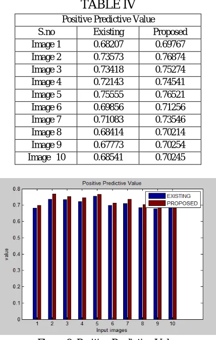
3) Positive Predictive Value: The positive predictive values PPV is the value of positive results in statistics and diagnostic tests that are true positive results, respectively. The PPV describe the performance of a diagnostic test or other statistical measure. A high result can be interpreted as indicating the accuracy of such a statistic.
[image:8.612.199.413.197.533.2]where PPV (positive predictive value), TP (true positives), FP (false positives), FN (false negatives), and TN (true negatives).
TABLE IV
Positive Predictive Value
S.no Existing Proposed
Image 1 0.68207 0.69767
Image 2 0.73573 0.76874
Image 3 0.73418 0.75274
Image 4 0.72143 0.74541
Image 5 0.75555 0.76521
Image 6 0.69856 0.71256
Image 7 0.71083 0.73546
Image 8 0.68414 0.70214
Image 9 0.67773 0.70254
Image 10 0.68541 0.70245
Figure 9. Positive Predictive Value
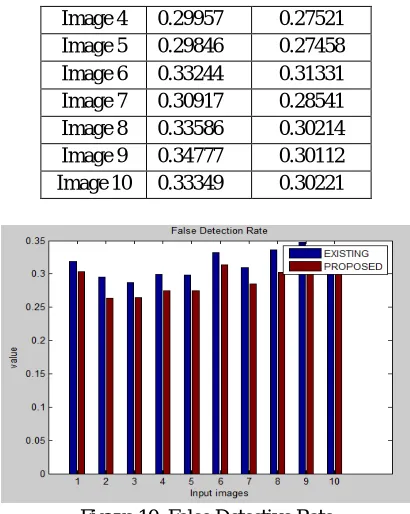
4) False Detection
RateThe
False Detection Rate refers to a procedure sometimes used when there is need to conduct many statistical tests, all of which correspond to an overlapping hypothesis. It is a method of conceptualizing the rate of errors in null hypothesis testing when conducting multiple comparisons.where FDR (false detection rate), PPV (positive predictive value), TP (true positives), FP (false positives), FN (false negatives), and TN (true negatives).
TABLE IV
False Detection Rate
S.no Existing Proposed
Image 1 0.31893 0.30370
Image 2 0.29527 0.26331
171
©IJRASET (UGC Approved Journal): All Rights are Reserved
Image 4 0.29957 0.27521
Image 5 0.29846 0.27458
Image 6 0.33244 0.31331
Image 7 0.30917 0.28541
Image 8 0.33586 0.30214
Image 9 0.34777 0.30112
[image:9.612.203.408.88.345.2]Image 10 0.33349 0.30221
Figure 10. False Detective Rate
5)
Specificity:
Specificity also called the true negative rate measures the proportion of negatives that are
correctly identified. It is calculated as
where Sp (Specificity) TN (true negative), FP (false positives).
TABLE V
Specificity
S.no Existing Proposed
Image 1 0.8860 0.9167
Image 2 0.8680 0.9245
Image 3 0.8861 0.9361
Image 4 0.8745 0.9596
Image 5 0.8888 0.9561
Image 6 0.8845 0.9554
Image 7 0.8641 0.9355
Image 8 0.8714 0.9471
Image 9 0.8965 0.9656
[image:9.612.227.385.461.619.2]172
[image:10.612.216.405.88.211.2]©IJRASET (UGC Approved Journal): All Rights are Reserved
Figure 11. Specificity
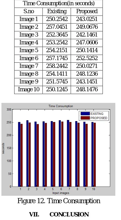
6)
Time Consumption:
Time consumption is total time consumed in computation. It is measured in seconds.
It is obtained by subtracting the end time from the starting time of computation.
TABLE V
Time Consumption(in seconds)
S.no Existing Proposed
Image 1 250.2542 243.0251 Image 2 257.0451 249.0676 Image 3 252.3645 242.1461 Image 4 253.2542 247.0606 Image 5 254.2151 250.1414 Image 6 257.1745 252.5252 Image 7 258.2442 250.0271 Image 8 254.1411 248.1236 Image 9 251.5745 243.1451 Image 10 250.1245 248.1476
Figure 12. Time Consumption
VII. CONCLUSION
Accurate vessel recognition performed in retinal images is a significant and tedious process. Automated segmentation of fundus image represents a significant role in detection of eye diseases. Detection regarding vessel as well as Retinal constructions mixed collectively may resolve the issue regarding highly accuracy in segmentation strategy. In this paper the effectiveness, designing and implementation of Adaptive Neighbourhood Estimator before Filling (ANEBF) is evaluated based on retina image segmentation
[image:10.612.203.416.262.648.2]173
©IJRASET (UGC Approved Journal): All Rights are Reserved
[1] Annunziata, Roberto, et al. "Leveraging Multiscale hessian-based enhancement with a novel exudate inpainting technique for retinal vessel segmentation." IEEE journal of biomedical and health informatics 20.4 (2016): 1129-1138.
[2] Wang, Chuang, Yongmin Li, and Ya Xing Wang. "Automatic choroidal layer segmentation using markov random field and level set method." IEEE journal of biomedical and health informatics (2017).
[3] Ram, Sundaresh, Forest Danford, Stephen Howerton, Jeffrey Rodriguez, and Jonathan Vande Geest. "Three-Dimensional Segmentation of the Ex-Vivo Anterior Lamina Cribrosa from Second-Harmonic Imaging Microscopy." IEEE Transactions on Biomedical Engineering (2017).
[4] Gou, Duoduo, Tong Ma, and Ying Wei. "A novel retinal vessel extraction method based on dynamic scales allocation." In Image, Vision and Computing (ICIVC), 2017 2nd International Conference on, pp. 145-149. IEEE, 2017.
[5] Guo, Jingyun, et al. "A framework for classification and segmentation of branch retinal artery occlusion in SD-OCT." IEEE Transactions on Image Processing (2017).
[6] Zhu, Shuxia, Fei Shi, Dehui Xiang, Weifang Zhu, Haoyu Chen, and Xinjian Chen. "Choroid Neovascularization Growth Prediction with Treatment Based on Reaction-Diffusion Model in 3D OCT Images." IEEE Journal of Biomedical and Health Informatics (2017).
[7] Bhuiyan, Alauddin, Akter Hussain, Ajmal Mian, Tien Y. Wong, Kotagiri Ramamohanarao, and Yogesan Kanagasingam. "Biometric authentication system using retinal vessel pattern and geometric hashing." IET Biometrics 6, no. 2 (2016): 79-88.
[8] Rani, Priya, N. Priyadarshini, E. R. Rajkumar, and Kumar Rajamani. "Retinal vessel segmentation under pathological conditions using supervised machine learning." In Systems in Medicine and Biology (ICSMB), 2016 International Conference on, pp. 62-66. IEEE, 2016.
[9] Xiao, Zhitao, Mengdie Wang, Fang Zhang, Lei Geng, Jun Wu, Long Su, and Jun Tong. "Retinal vessel segmentation based on adaptive difference of Gauss filter." In Digital Signal Processing (DSP), 2016 IEEE International Conference on, pp. 15-19. IEEE, 2016.
[10] Luo, Yuansheng, Hong Cheng, and Lu Yang. "Size-Invariant Fully Convolutional Neural Network for vessel segmentation of digital retinal images." In Signal and Information Processing Association Annual Summit and Conference (APSIPA), 2016 Asia-Pacific, pp. 1-7. IEEE, 2016.
[11] Lagarrigue-Charbonnier, Marthe, Florence Rossant, Isabelle Bloch, Marie-Hélène Errera, and Michel Paques. "Segmentation of retinal vessels in adaptive optics images for assessment of vasculitis." In Image Processing Theory Tools and Applications (IPTA), 2016 6th International Conference on, pp. 1-6. IEEE, 2016.
[12] Khan, Khan Bahadar, Amir A. Khaliq, and Muhammad Shahid. "B-COSFIRE filter and VLM based retinal blood vessels segmentation and denoising." In Computing, Electronic and Electrical Engineering (ICE Cube), 2016 International Conference on, pp. 132-137. IEEE, 2016.
[13] Li, Qiaoliang, Bowei Feng, LinPei Xie, Ping Liang, Huisheng Zhang, and Tianfu Wang. "A cross-modality learning approach for vessel segmentation in retinal images." IEEE transactions on medical imaging 35, no. 1 (2016): 109-118.
[14] Zhu, Chengzhang, Beiji Zou, Yao Xiang, Jinkai Cui, and Hui Wu. "An Improved Retinal Vessel Segmentation Method Based on Supervised Learning." In Computer-Aided Design and Computer Graphics (CAD/Graphics), 2015 14th International Conference on, pp. 216-217. IEEE, 2015.
[15] Zhu, Chengzhang, Beiji Zou, Yao Xiang, Jinkai Cui, and Hui Wu. "An Improved Retinal Vessel Segmentation Method Based on Supervised Learning." In Computer-Aided Design and Computer Graphics (CAD/Graphics), 2015 14th International Conference on, pp. 216-217. IEEE, 2015.