Georgia State University Georgia State University
ScholarWorks @ Georgia State University
ScholarWorks @ Georgia State University
Respiratory Therapy Theses Department of Respiratory Therapy
Summer 8-1-2011
The Independent Effect of Three Inline Suction Adapters and Lung
The Independent Effect of Three Inline Suction Adapters and Lung
Compliance change on Amplitude and delivered Tidal Volume
Compliance change on Amplitude and delivered Tidal Volume
during High Frequency Oscillatory Ventilation in an adult patient
during High Frequency Oscillatory Ventilation in an adult patient
with ARDS: Bench Model
with ARDS: Bench Model
Shreya Thacker
Follow this and additional works at: https://scholarworks.gsu.edu/rt_theses Part of the Medicine and Health Sciences Commons
Recommended Citation Recommended Citation
Thacker, Shreya, "The Independent Effect of Three Inline Suction Adapters and Lung Compliance change on Amplitude and delivered Tidal Volume during High Frequency Oscillatory Ventilation in an adult patient with ARDS: Bench Model." Thesis, Georgia State University, 2011.
https://scholarworks.gsu.edu/rt_theses/6
This Thesis is brought to you for free and open access by the Department of Respiratory Therapy at ScholarWorks @ Georgia State University. It has been accepted for inclusion in Respiratory Therapy Theses by an authorized administrator of ScholarWorks @ Georgia State University. For more information, please contact
ACCEPTANCE:
This thesis, The Independent Effect of Three Infine Suction Adapters and Lung Compliance change on Amplitude and delivered Tidal Volume during High Frequency Oscillatory Ventilation in an adult patient with ARDS: Bench Model, by Shreya J. Thacker was prepared under the direction ofthe Master's Thesis Advisory Committee of Respiratory Therapy
department at Georgia State University. It is accepted by the committee in partial fulfillment of
requirements for the Master's of Science degree in Respiratory Therapy at Byrdine F. Lewis
chool ofNursing and Health Professions, Georgia State University.
The Master's Thesis Advisory Committee, as representatives of the faculty, certifies that
this thesis has met all standards of excellence and scholarship as detennined by the faculty .
.. i ••• ,
~
.
~
-Robert Harwood, MS RRT
t{?~ ~
Ralph Zimmennan, MS RRT~0ULL
AUTHOR'S STATEMENT
In presenting this thesis as partial fu lfillment ofthe requirements for the Master's Degree in Respiratory Therapy from Georgia State University, I agree that the library of Georgia State University sha11 make it available for inspection and circulation in accordance with its regulation governing materials of this type. I agree that the permission to quote, to copy from or to publish thjs thesis may be granted by the professors under whose direction this thesis was written or by the Byrdine F. Lewis School ofNursing and Health Professions or by me. Such quoting, copying or publishing must be solely for scholarly purposes and will not involve potential fmancial gain.
It is understood that any copying from, quoting or publication of this thesis which involves potential financial gain will not be allowed without my written permission.
~~~
~ .Curriculum Vitae
S H R E Y A T H A C K E R
MS RRT (USA), BPT (India)Address: 489, Lindbergh Pl, NE Atlanta GA 30324 Date of birth: 10th November 1986
Email: [email protected] Mobile: (001) 770-837-4283
A C A D E M I C Q U A L I F I C A T I O N S
August 2009 – July 2011
Georgia State University, Atlanta, GA Master’s of Respiratory Therapy
GPA: 3.97
Awards: Academic Achievement Award
Dissertation: Effect of three inline closed suction systems on delivered tidal volume via High Frequency Oscillatory Ventilation (HFOV)
September 2004 – March 2009
Maharashtra University of Health Sciences, India Bachelor’s of Physical Therapy
GPA: 3.97
Awards: Best dissertation for the class of 2009
Dissertation: Comparing the effect of scalp massage vs. Acupressure in treatment of tension headaches
W O R K E X P E R I E N C E
August 2011 – Present Grady Memorial Hospital, Atlanta, GA
Work Areas: Adult and Neonatal ICU, Adult floors, Burns and Trauma units, Emergency department
January 2010 – April 2011 Intern, Respiratory Therapy
Northside and Crawford Long Hospital, GA
Work Areas: Neonatal ICU, Adult ICU, Adult floors, Pulmonary function laboratory
March 2009 – May 2009 Extern, Physical Therapy
Asian Heart Hospital, Mumbai, India Work Areas: Adult ICU, Physical therapy Unit, Pre-Operative physical therapy counselor
A D D I T I O N A L W O R K E X P E R I E N C E
January 2010 – April 2011
Teaching Assistant Georgia State University, Atlanta, GA
Undergraduate Biology
Duties: Preparing and presenting lectures, problem solving, preparing students for exam and grading papers
January 2010 – July 2011
Computer Laboratory
Duties: General Management, Communicating between departments for technical support. A D D I T I O N A L I N F O R M A T I O N
Registered Respiratory Therapist under the National Board of Respiratory Care (NBRC)
Respiratory Care Professional, State of Georgia # 8648
OTICE TO BORROWERS
All thesis deposited in Georgia State University library must be used in accordance with
stipulations prescribed by the author in the preceding statement. The author of this thesis is:
Shreya J. Thacker, B.P.Th (India)
Atlanta, Georgia 30324
The director of this thesis is:
Lynda T. Goodfellow Ed.D., RRT, FAARC
Associate Professor
Division of Respiratory Therapy
Byrdine F. Lewis School of Nursing and Health Professions
Georgia State University
ABSTRACT
The Independent Effect of Three Inline Suction Adapters and Lung Compliance change
on Amplitude and delivered Tidal Volume during High Frequency Oscillatory Ventilation in an
adult patient with ARDS: Bench Model
By
Shreya J. Thacker
Introduction: The use of high frequency oscillatory ventilation is increasing in treatment of acute respiratory distress syndrome over the past decade. The technique of HFOV of ventilating
the lungs at volumes less than the anatomical dead space calms the clinical concerns surrounding
ventilating stiff ARDS lungs with high pressures and volumes. This largely reduces the
probability of barotraumas and/or atelectrauma.
Purpose: The study was on an in vitro bench model that answered the following research questions: 1. The effect of three inline closed suction adapters on delivered tidal volume during
HFOV with varying lung compliance 2. The effect of varying compliance on the amplitude
delivered by HFOV; and 3. The effect of compliance on tidal volume delivered by HFOV.
Method: An in vitro bench model using high fidelity breathing simulator (ASL 5000, IngMar Medical) simulating an adult patient with ARDS was set up with 3100B SensorMedic high
frequency ventilator. The simulation included varying the compliance for each lung at 50, 40, 30
and 20cmH2O while maintaining fixed resistance of 15 cmH2O/L/sec. The ventilator was set to
the following parameters: power of 6, frequency (f) of 5, inspiratory time (Ti) of 33%, bias flow
(BF) of 30 LPM and oxygen concentration of 50%. The breathing simulator was connected with
the high frequency ventilator using a standard HFOV circuit and a size 8.0mm of endotracheal
tube. Fourteen French Kimberly Clark suction catheters (with T and Elbow adapters) and
Air-Life suction catheters (Y adapter) were placed in-line with the circuit successively to carry out
the study. Each run lasted for 1 minute after achieving stable state conditions. This
approximated to 300 breaths. The data was collected from the stimulator and stored by the host
computer.
Data Analysis: The data was analyzed using SPSS v.11 to determine the statistical significance. A probability value (P value) of ≤ 0.001 was considered to be statistically significant.
study also showed a direct relationship between amplitude and lung compliance i.e. an increase
in lung compliance caused an associated increase in amplitude (power setting remaining
unaltered). Lastly, the study did not show a statistically significant change in tidal volume with
changes in lung compliance. Future studies may be required to further evaluate the clinical
significance of the same.
Conclusion:
1. Many factors affect delivery of tidal volume during high frequency ventilation and thus it is
not constant. Choice of in-line suction system to be placed in line is one of the determinants of
the same.
2. Lung compliance changes lead to associated changes in amplitude delivery by HFOV. This
should be adjusted as patient condition improves by altering the power settings to ensure optimal
THE INDEPENDENT EFFECT OF THREE INLINE SUCTION ADAPTERS
AND
LUNG COMPLIANCE CHANGE ON
AMPLITUDE AND DELIVERED TIDAL VOLUME DURING
HIGH FREQUENCY OSCILLATORY VENTILTION IN AN ADULT PATIENT WITH
ARDS:
BENCH MODEL
By
Shreya J. Thacker
A thesis
Presented in Partial Fulfillment of Requirements for the
Degree of
Masters of Science
In
Health Sciences
In
Division of Respiratory Therapy
At
Byrdine F. Lewis School of Nursing and Health Professions
Georgia State University
Atlanta, Georgia
ACKOWLEDGEMETS
Firstly, I thank the almighty God for being my guide and showering his blessings on me
always.
I would like to thank Dr. Lynda T. Goodfellow, Ed.D., RRT, FAARC, chair person of my
thesis committee for teaching me to write a research paper and guiding me step-by-step through
this thesis. Without your assistance I would have not been able to complete my work.
I thank Mr. Robert Harwood, MS, RRT, for working with me in the respiratory therapy
laboratory. Your patience and support helped me learn and work on my data better.
I thank Ralph Zimmerman, MS, RRT, for being instrumental in reviewing my work and
providing with feedbacks.
I also thank John England, MS, RRT. You were extremely helpful by providing me with
the concept of this thesis, helping me understand the working of high frequency oscillatory
ventilation, providing me with raw material for the thesis and for lending me your research work
for reference. All of this formed the back bone for my work.
I would additionally want to thank Dr. Arzu Ari, Ph.D., RRT and Dr. Yong Tai Wang,
Ph.D., for teaching me and guiding me through the statistical analysis.
I am eternally grateful to my parents Mrs and Mr Thacker and my brother Ankit for their
love, support and personal sacrifices. May god continue to abundantly bless you all and may he
give me the chance to be the source of your happiness always.
TABLE OF COTETS
CONTENTS PAGE
Introduction 1
Review of Literature 6
Research Methodology 11
Results 14
Discussion 19
List of Tables 23
Abbreviations 24
Appendix 25
[image:11.612.73.547.163.558.2]1 | P a g e
Chapter 1 ITRODUCTIO
Mechanical ventilation forms the foundation of intensive care treatment for acute
respiratory diseases. For the majority of patients requiring assistance, conventional mechanical
ventilation (CMV) ensures adequate gas exchange by typically delivering tidal volumes that
approximate 1 to 1.5 times the spontaneous tidal volume for a given patient. However, often high
pressures and larger tidal volumes are required that culminate in undesirable effects of cardiac
depression, hemodynamic instability and pulmonary air leaks. Thus to overcome the deleterious
effects of high pressure and larger tidal volumes, high frequency oscillatory ventilation (HFOV)
was developed. Apart from instituting HFOV to avoid high alveolar pressures, pulmonary
hygiene is equally important and forms the cornerstone in maintaining appropriate pulmonary
pressures. Suctioning has been studied extensively to cause loss of tidal volume on patients
receiving mechanical ventilation (Fernandez, Piacentini et al. 2004). This can be partly owed to
the dead space added by the suctioning apparatus to the ventilatory circuit. Since HFOV
ventilates the lungs at tidal volumes that are lesser than the anatomical dead space, suctioning
procedures have been avoided for long periods of time in these patients to prevent derecruitment
of alveoli. This often culminates into secretion retention and high alveolar pressures (Pillow
2005). Thus to maintain adequate tidal volumes and to prevent the suboptimal use of suctioning
during HFOV, research is required to understand the mechanics between the ventilator and the
ventilatory circuit. In lieu of this, the presenting thesis studied The Independent Effect of Three Inline Suction Adapters and Lung Compliance change on Amplitude and delivered Tidal Volume
during High Frequency Oscillatory Ventilation: Bench model.
After an initial enthusiasm for use of HFOV in the 1970’s and 1980’s, many clinical
2 | P a g e
been a renewed interest in use of HFOV because of two main reasons: (1) the literature supports
the finding that CMV can intensify lung injury in patients with acute lung injury (ALI) or acute
respiratory distress syndrome (ARDS); (2) Modification of ventilation techniques in terms of
manipulating tidal volumes and airway pressure can provide a safer method of treating patients
with ALI/ARDS (Krishnan and Brower 2000).
HFOV was developed to treat refractory hypoxemia in patients with ARDS/ALI. Hence
HFOV works mainly for oxygenation rather than ventilation. The major advantage that HFOV
provides is that it works at a respiratory rate (RR) of 60 – 900 breaths/min but at low tidal
volumes which are often smaller than the anatomical dead space. This allows the maintenance of
high mean airway pressures (mPaw). Therefore, when high respiratory rates maintain efficient
gas exchange, alveolar derecruitment and overdistension are prevented by higher mPaw and
lower tidal volumes respectively (Chan, Stewart et al. 2007).
Technique of HFOV:
While using HFOV in adults, a piston pump oscillates to maintain the frequency between
3-10Hz i.e. 180 – 600 breaths/min. mPaw is maintained via a resistance valve and the bias flow
of gas between 30L – 60L/min present within the circuit. A distinctive feature of HFOV is that
the back stroke of the piston brings about active expiration of gas. This is debatably thought to
reduce air trapping. Another distinctive feature of HFOV is that it provides separate controls for
oxygenation and ventilation. Fraction of inspired oxygen (FiO2) and mPaw dictate the
oxygenation status whereas ventilation is dictated by pressure amplitude (∆P) and respiratory
3 | P a g e
Mechanisms of Gas exchange during HFOV:
There are seven plausible mechanisms thought to enhance gas exchange during HFOV.
These include: turbulence in large airways; direct ventilation of closed alveoli; turbulent flow
with lateral convective mixing; pendelluft; gas mixing due to velocity profiles that are axially
asymmetric; laminar flow with lateral transport by diffusion (taylor dispersion); collateral
ventilation through non-airway connection between adjacent alveoli (Slutsky and Drazen 2002).
A short description is given on these mechanisms:
Direct bulk flow: direct flow of inspired gas is received by the alveoli in the proximal
tracheobronchial which leads to gas exchange by convective or bulk flow. Taylor dispersion:
when diffusion is overlapped by convective flow, turbulent eddies and swirling occurs thereby
increasing the amount of gas exchange that would occur from simple bulk flow. Pendelluft:
variation in the resistance and compliance causes a difference in the pattern of airflow between
lung regions and within these regions. Thus gas flows occurs between regions if these regions
with variations in flow pattern are in close proximity. Asymmetric velocity profiles: air in the peripheral regions of the airway has a velocity lower than that in the center of the
tracheobronchial tree. Owing to the difference in the flow rates, this parabolic velocity profile is
pronounced during inspiration. With repetition of such cycles, air in the central part tends to
move deep in the alveoli while that in the periphery moves out towards the mouth. Cardiogenic
mixing: heart beats lead to mechanical stirring which is more evident in the lung units close to
the heart. Molecular diffusion: this mechanism is similar to the other modes of ventilation which plays an important role in mixing of gas near the alveolar-capillary membrane (Chang 1984;
4 | P a g e
Acute Respiratory Distress Syndrome
ALI and ARDS are defined by the American-European consensus committee. They
describe ALI and ARDS as acute onset respiratory distress that presents clinically as refractory
arterial hypoxemia and radiologically as diffuse pulmonary infiltrates. Both these diseases have
pulmonary capillary wedge pressure of ≤ 18mmHg with absence of clinical evidence of left atrial
hypertension. The difference between the two diseases can be given by the difference in the
PaO2/FiO2 ratio, which when ≤ 300 signifies ALI and when ≤ 200 signifies ARDS.
Etiologically, the risk factors for ARDS can be categorized into direct and indirect
categories. Direct risk factors include pulmonary contusions, diffuse pulmonary infections, near
drowning, inhalation of toxic fumes and aspirations. Indirect risk factors includes sepsis
syndrome, severe non-thoracic trauma and cardio-pulmonary bypass (Kane and Galanes 2004).
Pathophysiologically, patients with ARDS present with acute inflammation of the lung
parenchyma. Inflammation causes increased capillary permeability leading to extravasation of
proteinaceous fluid into the alveoli. Protein molecules thus released inactivate the already
existing surfactant. More so, the production of surfactant by type II pneumocytes is also reduced.
The effect of same is an increase in surface tension and microatelectasis of alveoli. Worsening
arterial oxygenation ensues due to increased dead space, pulmonary shunting and altered
ventilation-perfusion mismatch. Minute ventilation is increased as a compensation to maintain
the desired PaCO2 and pH. Mechanical ventilation is generally required to support the
respiratory system (Krishnan and Brower 2000).
To summarize, this chapter covered the need to introduce HFOV in management of
5 | P a g e
avoid deleterious effects of secretion retention. Thus the following research questions were
intended to be answered:
1. The effect of three inline closed suction adapters on delivered tidal volume during HFOV
with varying lung compliance
2. The effect of varying compliance on the amplitude delivered by HFOV
3. The effect of compliance on tidal volume delivered by HFOV
The answers to these questions would help prevent suboptimal use of suctioning in
patients placed on HFOV and would also enhance the understanding of mechanics between
ventilator and the ventilatory circuits. The study would help identify the most efficient suction
adapter in terms of the amount of loss of tidal volume caused by its placement in-line with the
6 | P a g e
Chapter 2
REVIEW OF LITERATURE
The articles for literature review were collected by using the online database system
provided by the Georgia State University’s library services. The databases that were used were:
CINHAL plus with full text, PubMed, Health source: Nursing, Nursing and Allied Health Source
and Web of Science. The articles presented in this chapter would highlight the use of HFOV in
the treatment of patients with ARDS. The chapter would also present the very few studies
relating to evaluating the effects of placement of inline suction catheters on the tidal volume
delivered via HFOV.
The research questions intended to be answered at the end of the study were:
1. The effect of three inline closed suction adapters on delivered tidal volume during HFOV
with varying lung compliance
2. The effect of varying compliance on the amplitude delivered by HFOV
3. The effect of compliance on tidal volume delivered by HFOV
High Frequency Oscillatory Ventilation in ARDS
Derak and colleagues carried out a multicenter randomized controlled trial on 148 adult
patients suffering from ARDS with the aim to compare the efficiency and safety of employing
HFOV with conventional mechanical ventilation (CMV). ARDS was defined as a PaO2/FiO2
(P/F) ratio of ≤ 200 on 10 or more cmH2O of positive end-expiratory pressure (PEEP). All
patients included in the study had a mean age of 48 years, mean PaO2/FiO2 ratio of 112.5, mean
oxygenation index (OI) of 25.2 and a mean Acute Physiology and Chronic Health Evaluation
7 | P a g e
early improvement in PaO2/FiO2 ratio but this improvement waned after the first 24 hours (P =
0.008). Both the groups showed an improvement in oxygenation index over the first 3 days.
When 30-day mortality was assessed, HFOV had a mortality rate of 37% against 52% in CMV
(P = 0.102). Nevertheless, there was no significant difference between these groups in causation
of adverse reactions like hemodynamic instability, ventilation or oxygenation failure, mucus
plugging and barotraumas. Conclusions revealed that HFOV was safe and efficient but showed
no difference in incidence and intensity of complication compared to CMV (Derdak, Mehta et al.
2002).
Another randomized controlled trial was carried out by Bollen and colleagues on 61
patients with a mean OI of 22 and APACHE II score of 21 comparing the effects of HFOV vs.
CMV. The study was mainly focused on the complications and mortality rate associated with
HFOV. The study documented that of the 37 patients receiving HFOV, four patients (10.8%)
developed pneumothorax, and one patient (2.7%) had an air leak. Of the 24 patients receiving
CMV, one (4.2%) developed an air leak and one (4.2%) developed hypotension. However,
baseline OI was higher in HFOV (25 vs. 18) than CMV. The 30-day mortality revealed a higher
mortality in HFOV (43%) and a lower mortality in CMV (33%). The results of this study were
contradicting the belief that HFOV has an effect in reducing mortality rates (Bollen, van Well et
al. 2005).
On the other hand, a study conducted by Finkielman and colleagues showed somewhat
different results. The retrospective study was carried out on 14 patients suffering for ARDS with
a mean age of 56-years, APACHE II score of 35 and Sequential Organ Failure Assessment score
(SOFA score) of 11.5. The authors reported that the data analysis showed an improvement in P/F
8 | P a g e
rate was documented to be 57%. No mention was made of complications after implementation of
HFOV except in one patient in whom HFOV was discontinued due to development of refractory
severe hypotension. The authors concluded that HFOV is an acceptable alternative to CMV in
treatment of ARDS when protective lung strategies are required (Finkielman, Gajic et al. 2006).
Since the sample size of this study was comparatively less and the authors mentioned the lack of
use of a standardized protocol, the findings of the study are arguable.
Unlike the results of these studies, Mehta and colleagues reported a significant
improvement in P/F ratio and OI which lasted for 72-hour in patients with ARDS receiving
HFOV. They conducted a retrospective study on 156 patients with a mean age of 47.8years, P/F
ratio of 91, OI of 31.2 and APACHE II score of 21. Apart from the findings mentioned, other
important findings presented were that HFOV was discontinued in 19 patients within four hours
due to difficulty with maintaining oxygenation and ventilation; or deleterious hemodynamic
changes like a significant rise in central venous pressure (CVP), reduced cardiac output and/or
increase in pulmonary artery occlusion pressure. Additionally, 21.8% of the patients developed
pneumothorax. Thirty-day mortality was assessed to be 61.7% and is highest amongst the studies
mentioned (Mehta, Granton et al. 2004).
Another novel comparison was made to assess the effects of normocapnic HFOV in
pulmonary (ARDSp) and extra-pulmonary (ARDSexp) forms of ARDS. It was a prospective
study on 30 adult patients with mean age of 56 years, P/F ratio of 121 and SOFA score of 9.6.
Assessments were made at six hours to evaluate improvements in P/F ratio. ARDSexp showed
significant improvement in P/F ratio (114 to 200 torr, P < 0.01) against no significant
improvement in ARDSp. It was also mentioned that a higher continuous distending pressure was
9 | P a g e
was documented to be 46% overall. It was inferred that these findings were suggestive of HFOV
being more effective in extra-pulmonary ARDS compared to the pulmonary forms of ARDS
(Pachl, Roubík et al. 2006).
Closed suction systems and High Frequency Oscillatory Ventilation
A bench model study was carried out in a neonatal lung model to assess for the loss of
tidal volume and change in pressures associated with insertion of closed tracheal suction system
(CTSS) with and without suctioning. An 8-Fr and a 6-Fr catheter were tested with a 4.5, 4.0 and
3.5 and a 3.5, 3.0 and 2.5mm ID ETT respectively. Measurements of pressures were done
proximal and distal to the ETT along with measurements for inspiratory and expiratory tidal
volumes. When 8-Fr catheter was inserted without the application of suctioning, no change was
seen in proximal airway pressures but a decrease in distal peak inspiratory pressure was noted
and no change in distal end-expiratory pressure with an increasing ratio of suction catheter to
ETT size. Tidal volumes were also documented to be reduced. With the use of a 6-Fr catheter,
though the proximal pressures continued to remain unchanged, a decrease in both distal pressures
was seen owing to the reduction in size of the ETT. Thus, it was concluded that CTSS neither
maintains the constant flow of gas nor does it maintain adequate pressures with its mere presence
in the circuit even when the suction pressures are not applied (Monaco 1992).
The recommendation that with the use of HFOV, it is vital to maintain a smooth circuit
with minimal interference between patient and the ventilator lead to the study that tested for
pressure changes during HFOV with presence of inline suction catheters. Gaudet, et al.
performed a bench model study on neonatal test lung using 3100A ventilators testing for 6-Fr
sized catheters with an ETT size of 2.5, 3.0, and 3.5 mm ID. A ‘Y’ (45°), an elbow (90°) or no
10 | P a g e
measurement mean airway pressure and amplitude were measured at distal ETT. Data analysis
revealed a statistically significant but clinically insignificant difference in pressure changes with
the use of the different catheters. Thus, the study gives further scope for research to ascertain if
placement of inline catheters effect the pressure as well as tidal volumes in HFOV (Gaudet,
Branconnier et al. 2005).
Thus from the above presented literature, one can conclude that studies conducted to
evaluate the use of HFOV in treatment of patients with ARDS does show promising results. It is
also evident that the placement of inline suction catheters alters the delivered tidal volume and
11 | P a g e
Chapter 3
RESEARCH METHODOLOGY
This chapter describes the method used to answer the following research
questions:
1. The effect of three inline closed suction adapters on delivered tidal volume during HFOV
with varying lung compliance
2. The effect of varying compliance on the amplitude delivered by HFOV
3. The effect of compliance on tidal volume delivered by HFOV
Lung Model
An adult patient with ARDS was simulated using an in-vitro test lung model with
high fidelity breathing stimulator Active Servo Lung (ASL) 5000, {Ingmar Medical, Pittsburgh,
PA (USA)}. The ASL 5000 is digitally controlled, piston driven and simulates real time
breathing. The following research question would be answered: the effect of three inline closed
suction systems on delivered tidal volume with varying lung compliance (20, 30, 40 and 50
mL/cmH2O) at predetermined airway resistance (Raw) of 15 cmH2O/L/sec.
Ventilator Settings
A standard HFOV adult ventilator circuit and a size 8.0mm internal diameter (I.D)
(Portex) cuffed endotracheal tube was used with the SensorMedics 3100B ventilator. Fourteen
French Ballard suction systems (Kimberly Clark Roswell, Georgia (USA)) with Ballard T (T
adapter), Ballard Elbow (E adapter) and AirLife-Y adapters (Y adapter) were used in-line with
the ventilator. The ventilator was set to the following parameters: power of 6, frequency (f) of 5,
12 | P a g e
mean airway pressure was variable reflecting the change in compliance set on ASL 5000
software (Fessler and Hess 2007). The above mentioned settings were used in accordance to the
guidelines dictated by Fessler et al., (2007).
Dead Space Measurement
Each of the T, Elbow and Air-Life Y adapters were filled with water. The water was then
aspirated with a syringe and measurements were made to denote the dead space volume.
Protocol
The scenario of an adult passive patient was selected on the ASL 5000 software to
simulate an adult patient on HFOV with ARDS. The scenario allowed the resistance to be fixed
while allowing compliance change throughout the study. The resistance of 15 cmH2O/L/sec was
maintained with a changing compliance of 20, 30, 40 and 50 mL/cmH2O (protocol is included in
the Appendix A). Ballard suction catheters sized 14-Fr with T/Elbow/Y adapters were used
in-line with the circuit with each of the above mentioned permutations to measure the tidal volumes
inside the ASL 5000 test lung.
The 3100B ventilator was set at the previously mentioned settings after an initial
calibration performed as per the company manual. The test lung was connected to the ventilator
using a standard adult HFOV circuit, 8.0 mm I.D endotracheal tube and an inline suction
catheter. The test lung was then connected to the ASL 5000 software in the host computer using
the RS-232 cable. The run time for each reading was 90 seconds that approximated to 450
breaths. The tidal volumes were measured in the test lung by the ASL 5000 software in the host
13 | P a g e
Data Collection
Tidal volume was measured and recorded for each change in compliance with
each of the three different suction systems. The tidal volume and number of breaths was
measured and recorded by the ASL 5000 on the previously mentioned ventilatory settings. A run
time of 90 seconds was used with the first 30 seconds used to achieve steady state and the next
60 seconds for recording of the relevant data. Approximately 450 breaths were used over the
period of 90 seconds to measure an averaged expired tidal volume.
Data analysis
Statistical analysis was carried out using SPSS v.11. A three by four factorial analysis
was done. Post hoc Bonferroni and descriptive statistic was also carried out to answer the
following research questions: 1. the effect of three inline closed suction adapters on delivered
tidal volume during HFOV with varying lung compliance; 2. The effect of varying compliance
on the amplitude delivered by HFOV; and 3. The effect of change of compliance on tidal volume
delivered by HFOV. A probability value (P ≤ 0.001) was accepted as statistically significant.
To summarize, this chapter described the methods used to answer the research questions
listed above. Data was collected using a 3100B HFOV on adult lungs simulating ARDS. SPSS
14 | P a g e
Chapter 4
RESULTS
The results and tables presented in this chapter are provided to help answer the following
research questions:
1. The effect of three inline closed suction adapters on delivered tidal volume during HFOV
with varying lung compliance
2. The effect of varying compliance on the amplitude delivered by HFOV
3. The effect of compliance on tidal volume delivered by HFOV
Dead Space Measurements of Suction Adapters
Ballard T-adapter: 19.4 mL
Ballard Elbow-adapter: 7.0 mL
AirLife Y-adapter: 11.0 mL
Tidal volumes as it relates to Adapter changes (T, Elbow and Y adapters)
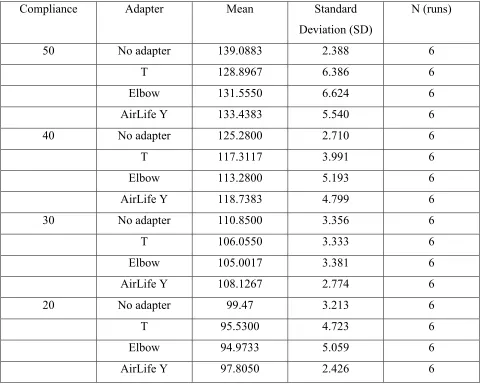
Table 1 and 2 show the statistical analysis carried out to ascertain the effect of different
adapters on tidal volume change while placing suction catheters in-line with HFOV circuit. The
15 | P a g e
Table 1: Descriptive Statistics – tidal volume and compliance changes as related to adapter changes
Compliance Adapter Mean Standard
Deviation (SD)
N (runs)
50 No adapter 139.0883 2.388 6
T 128.8967 6.386 6
Elbow 131.5550 6.624 6
AirLife Y 133.4383 5.540 6
40 No adapter 125.2800 2.710 6
T 117.3117 3.991 6
Elbow 113.2800 5.193 6
AirLife Y 118.7383 4.799 6
30 No adapter 110.8500 3.356 6
T 106.0550 3.333 6
Elbow 105.0017 3.381 6
AirLife Y 108.1267 2.774 6
20 No adapter 99.47 3.213 6
T 95.5300 4.723 6
Elbow 94.9733 5.059 6
[image:26.612.66.549.126.511.2]16 | P a g e
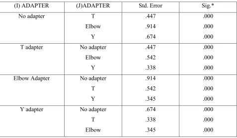
Table 2: Multiple comparisons dependent variable: Bonferroni analysis
(I) ADAPTER (J)ADAPTER Std. Error Sig.*
No adapter T
Elbow Y .447 .914 .674 .000 .000 .000
T adapter No adapter
Elbow Y .447 .542 .338 .000 .000 .000
Elbow Adapter No adapter
T Y .914 .542 .345 .000 .000 .000
Y adapter No adapter
T Elbow .674 .338 .345 .000 .000 .000
.* P value of ≤ 0.001 is significant
Tidal volumes as it relates to Compliance changes
Tables 1 and 3 show the tidal volume changes as it relates to the changes in compliance
(20, 30, 40 and 50 cmH20). The analysis showed that the changes in tidal volumes were
17 | P a g e
Table 3: Multiple Comparisons Dependent variable: Bonferroni Analysis
(I)COMP (J)COMP Std. Error Sig.*
50 40
30 20 .882 .975 .575 .004 .004 .005
40 50
30 20 .882 .619 .568 .004 1.000 .037
30 50
40 20 .975 .619 .601 .004 1.000 .016
20 50
40 30 .575 .568 .601 .005 .037 .016
Amplitude as related to compliance changes
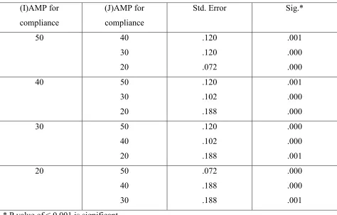
Table 4 shows the changes in amplitude as related to the changes in compliance. The
analysis of data showed that the changes in amplitude were statistically significant when
18 | P a g e
Table 4: Amplitude changes as related to compliances changes
(I)AMP for
compliance
(J)AMP for
compliance
Std. Error Sig.*
50 40
30 20 .120 .120 .072 .001 .000 .000
40 50
30 20 .120 .102 .188 .001 .000 .000
30 50
40 20 .120 .102 .188 .000 .000 .001
20 50
40 30 .072 .188 .188 .000 .000 .001
.* P value of ≤ 0.001 is significant
In summary, there was a statistically significant decrease in tidal volumes as compared to
the suction adapter changes. A statistically significant increase in amplitude was also seen as the
lung compliance improved. There was no significant change seen in tidal volume as compared to
19 | P a g e
Chapter 5
DISCUSSIO
This chapter discusses the results presented in the preceding section for the following
research questions:
1. The effect of three inline closed suction adapters on delivered tidal volume during HFOV
with varying lung compliance
2. The effect of varying compliance on the amplitude delivered by HFOV
3. The effect of compliance on tidal volume delivered by HFOV
In clinical practice, efforts have been directed towards maintaining a circuit between
ventilator and patient with minimal angulations so as to prevent its interference with
transmission of oscillatory waves and pressure during HFOV. This ensures the maintenance of
optimal ventilation. In this respect, the combined impedance of the ventilator, the ventilatory
circuit, endotracheal tube and patient’s respiratory system plays prime importance in deciding
the efficiency of ventilation during HFOV. Impedance is described as mechanical barrier to flow
of gas (Pillow 2005). With the use of suction adapters that have larger angles and bends, larger
mechanical interference with the oscillatory waves is caused creating a dampening effect on the
same. Additionally, angulations cause hindrance in the flow of gas through the circuit.
Dampening of the oscillatory waves combined with the hindrance to the flow of gas leads to an
associated loss of tidal volume. Larger pressure swings are thus required to restore the flow of
gas and generate an equivalent tidal volume. Pressure swings are transmitted to the lung tissue
causing it to distend thus increasing the chances of barotrauma and/or pneumothoraces. This
negates one of the main advantages of HFOV of ventilating the lungs at low tidal volumes and
20 | P a g e
This thesis used three different adapters namely the T, Elbow and Air-life Y adapters to
test for the loss of tidal volume during HFOV due to dampening of the oscillatory waves. The
results revealed that the Air-Life Y adapter caused the least loss of tidal volume. The T and the
Elbow adapters have 90° angulations between the ports for the ETT and the suction catheters. On
the other hand, the Air-Life Y adapter has 45° angulations between these ports. Based on these
facts, the lesser degree of bend leads to a decreased intensity of dampening effect and hence
lesser loss of pressure and tidal volume.
Another important observation was the differences in dead space each of these adapters
added when placed in-line with the ventilatory circuit. The T, the Elbow and the Air-Life Y
adapters added 19.4, 7.0 and 11.0 mL respectively of dead space respectively. It is undeniable
that the greater the dead space the greater the loss of tidal volume and hence greater the dead
space ventilation. It should be noted that these changes in tidal volume cause exponential
changes in minute volume owing to minute volume during HFOV being the product of
respiratory rate and squared tidal volume. Hence efforts should be taken to choose a closed
suction system adapter that has minimal dead space to minimize the loss of volumes while
placing suction systems in-line with HFOV circuit.
Additionally these Y-adapters offer an advantage of providing a versatile port that allows
performance of multiple procedures like mini- broncho-alveolar lavage (BAL), closed
suctioning, bronchoscopic procedures and instilled drug delivery without disconnecting the
circuit. This helps prevents lung derecruitment, loss of oxygenation and cross contamination
during these procedures. There is no doubt that a suction system which is placed in-line that
prevents patient disconnection from the ventilator provides a distinct advantage over a suction
21 | P a g e
This study also observed the changes in amplitude with changes in lung compliance. It
was seen that as lung compliance improved, there was a statistically significant decrease in
amplitude with no statistically significant change in tidal volume. It should be understood that
these changes are normal. With an improvement in lung compliance, less pressure is required to
deliver the set tidal volume and hence the piston moves less of a distance. This in turn correlates
to the decreased amplitude with improved lung compliance. Clinicians should understand that
the power settings should not be altered at this point to increase the amplitude as this leads to an
increase in the delivered tidal volume to the lungs which inadvertently causes increase in
alveolar pressures and increased risk of barotraumas. Further, as explained previously, a small
increase in tidal volume due to altered power settings will lead to an exponential increase in
minute volume creating mismatched ventilation. Modifying the tidal volume and power settings
abrogates the low tidal volume, high PEEP and low alveolar pressure benefit of HFOV(England
2009).
Limitations: the study did not look for the effects of the procedure of suctioning i.e. the
application of negative pressure. This can be a potential for further studies.
Clinical Applications: the study would help prevent suboptimal use of suctioning in patients
placed on HFOV and would also enhance the understanding of mechanics between ventilator and
the ventilatory circuits. The study would help identify the most efficient suction adapter in terms
of the amount of loss of tidal volume caused by its placement in-line with the ventilatory circuit.
In conclusion, this study revealed that:
1. The AirLife closed suction adapters caused the least loss of tidal volume when placed
in-line with a HFOV circuit and hence were the most appropriate for use with the
22 | P a g e
2. The study also showed that as lung compliance improves, amplitude decreases
(without a manual change made in the power settings).
3. The study did not show any significant change in tidal volumes correlating to
23 | P a g e
LIST OF TABLES
Table 1: Statistical analysis (descriptive statistics) – tidal volume and compliance changes as
related to adapter changes
Table 2: Multiple comparisons dependent variable: Bonferroni analysis
Table 3: Multiple Comparisons Dependent variable: Bonferroni Analysis
24 | P a g e
ABBREVIATIOS
ALI: Acute Lung Injury
APACHE II: Acute Physiology and Chronic Health Evaluation II
ARDS: Acute Respiratory Distress Syndrome
ASL: Active Servo Lung
BAL: Broncho - Alveolar Lavage
BF: Bias Flow
CMV: Conventional Mechanical Ventilation
CTSS: Closed Tracheal Suction System
CVP: Central Venous Pressure
ETT: Endo Tracheal Tube
FiO2: Fraction of Inspired Oxygen
HFOV: High Frequency Oscillatory Ventilation
I.D: Internal Diameter
OI: Oxygen Index
PEEP: Positive End Expiratory Pressure
P/F ratio: PaO2/FiO2 ratio, partial pressure of arterial oxygen/fraction of inspired oxygen
SOFA Score: Sequential Organ Failure Assessment score
Ti: Inspiratory time
25 | P a g e
APPEDIX Protocol
High Frequency and Tidal Volume Goals:
1. The effect of three inline closed suction adapters on delivered tidal volume during HFOV
with varying lung compliance
2. The effect of varying compliance on the amplitude delivered by HFOV
3. The effect of compliance on tidal volume delivered by HFOV
Methods:
1. Dead space (VD) was measured for each of the adapters of suction catheters i.e. for the
Ballard elbow adapter, the Ballard T-adapter and the AirLife Y-adapter.
2. Initiation, calibration and setup of the equipment is as follows:
a. Test Lung:
i. Use ASL 5000 breathing stimulator
ii. Connect the ASL 5000 stimulator to line power and switch it “on”
1. The motor red light is enabled
2. Wait for the motor red light to turn “off”
3. ASL 5000 is calibrated and ready to be connected to the ventilator
and computer
iii. Connect the ASL 5000 to the host Ethernet via networking cable
iv. Connect ASL 5000 to high frequency oscillatory ventilator (HFOV)
through standard high frequency breathing circuit and sized 8.0 I.D cuffed
endotracheal tube (ETT)
26 | P a g e
i. Use sensor medics 3100B ventilator
ii. Calibrate the 3100B with capped standard high frequency breathing circuit
in the following way:
1. Set the bias flow at 30 L/min
2. Mean Airway Pressure (mPaw) to maximum
3. Pressure amplitude (delta-P) to 60 cmH2O
4. Set the inspiratory time (I-time) to 33%
5. Start the 3100B ventilator and wait for the mPaw to reach between
40 & 45 cm H2O
iii. Set the 3100B ventilator to the following:
1. Set the power to 6.0. The pressure amplitude reading will be
variable
2. Frequency to 5Hz
3. Mean airway pressure will be variable
4. The bias flow will be set at 30 L/min
5. I-time would be 33%
6. Oxygen to 50%
iv. Connect the Ballard suction system (with T-adapter) to the 8.0 mm I.D
cuffed ETT (Portex, Hythe Kent, England) and to the standard high
frequency breathing circuit. This assembly is then connected to the ASL
5000
27 | P a g e
i. After all the cable connections have been completed, launch the lab view
software on the host PC
ii. Working page (welcome window) is presented
1. From the welcome window choose run the software with Ethernet
mode
2. Welcome window will disappear and several screens will stack up
on computer screen
3. Host computer will attempt to synchronize with ASL 5000
4. Wait for the ASL 5000 to respond (stimulator piston will start
moving)
iii. From the script editor window choose (Adult_apnea.sct) file to edit its
lung model parameter files
iv. From the script file box, double click on the file name (Adult_apnea.sct)
v. A window titled “Step 1: select simulation parameter set” will open
1. Click “edit” to proceed to next step
vi. A window titled “step 2: choose a lung model” will open
1. Choose the two compartment lung model
2. Set the resistance (Raw 1) to 15 cm H2O/L/sec
3. Set the resistance (Raw 2) to 15 cm H2O/L/sec
4. Set compliance (CL 1) to 20 mL/cm H2O
5. Set compliance (CL 2) to 20 mL/cm H2O
6. Click “next”
28 | P a g e
1. From the pop-up menu of the chest wall model, choose passive
model
2. Set the passive cycle to 12 breaths/min
3. Click next
viii. A window titles “step 4: save stimulation parameter set” will open
1. Click “save” to save the new variables in the file
ix. From the “central run time” window do the following while the ventilator
is running and connected to ASL 5000
1. Start the simulation by moving the slide switch from “off to on”
position.
2. From the file window choose a name for the data file path to store
the data
3. Click OK
x. Go to breath detection/real-time analysis window
1. Check the “save data to high resolution file” checkbox
2. Wait for the data of 300 breaths to be collected
xi. Turn “off” the simulation by moving the slide switch from “on to off”
position
d. Data collection:
i. Launch the Lab VIEW software on the host computer
ii. Go to post-run window
29 | P a g e
2. Open the “multi parameter trend” from the “green box” under the
display data list
iii. Check the tidal volume waveform to establish steady state
iv. Record the tidal volume and use it in data analysis
3. With a compliance of 20 mL/cm H20, do three trails for Ballard suction systems with T
-adapter as described above but at step (c-vi) set the compliance settings to 20 mL/cm
H20.
4. With a compliance of 30 mL/cm H20, do three trails for Ballard suction systems with T
-adapter as described above but at step (c-vi) change the compliance settings to 30 mL/cm
H20.
5. With a compliance of 40 mL/cm H20, do three trials for Ballard suction systems with T
-adapter as described above but at step (c-vi) change the compliance settings to 40 mL/cm
H20
6. With a compliance of 50 mL/cm H20, do three trials for Ballard suction systems with T
-adapter as described above but at step (c-vi) change the compliance settings to 50 mL/cm
H20 (the readings obtained from this settings would act as a ‘control’ against which the
other reading would be compared)
7. Similar procedure will be repeated while testing for tidal volume changes with inline
Ballard suction systems with Elbow-adapters or Air Life suction systems along with each
compliance change of 20 ml/cm H2O, 30 mL/cm H20 and 40 mL/cm H20 and
30 | P a g e
The layout of the data table:
Compliance (mL/cmH2O)
Tidal Volume (Vt) in mL
No Catheter Ballard with “T” adapter
Ballard with “Elbow” adapter
AirLife with “Y” adapter
50 1 2 3 Mean 1 2 3 Mean 1 2 3 Mean 1 2 3 Mean
20 1 2 3 Mean 1 2 3 Mean 1 2 3 Mean 1 2 3 Mean
30 1 2 3 Mean 1 2 3 Mean 1 2 3 Mean 1 2 3 Mean
31 | P a g e
REFERECES:
Bollen, C., G. van Well, et al. (2005). "High frequency oscillatory ventilation compared with conventional mechanical ventilation in adult respiratory distress syndrome: a randomized controlled trial [ISRCTN24242669]." Critical Care 9(4): R430 - R439.
Chan, K., T. E. Stewart, et al. (2007). "High Frequency Oscillatory Ventilaiton for Adult Patients With ARDS." CHEST 131(6): 1907-1916.
Chang, H. K. (1984). "Mechanisms of gas transport during ventilation by high-frequency oscillation." J Appl Physiol 56(3): 553-563.
Derdak, S., S. Mehta, et al. (2002). "High-frequency oscillatory ventilation for acute respiratory distress syndrome in adults: a randomized, controlled trial.[see comment]." American Journal of Respiratory & Critical Care Medicine 166(6): 801 - 808.
England, J. A. (2009). "The Effect of Lung Compliance Changes on Delivered Tidal Volume and Amplitude in an Adult Patient Ventilated with High Frequency Oscillatory Ventilation: A Bench Model."
Fernandez, M. D., E. Piacentini, et al. (2004). "Changes in lung volume with three systems of endotracheal suctioning with and without pre-oxygenation in patients with mild-to-moderate lung failure." Intensive Care Medicine 30(12): 2210-2215.
Fessler, H. E. and D. R. Hess (2007). "Does high-frequency ventilation offer benefits over conventional ventilation in adult patients with acute respiratory distress syndrome?" Respiratory Care 52(5): 595-608.
Finkielman, J., O. Gajic, et al. (2006). "The initial Mayo Clinic experience using high-frequency oscillatory ventilation for adult patients: a retrospective study." BMC Emergency Medicine 6(1): 2.
Gaudet, D., M. P. Branconnier, et al. (2005). "The Effect of An Inline Suction Catheter On Pressure Delivery During High Frequency Oscillatory Ventilation (HFOV)." The Science Journal of the American Association for Respiratory Care.
Kane, C. and S. Galanes (2004). "Adult respiratory distress syndrome." Critical Care Nursing Quarterly 27(4): 325-335.
Krishnan, J. A. and R. G. Brower (2000). "High-Frequency ventilation for Acute Lung Injury and ARDS*." CHEST 118(3): 795-807.
32 | P a g e
Monaco, F. J. (1992). "bench test evaluation of a neonatal closed tracheal suction system." Pediatr Pulmonol 13(2): 121.
Pachl, J., K. Roubík, et al. (2006). "Normocapnic high-frequency oscillatory ventilation affects differently extrapulmonary and pulmonary forms of acute respiratory distress syndrome in adults." Physiological Research / Academia Scientiarum Bohemoslovaca 55(1): 15-24.
Pillow, J. J. (2005). "High-frequency oscillatory ventilation: mechanisms of gas exchange and lung mechanics." Critical Care Medicine 33(3): S135-141.