AN EFFICIENT AND FAST BRAIN CT IMAGE CLASSIFICATION
USING HYBRID TECHNIQUE
A. Veeramuthu1, S. Meenakshi2 and Yalavarthi Dharma Tejaswi1
1Department of IT, Sathyabama University, Chennai, India
2Department of IT, SRR Engineering College, Chennai, India
E-Mail: [email protected]
ABSTRACT
Nowadays, brain tumor is a standout amongst the most hazardous maladies happening regularly among people. The shots of survival can be expanded if the tumor is located accurately at its initial stage. A CT brain imaging technique is extensively used to conceptualize the perusal and structure of the brain. The images created by CT are high in tissue variance and have fewer artifacts. It has a few points of interest over other imaging procedures, giving high differentiation between delicate tissues. Nonetheless, the measure of information is a great deal excessively for manual examination, which has been one of the greatest deterrents in the compelling utilization of CT image. The recognition and forecast of tumor obliges a few courses of action on CT image which incorporates image preprocessing, segmentation, feature extraction, feature selection and classification. The concluding classification process using hybrid technique concludes that a status of CT image like normal, begnin, moderate or malignant. Finally, we shown experimentally our proposed framework is very effective and efficient prediction of tumor disease rather than other frameworks.
Keywords: brain tumor, CT, classification, hybrid technique, prediction.
1. INTRODUCTION
The field of medicinal imaging picks up its significance with increment in the need of mechanized and productive determination in a brief time of time. Machine and Information Technology are all that much helpful in medicinal image processing, analyses of image and prediction. The restorative pictures information acquired from Bio-therapeutic Devices which utilize imaging strategies like Magnetic Resonance Imaging (MRI), Computed Tomography (CT) and mammogram, which demonstrates the vicinity or unlucky deficiency of the sore alongside the patient history, is an imperative variable in the determination [1]. Attractive Computed Tomography (CT) image is an examining gadget that uses attractive fields and machines to catch pictures of the mind on film. It doesn't utilize x-beams. It gives pictures from different planes, which allow specialists to make a three-dimensional picture of the tumor. X-ray locates signs transmitted from typical and strange tissue, giving clear pictures of most tumors [2]. It has turned into a generally utilized system for great therapeutic imaging, particularly in mind imaging where delicate tissue contrast and non-obtrusiveness are clear points of interest. X-ray is analyzed by radiologists focused around visual translation of the movies to recognize the vicinity of unusual tissue. Mind pictures have been chosen for the picture reference for this examination on the grounds that the wounds to the cerebrum have a tendency to influence substantial ranges of the human brain part.
The cerebrum controls and organizes most conduct, development and homeostasis body capacities, for example, pulse, circulatory strain, liquid adjust and body temperature. Capacities of the mind are in charge of insight, feeling, memory, engine training and different sorts of training. The classification of cerebrum CT information as typical and irregular are vital to prune the
ordinary patient and to consider just the individuals who have the likelihood of having anomalies or tumor [3]. The deficiency of radiologists and the huge magnitude of CT image to be broke down make such readings work escalated and cost extravagant. This requires a mechanized framework to break down and order all the restorative pictures. In managing human life, the aftereffects of human investigation including false negative cases must be at low rate. A twofold perusing of medicinal imaging could prompt better tumor arrangement. Examination work has demonstrated that order of human cerebrum in attractive reverberation (CT) pictures is conceivable through regulated systems, for example, simulated neural systems and unsupervised classification methods, for example, self association map (SOM) and fluffy c-implies [4].
However, remaining of the paper is organized as follows: In section II is related work, we depict the tumor classification using Hybrid technique for preprocessing, segmentation, features extraction, feature selection, similarity measures, and classification. The proposed architecture and the algorithm for classification are explained in detail, in section III. In section IV, we depict about the experimental results with discussion. In section V, we depict about the performance evaluation about execution with proposed approach in terms of sensitivity, specificity and accuracy. Finally, in section VI, we conclude the work with future enhancement.
2. RELATED WORK
prediction of tumor was done uniquely. As well as, another author in [9] has proposed a computer-based method for identifying the tumor region correctly in the brain via MRI images. Here, the classification has been performed on a tumor affected brain image for identifying whether the tumor is a benign stage or malignant stage. Research work [10] have presented a unity based self merging (CSM) algorithm for the segmentation and classification of brain MRI in order to find the exact area of brain tumor. CSM has drawn much consideration because it gives a reasonable result when compared to other merging processes. Here, the cause of noise has been reduced significantly and found that the ability of obtaining the exact region of tumor was more and the computation time was considerably less. Their algorithm was much simpler and computationally less complex.
Exploration work [11] proposed another technique for division of neurotic cerebrum structures. This technique consolidates former data of structures and picture data (region and edge) for segmentation. The robotized brain tumor segmentation technique that we have created comprises of two fundamental segments: preprocessing and segmentation. The inputs of this framework are two separate modalities of MR pictures: CE-T1w and FLAIR that we accept are sufficient for cerebrum tumor segmentation [12]. The Graph Cut [13] strategy endeavors to settle the min cut/max stream issue. Snakes and Level Sets are dynamic form systems that advance a bend based upon geometric and image constrains. For the issue of cerebrum tumor segmentation, exploration work [14] actualized a level set solver on the GPU. Quantitative consequences of this level set detailing contrast well and hand molding results. The creator in [15] utilized a chart book and factual data to section cerebrum tumors. Exploration work [16] proposed a division strategy, which uses factual seed disseminations to conquer the neighborhood predisposition seen in the conventional cell automation frame work.
Several techniques have been used to detect and classify brain tumors in CT images. The author in [5] developed a system for detection of brain tumors and classify them base on artificial neural network. They use grey-level co-occurrence matrix for texture feature selection and Neuro-Fuzzy classifier to classify the tumors. Research work [6] proposed a method to classify primary tumors and metastatic, which are originated outside brain. They applied non-linear least square feature transformation and combined it with a probabilistic neural network (PNN). The author in [7] used SVM with recursive feature elimination for classification of tumors grades in CT images using texture, shape and rotation invariant texture features. They suggested one-versus-all SVM classification and majority voting for multi-class problem.
3. PROPOSED WORK
A. Problem description
The proposed system develop a novel classification mechanism for classify the brain tumor images using hybrid technique. The bilateral filter removes the noises in brain CT image. ROI (Region of Interest) segmentation method used for segment brain CT image. The segmented image’s features are extracted based on DWT. After that, the extracted features are taken as input for PCA to reduce the dimensionality of feature extracted image. Then the image was trained with hybrid classifier technique. Finally based on the training, the classifier predicts the tumor stage such as normal, begnin, moderate or malignant. Hybrid technique detects the object and classify whether the tested image is normal or abnormal.
B. System architecture
In Figure-1, shown the system architecture which consist of the following components, Preprocessing, Segmentation, Feature Extraction, Feature Selection, Similarity Measure and Classification?
1) Image preprocessing
Preprocessing is the preliminary process of our proposed framework. Initially, the number of brain CT tumor images is collected from any of the healthcare organization. The brain tumor images may be taken by CT or MRI scan device. CT image data consists of multiple channels of independent but geometrically registered medically significant data, it analogous to multispectral remote sensing data. Many preprocessing techniques are used to remove the unwanted contents such as noise, highlights, and background in the given image. A noise reduction procedure is needed to improve the quality of CT images, because there are plenty of artifacts (e.g. attenuations, speckles, shadow and signal dropout) in brain tumor images. Bilateral filter which has been proved to be an efficient and effective method for noise reduction.
2) Segmentation
distinguished by the neurons that fire amid essential terminating and the region developing is refined by catching spatially associated neighboring neurons through auxiliary terminating.
3) Feature extraction
This stage portrays the insights about feature extraction process. Features are said to be properties that portrays the entire image. It can likewise allude as a vital
bit of data which is important for settling the computational errand identified with particular application. The reason for feature extraction is to decrease the first dataset by measuring certain features. The concentrated feature goes about as info to classifier by considering the portrayal of applicable properties of image into feature space. DWT is an as often as possible utilized image transforming method which performs the capacity of hanging images from the spatial area into the frequency
Figure-1. System architecture.
domain. By applying DWT, we have the capacity deteriorate an image into the relating sub-groups with their relative DWT coefficients. The DWT is executed utilizing bilateral filter banks as a part of which the low pass and high pass channels fulfill certain particular constrains. After feature extraction procedure, lessen the dimensionality of CT image can happens utilizing Principal Component Analysis. The principle thought behind utilizing PCA as a part of our methodology is to lessen the dimensionality of the wavelet coefficients which brings about a more proficient and precise classifier. PCA is a proficient methodology for changing the current information features of an information set comprising of an expansive number of interrelated variables into another lower-measurement features space while holding a large portion of the varieties.
4) Feature selection
Feature extraction phase, all the features of given input image is extracted. The extracted features are high in dimensionality in nature, it means that it may contain relevant and irrelevant feature with respect to mutual
information. It is a time consuming process if contains all the features for classification. Hence it needs to select highest mutual information into the feature subset by using PCA algorithm for efficient classification process.
5) Classification
In classification here the act of forming the images will classify into which stage the tumor is present, either in normal, begin or malignant.
a) DWT
decomposition of next scale. The LL sub merge at the last level is used as output feature.
b) K-Nearest neighbor
The K-Nearest Neighbor is one of the most simple pattern recognition algorithms [17]. Existing technique used KNN classifier for classification of brain images as normal or abnormal. The KNN algorithm is mainly based on wavelet transform, which utilize the information hiding in the transform to reduce the computerized tomography (CT) image is determined then no more processing is required. The KNN algorithm is a slow running technique.
c) SVM training and testing
SVM is one of the best known routines in example characterization and picture order. The concentrated peculiarities are taken as information to the preparation stage. Help vector machine prepares a picture utilizing concentrated DWT characteristic. Utilizing aforementioned courses of action, prepare the picture for different tumor levels like low, direct and high. In the wake of finishing the preparation procedure, test the forecast exactness of our proposed characterization instrument. In every CT image, we apply a classifier to figure out if it is ordinary or strange. The utilization of SVM includes preparing and testing the SVM, with a specific portion capacity, which thusly has particular piece parameters. Preparing a SVM is the most vital piece of the machine learning procedure. The standard preparing and testing sets are made. Second request surmised wavelet coefficients of typical and anomalous pictures utilized for preparing and testing the SVM. The RBF and polynomial capacities have been utilized for non-straight preparing and testing with degrees 2, 3 and 4.the direct part was likewise utilized for SVM preparing and testing, however it shows lower grouping rate than the polynomial and RBF portions. The precision of arrangement is high in RBF portion contrasted and polynomial and straight parts.
6) Hybrid technique
SVM maps info vectors to a highly dimensional vector space where, an ideal hyper plane is developed. Among the numerous hyper planes accessible, there is one and only hyper plane that augments the separation in the middle of itself and the closest information vectors of every class. This hyper plane which augments the edge is known as the ideal dividing hyper plane and the edge is characterized as the whole of separations of the hyper plane to the closest preparing vectors of every class. Representation for hyper plane,
(1)
q = Set of preparing vectors
p = Vectors perpendicular to the dividing hyper plane r = Offset parameter which permits the increment of the edge and the edge is d1+d2,
Part capacity is utilized when choice capacity is not a straight capacity of the information and the information will be mapped from the info space through a non direct change as opposed to fitting non-straight bends to the vector space to independent the information.
With an ideal piece capacity executed in SVM model, the characterization assignment has the capacity scale in high dimensional information generally well; exchange among classifier unpredictability and order blunder can be restricted expressly.
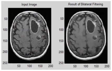
4. EXPERIMENTAL RESULTS AND DISCUSSION Mostly the filters will be used for filtering the image. Here we used efficient filter technique called Bilateral filter, which will gives a highly purified filtered images by reducing all type of noises like salt and pepper noises and preserving the edge of an image, which is shown in Figure-2.
[image:4.612.320.535.511.648.2]
Figure-2. Result of bilateral filter.
Figure-3. Result of segmented image.
After Segmentation technique, the image’s features are extracted from the segmented image, for feature extraction using Discrete wavelet transform (DWT), when the image have the high pass filter yielding image, the DWT will helps in reduce the higher yielding image to get the image into low pass filter to view clearly for extracting the features, which is shown in Figure-4.
Figure-4. Result of feature extraction.
In Table-1, shows the lists of features are extracted from the segmented input image and highly relevant features are selected for classification process.
Table-1. List of features.
S. No. Extracted
features S. No.
Selected features 01 Contrast
01 Coarseness
02 Entropy
03 Coarseness
02 Color-gray
04 Color-gray
05 Intensity
03 Intensity
06 Gabour
07 Invariant
04 Gabour
08 Prominence
5. PERFORMANE ANALYSIS
We assess the performance of the proposed method is measured in terms of sensitivity, specificity and accuracy. In equation (2) - (4), the three terms are defined:
(2)
(3)
(4)
Where,
True Positives (TP) = properly classified positive cases, True Negative (TN) = properly classified negative cases, False Positives (FP) = imperfectly classified negative cases,
Table-2. Different classifier comparison.
Classifiers Classified Images (Total:75) Classification
accuracy (%) Normal Begnin Malignant
Neural
classifier 22 23 24 92.00
Statistical
classifier 22 21 23 88.00
Hybrid
classifier 24 24 25 97.33
In performance comparison, shows that proposed classifier gives 97.33% accuracy, where as neural classifier gives 92% accuracy, and statistical classifier gives 88% accuracy, which is given in Table-2. Hence proposed Hybrid classifier, predict the tumor images accurately as early as possible which helps for proper medication.
Figure-5. Performance comparison.
6. CONCLUSIONS
Our proposed system implements a novel classification mechanism for efficiently analyze the brain tumor images using hybrid classifier. We utilized ROI (Region of Interest) segmentation method for CT image. Using DWT, the key features are extracted; the extracted features are taken as input for PCA to reduce the dimensionality of features. Then the images were trained with hybrid classifier. Finally, the proposed algorithm is significantly efficient for classification of the human brain image is benign and malignant with high sensitivity, specificity and accuracy rates. The performance of this study appears some advantages of this technique: it is accurate, robust easy to operate, non-invasive and inexpensive.
In future work, we have a plan to explore different types of medicinal images as well as some other application domains and study some formal properties of image features.
REFERENCES
[1] T. Kesavamurthy, S. SubhaRani. 2006. “Pattern Classification using imaging techniques for Infarct
and Hemorrhage Identification in the Human Brain”, Calicut Medical Journal.
[2] http://www.braintumor.org/TumorTypes.
[3] D. Selvathi, R.S. Ram Prak ash, Dr. S.Thamarai Selvi. 2007. Performance Evaluation of Kernel Based Techniques for Brain MRI Data Classification, International Conference on Computational Intelligence and Multimedia Applications.
[4] Mohd FauziBin Othman, Noramalina Bt Abdullah, Nurul Fazrena Bt Kamal. 2010. MRI Brain Classification Using Support vector machine, Cairo University, Malaysia.
[5] D. Bhattacharyya, Tai-hoon Kim. 2011. Brain Tumor Detection Using MRI Image Analysis, Communications in Computer and Information Science, Vol: 151, pp: 307-314.
[6] E.F. Badran, E.G. Mahmoud, N. Hamdy. 2010. An algorithm for detecting brain tumors in MRI images, Proceedings of the International Conference on Computer Engineering and Systems (ICCES), Cairo, pp: 368-373.
[7] S.Koley, A. Majumder. 2011. Brain MRI
segmentation for tumor detection using cohesion based self merging algorithm, Proceedings of the IEEE 3rd International Conference on Communication Software and Networks (ICCSN), Xi'an, pp: 781-785.
[8] D. M. Joshi, N. K. Rana, V. M. Misra. 2010. Classification of brain cancer using artificial neural network, Proceedings of the 2010 2nd International
Conference on Electronic Computer Technology, 112-116, Kuala Lumpur, Malaysia, May.
[10]E. I. Zacharaki, S. Wang, S. Chawla, D. Soo Yoo, R. Wolf, E. R. Melhem, C. Davatzikos. 2009. Classification of brain tumour type and grade using MRI texture and shape in a machine learning scheme, Magnetic Resonance in Medicine, 62(6): 1609-1618.
[11]P. Narendran, V.K. Narendira Kumar, K.
Somasundaram. 2012. 3D Brain Tumors and Internal Brain Structures Segmentation in MR Images, I.J. Image, Graphics and Signal Processing, 1, 35-43.
[12]Dou, W., Ruan, S., Chen, Y., Bloyet, D., and Constans, J. M. 2007. A framework of fuzzy information fusion for segmentation of brain tumor tissues on MR images, Image and Vision Computing, 25: 164-171.
[13]Y. Boykov and V. Kolmogorov. 2004. An
experimental comparison of min-cut/max-flow algorithms for energy minimization in vision, IEEE Transactions on Pattern Analysis and Machine Intelligence, pages 1124-1137.
[14]A. Lefohn, J. Cates, and R. Whitaker. 2003. Interactive, gpu-based level sets for 3d segmentation, MICCAI, pp. 564-572.
[15]M. Kaus, S. K. Warfield, A. Nabavi, P. M. Black, F. A. Jolesz, and R. Kikinis. 2001. Automated Segmentation of MRI of Brain Tumors, Radiology, 218(2) (586-91).
[16]Edward Kim, Tian Shen, Xiaolei Huang. 2010. A Parallel Cellular Automata with Label Priors for Interactive Brain Tumor Segmentation, Lehigh University, Department of Computer Science and Engineering, Bethlehem, PA, USA.