Strabismus in Cases of Cataract in Pediatric Age Group
*
Shreya M. Shah, Mehul A. Shah, Pramod R. Upadhyay, Geetopam B. Bardoloi, Drashti Netralaya#
Drashti Netralaya, Dahod, India. Email: #[email protected]
Received October 21st, 2012; revised November 21st, 2012; accepted November 28th, 2012
ABSTRACT
Purpose: To investigate the epidemiology of strabismus in cases of pediatric cataracts. To assess visual outcome fol-
lowing orthoptic treatment for amblyopia in cases of cataracts in the pediatric age group. Methods: This was a
retrospective cohort study. We investigated a consecutive series of pediatric patients with congenital, developing, or traumatic cataracts who underwent surgery between January 1999 and April 2012 at our center. Patient demographics, cataract type, presenting symptoms, surgical intervention, postoperative visual acuity, and follow-up refractive changes were recorded. Results: In total, 1331 eyes of 1043 children were included: unilateral cataracts were present in 785
(59%) eyes. There were 605 (45.5%) traumatic and 726 (54.5%) non-traumatic cases. Ages at surgery ranged from 1 to 215 months. All eyes were examined for ocular alignment; 66 (5%) were found to manifest strabismus. Deviation was significantly associated with age at intervention (p < 0.001), sensory nystagmus (p < 0.001), and etiology of cataracts (p
< 0.001). We found significant differences in visual outcome following amblyopia therapy (p < 0.001). Conclusions:
Surgical treatment with intraocular lens implantation in children with congenital, developmental, or traumatic cataracts is effective for visual rehabilitation. Orthoptic treatment made a significant difference in visual outcome (p < 0.001).
Keywords: Pediatric Cataract; Visual Outcome; Traumatic Cataract; Developmental Cataract; Congenital Cataract
1. Introduction
Childhood cataracts are responsible for 5% - 20% of blindness in children worldwide and for an even higher percentage of childhood visual impairment in developing countries [1-5]. The overall incidence of clinically sig- nificant cataracts (unilateral or bilateral) in childhood is unknown, but has been estimated to be as high as 0.4%. [6,7]. The prevalence of childhood cataracts varies from 1.2 to 6.0 cases per 10,000 infants. Pediatric cataracts are responsible for more than 1 million cases of childhood blindness in Asia. In developing countries, such as India, 7.4% - 15.3% of childhood blindness is due to cataracts. [8,9] Internationally, the incidence is unknown. Although the World Health Organization and other health organi- zations have made great progress in vaccination and dis- ease prevention, the rate of congenital cataracts remains much higher in underdeveloped countries.
The visual results of cataract surgery in children have generally [10,11] been poorer than in adults [1-3,6,11,12]. This difference is due, in part, to the various types of am- blyopia that develop in children with cataracts, the asso-ciation of nystagmus with early onset cataracts, and the
presence of other ocular abnormalities that adversely affect vision in eyes with developmental lens opacities. Since the introduction of the aspiration technique of ca- taract removal by Scheie in 1960 [13], surgical proce- dures for the removal of the lens in childhood have im- proved [14,15], and earlier surgery for congenital cata- racts has been encouraged [16-18]. Strabismus and am- blyopia are important causes of poor visual outcome. [18].
Any opacification of the lens and its capsule in chil- dren is defined as a pediatric cataract. Pediatric cataracts can be unilateral or bilateral. They can be subdivided ba- sed on morphology, as well as etiology. Morphologically, the most common type of pediatric cataract is the zonular cataract, characterized by opacification of a discrete re- gion of the lens. This type includes nuclear, lamellar, su- tural, and capsular cataracts [6,10].
Congenital cataracts are one of the most common cau- ses of treatable blindness in children, particularly in de-veloping countries [1]. A recent report indicated that in-fants with bilateral congenital cataract who underwent early surgery (within 1 month of birth) and received ap- propriate optical rehabilitation could obtain visual acuity of better than 0.4 and could even achieve stereopsis [2]. However, because of typically relatively late detection and diagnosis, the lack of availability of facilities for
*No financial support received from any company or institution, This
study is not presented at any conference or meeting, Authors do not have any financial interest in any aspect of this study
ininfant anesthesia, and poor compliance with long-term follow-up, the visual prognosis for infants with conge- nital cataract in developing countries differs markedly from that in industrialized nations. Visual loss is primar- ily attributable to amblyopia, most importantly, to “stimu- lus-form deprivation amblyopia” with the additional fac- tor of ocular rivalry in unilateral disease. Thus, improved understanding of the critical periods of visual develop- ment has resulted in surgical intervention for dense cata- racts being deemed necessary within the first 3 months of life, possibly as early as the first 6 weeks in unilateral disease. Clinical factors believed to be important to vis- ual outcome in children include age at diagnosis and sur- gery, type of refractive correction, type of cataract sur- gery, compliance with an occlusion regimen, etiology of the cataract, presence of non-ophthalmic disorders, deve- lopment of capsular opacity or secondary membrane, and serious ocular postoperative complications.
2. Materials and Methods
The study was approved by the hospital ethics committee. This was a prospective hospital-based study at a tertiary care eye hospital in western India over 20 years, from January 1992 to April 2012. All pediatric patients (0 - 18) with cataracts presenting to our department during this period were enrolled in the study. Primary patient details and history were documented using a pre-tested online format. Ocular trauma details were documented with an online world eye injury registry form.
Gifting Vision was checked according to the American Academy of Pediatrics vision check protocol. Both eyes were assessed [19]. Anterior segment examinations were conducted using a slit lamp bio-microscope. The pupils were dilated.
Ocular pressure was measured using a Perkin’s hand- held tonometer. If this was not possible, the pressure was measured under general anesthesia. This procedure was omitted for eyes with open globe injuries. The posterior segment of the eye was evaluated with the help of an indirect ophthalmoscope, a +20 D lens, and an ultrasound ‘B’ scan if the media was not clear. The surgical tech- nique was decided based on etiology, cataract morphol- ogy, and the position of the lens. Surgery was conducted by the anterior or pars plana route. Anterior route surger- ies were performed using a phacoemulcifier or manual suction. Membranectomies and lensectomies were per- formed using a pneumatic cuter. Intraocular lenses were not implanted in patients younger than 1.5 years. Chil- dren below this age underwent lensectomies/membranec- tomies; secondary implant placement was conducted la- ter. Patients were rehabilitated using glasses or contact lenses in-between. For IOL power calculations, we fol- lowed published guidelines [20,21].
In cases of globe rupture, open globe injury wound repair was done as a first stage and the cataract was op- erated on at a second sitting. All steps of the surgical techniques were documented using a pretested online for- mat.
All traumatic cataract patients without infection were treated with systemic corticosteroids. In all patients with inflammation and membranous cataracts, a primary po- sterior capsulotomy and anterior vitrectomy were per- formed.
Postoperative follow-up was performed according to a pretested online format, including vision, anterior and posterior segment findings, and intraocular pressure, over an appropriate follow-up schedule. Glasses were pre- scribed when the media were clear and the final pre- scription was at 6 weeks post-operation. Patients under- went orthoptic evaluations and amblyopic patients were treated with appropriate patching. Aphakic patients were rehabilitated using glasses or contact lenses. Patients were evaluated for stereopsis and contrast sensitivity using a Titmus vision tester or a Titmus fly test.
Patients developing later cataracts underwent mem- braneectomies and vitrectomies as required. For children operated on below the age of 1.5 years, secondary lens implantation was performed after they reached 2 years of age.
Data were analysed using the SPSS software (ver. 19.0; SPSS Inc., Chicago, IL, USA). Univariate parametrical analyses were used. A P value of <0.05 was considered to indicate statistical significance.
3. Results
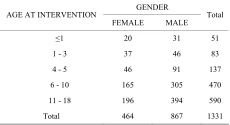
The enrolled patient group consisted of 1331 eyes in 1043 pediatric patients with cataracts. There were 867 (65.1%) males and 464 (34.9%) females (Table 1). The
[image:2.595.309.539.612.737.2]mean patient age was 4.1 ± 1.06 (range, 0 - 18) years. Of the cataracts, 605 (45.5%) were traumatic and 726 (54.4%) were congenital or developmental. Of the eyes, 1145 (86%) had diminished vision and 188 (14.0%) present- ed with leucokoria. The follow-up period was 1 - 3084 (mean, 117.4) days.
Table 1. Age and gender distributions.
GENDER AGE AT INTERVENTION
FEMALE MALE Total
≤1 20 31 51
1 - 3 37 46 83
4 - 5 46 91 137
6 - 10 165 305 470
11 - 18 196 394 590
We found strabismus in 66 (5%) cases (Tables 2 and 3). We compared strabismus according to age of inter-
vention. A significant association with strabismus was found in the younger age group (Table 2; P < 0.001).
In the non-traumatic group, eyes were further subdi- vided into congenital (282, 21.2%), developmental (415, 31.2%), and secondary cataracts (29, 2.2%). According to the statistical analysis, the demographic factors ana- lyzed, including socioeconomic status (74.5% were of lower socioeconomic status) and residence (92% were from rural areas), had no significant relationship with final visual acuity. When compared with category, stra- bismus was less commonly associated with traumatic cataracts (Table 4; P < 0.001).
Regarding patient entry, 9.2% of the patients had re- ceived primary treatment prior to reaching our center; this was not associated with a significant difference in the final visual outcome (P = 0.2). Of the total patients enrolled, 26.4% entered via an outreach department, and 71% were self-referred.
Table 2. Strabismus according to age.
SQUINT AGE AT INTERVENTION
NO YES Total
≤1 44 7 51
1 - 3 70 13 83
4 - 5 131 6 137
6 - 10 455 15 470
11 - 18 565 25 590
Total 1265 66 1331
P < 0.001.
Table 3. Amblyopia according to age.
AMBLYOPIA AGE OF INTERVENTION
−1.00 0.00 Total
≤1 9 42 51
1 - 3 17 66 83
4 - 5 11 126 137
6 - 10 19 451 470
11 - 18 18 572 590
Total 74 1257 1331
P < 0.001.
Table 4. Squint according to cataract etiology.
SQUINT CATEGORY
NO YES Total
TRAUMATIC 595 10 605
NON TRAUMATIC 670 56 726
Total 1265 66 1331
P < 0.001.
Among the injuries, 30% were reported within the first 24 h, 30% were reported within 3 days, and 33.9% were reported within 1 month. A wooden stick was the most common object causing eye injury (51.4%). Neither the injury-causing object (P = 0.3) nor the activity at the time of injury (P = 0.3) was significantly associated with the final visual acuity.
A comparison of pre- and post-operative visual acui- ties showed that treatment significantly improved visual acuity (Table 5; P < 0.001, Pearson’s χ2 test; P = 0.001,
ANOVA).
We examined unilateral versus bilateral cataracts (Ta- ble 6; P < 0.001) and found a significant association with
strabismus in bilateral cases.
Sensory nystagmus was present in 250 (18.8%) eyes and was significantly associated with strabismus (Table 7; P < 0.001).
An intraocular lens was implanted in 1210 cases (91.1%) and was not significantly associated with stra- bismus (Table 8; P= 0.137).
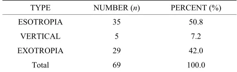
When we studied the strabismus, we found that 35 (50.8%) had esotropia, 29 (42%) had exotropia, and five (7.2%) had vertical strabismus with esotropia or exo- tropia (Table 9). In total, 36% cases had “special” forms
Table 5. Visual outcome pre–post-patching.
POST PATCH VISION PRE PATCH
VISION <1/60 1/60 TO
3/60
6/60 TO 6/36
6/24 TO 6/18
Total
<1/60 7 0 0 1 8
1/60 TO 3/60 2 21 0 0 23
6/60 TO 6/36 0 4 4 0 8
6/24 TO 6/18 1 0 2 27 30
6/12 TO 6/9 0 0 3 1 4
Total 10 25 9 29 73
Table 6. Strabismus compared with laterality.
SQUINT LATERALITY
NO YES Total
UNILATERAL 763 22 785
BILATERAL 502 44 546
Total 1265 66 1331
P < 0.001.
Table 7. Strabismus compared with sensory nystagmus.
SQUINT SENSORY
NYSTAGMUS NO YES Total
YES 1051 30 1081
NO 214 36 250
Total 1265 66 1331
Table 8. Strabismus compared with the presence of IOL.
SQUINT IOL
NO YES Total
YES 1153 57 1210
NO 112 9 121
Total 1265 66 1331
[image:4.595.55.287.213.284.2]P = 0.137.
Table 9. Type of strabismus.
TYPE NUMBER (n) PERCENT (%)
ESOTROPIA 35 50.8
VERTICAL 5 7.2
EXOTROPIA 29 42.0
Total 69 100.0
of strabismus, like A pattern, V pattern, or dissociated ver- tical deviation. In cases with strabismus, 37 (53.6%) had sensory nystagmus and 56 (81.2%) had amblyopia.
4. Discussion
The enrolled patient group consisted of 1331 eyes in 815 pediatric patients with cataracts. There were 867 (65.1%) males and 464 (34.9%) females (Table 2). The mean
patient age was 4.1 ± 1.06 (range, 0 - 18) years. Of the cataracts, 605 (45.5%) were traumatic and 726 (54.4%) were congenital or developmental. The mean patient age was 4.1 ± 1.6 years. The mean age in another report was 7.1 [22]. Age at intervention had a significant effect on visual outcome (Table 2). Other investigators have re-
ported similar findings [23].
The incidence of traumatic cataracts in children was higher than that reported previously [24]. In a compare- son of the traumatic and non-traumatic groups, the group with traumatic cataracts had a significantly lower inci- dence of strabismus, likely attributable to the more fully developed visual system in children in traumatic cases. Spanou et al. reported strabismus in 23% and Magli et al. reported a 34% incidence of strabismus in contrast to only 5% in our study [25,26], perhaps because of the larger number of traumatic cataracts. Regarding unila- teral and bilateral cases, we found a higher incidence of strabismus in bilateral cataracts in contrast to another re- port [25].
A retrospective study of the outcome of surgery for ca- taracts in the pediatric age group has several limitations. Although we believe that all patients included in the stu- dy had congenital, developmental, or traumatic lens opa- cities, not all patients were seen and followed by us from the time of birth. In particular, some patients with lamel- lar cataracts were not seen by us until they were several years old.
Treatment of strabismic amblyopia following bilateral congenital cataract surgery is useful, although the ocular misalignment is sometimes hard to identify, and the am- blyopia may be profound by the time it is recognized [10, 27]. Deprivational amblyopia due to asymmetry of cata-racts from the outset is very difficult to reverse, similar to the situation in patients with monocular congenital cata- racts. An early start of treatment would seem to be the only hope of success in these asymmetric cases [10].
In conclusion, the incidence of strabismus in cases of pediatric cataract was only 5%, was more common in the non-traumatic group and bilateral cases in the younger age group. Orthoptic treatment had a significant effect on visual outcome.
REFERENCES
[1] V. Long and S. Chen, “Surgical Interventions for Bilate- ral Congenital Cataract,” Cochrane Database of Systema- tic Reviews, Vol. 19, No. 3, 2006, Article ID: CD003171. doi:10.1002/14651858.CD003171.pub2
[2] M. A. Zimmermann-Paiz, and C. R. Quiroga-Reyes, “Pe- diatric Cataract in a Developing Country Retrospective Re- view of 328 Cases,” Arquivos Brasileiros de Oftalmolo- gia, Vol. 74, No. 3, 2011, pp. 163-165.
[3] P. Gogate and K. Kalua, “Blindness in Childhood in De- veloping Countries Time for a Reassessment?” PLOS Me- dicine, Vol. 6, No. 12, 2009, Article ID: e1000177. [4] M. E. Wilson and S. K. Pandey, “Paediatric Cataract Blind-
ness in the Developing World Surgical Techniques and Intraocular Lenses in the New Millennium,” British Jour- nal of Ophthalmology, Vol. 87, No. 1, 2003, pp. 14-19. [5] K. M. de Carvalho and N. Minguini, “Characteristics of a
Pediatric Low-Vision Population,” Journal of Pediatric Ophthalmology and Strabismus, Vol. 35, No. 3, 1998, pp. 162-165.
[6] W. C. Owens and W. F. Hughes, “Results of Surgical Treatment of Congenital Cataract,” Archives of Ophtha- lmology, Vol. 39, No. 3, 1948, pp. 339-350.
doi:10.1001/archopht.1948.00900020346009
[7] G. Msukwa and M. Njuguna, “Cataract in Children At- tending Schools for the Blind and Resource Centers in Eastern Africa,” Ophthalmology, Vol. 116, 2009, pp. 1009- 1012. doi:10.1016/j.ophtha.2008.12.020
[8] R. Dandona and L. Dandona, “Review of Findings of the Andhra Pradesh Eye Disease Study: Policy Implications for Eye-Care Services,” Indian Journal of Ophthalmology, Vol. 49, No. 4, 2001, pp. 215-234.
[9] A. R. Vasavada, S. A. Vasavada, N. Bobrova, M. R. Pra- veen, S. K. Shah, V. A. Vasavada, et al., “Outcomes of Pe- diatric Cataract Surgery in Anterior Persistent Fetal Vas- culature,” Journal of Cataract & Refractive Surgery, Vol. 38, No. 5, 2012, pp. 849-857.
doi:10.1016/j.jcrs.2011.11.045
183-194
[11] J. Francois “Late Results of Congenital Cataract Surge- ry,” Ophthalmology, Vol. 86, No. 9, 1979, pp. 1586-1598. [12] D. A. Hiles and P. H. Wallar, “Visual Results Following
Infantile Cataract Surgery,” International Ophthalmology Clinics, Vol. 17, No. 4, 1977, pp. 265-282.
[13] H. G. Scheie, “Aspiration of Congenital or Soft Cataracts: A New Technique,” American Journal of Ophthalmology, Vol. 50, 1960, pp. 1048-1056.
[14] D. Taylor, “Choice of Surgical Technique in the Mana- gement of Congenital Cataract,” Transactions of Ophtha- lmological Societies of United Kingdom, Vol. 101, No. 1, 1981, pp. 114-117.
[15] M. M. Parks, “Posterior Lens Capsulectomy during Pri- mary Cataract Surgery in Children,” Ophthalmology, Vol. 90, No. 4, 1983, pp. 344-345.
[16] G. L. Rogers, C. L. Tishler and B. H. Tsou, “Visual Acui- ties in Infants with Congenital Cataracts Operated on Pri- or to 6 Months of Age,” Archives of Ophthalmology, Vol. 99, No. 6, 1981, pp. 999-1003.
doi:10.1001/archopht.1981.03930010999002
[17] S. S. Gelbart, C. S. Hoyt, G. Jastrebski and E. Marg, “Long-Term Visual Results in Bilateral Congenital Cata- racts,” American Journal of Ophthalmology, Vol. 93, No. 5, 1982, pp. 615-621.
[18] D. Taylor, Vaegan and J. A. Morris, “Amblyopia in Bi- lateral Infantile and Juvenile Cataract, Relationship to Ti- ming of Treatment,” Transactions of Ophthalmological Societies of United Kingdom, Vol. 99, No. 1, 1979, pp. 170-175.
[19] M. Yamamoto and M. Dogru, “Visual Function Fol- low- ing Congenital Cataract Surgery,” Japanese Journal of Ophthalmology, Vol. 42, No. 5, 1998, pp. 411-416. doi:10.1016/S0021-5155(98)00039-2
[20] R. H. Trivedi and M. E. Wilson, “Accuracy of the Holla-
day 2 Intraocular Lens Formula for Pediatric Eyes in the Absence of Preoperative Refraction,” Journal of Cataract & Refractive Surgery, Vol. 37, No. 7, 2011, pp. 1239- 1243. doi:10.1016/j.jcrs.2011.01.021
[21] S. K. McClatchey, “Choosing IOL Power in Pediatric Ca- taract Surgery,” International Ophthalmology Clinics, Vol. 50, No. 4, 2010, pp. 115-123.
doi:10.1097/IIO.0b013e3181f0f2e0
[22] N. G. Congdon and S. Ruiz, “Determinants of Pediatric Cataract Program Outcomes and Follow-Up in a Large Series in Mexico,” Journal of Cataract & Refractive Sur- gery, Vol. 33, No. 10, 2007, pp. 1775-1780.
doi:10.1016/j.jcrs.2007.06.025
[23] M. L. Yang and C. H. Hou, “Clinical Characteristics and Surgical Outcomes of Pediatric Cataract in Taiwan,” Gra- efe’s Archive for Clinical and Experimental Ophthalmo- logy, Vol. 244, No. 11, 2006, pp. 1485-1490.
doi:10.1007/s00417-006-0308-4
[24] R. Khandekar and A. Sudhan, “Pediatric Cataract and Sur- gery Outcomes in Central India a Hospital Based Stu- dy,” Indian Journal of Medical Sciences, Vol. 61, No. 1, 2007, pp. 15-20. doi:10.4103/0019-5359.29593
[25] A. Magli and A. Iovine, “Strabismus in Developmental Cataract,” European Journal of Ophthalmology, Vol. 18, No. 4, 2006, pp. 540-543.
[26] N. Spanou and L. Alexopoulos, “Strabismus in Pediatric Lens Disorders,” Journal of Pediatric Ophthalmology and Strabismus, Vol. 48, No. 3, 2011, pp. 163-166. doi:10.3928/01913913-20100618-05
[27] P. Hochstrasser and B. Gloor, “Surgical Results of Uni- and Bilateral Congenital and Traumatic Cataract in In- fancy to Adolescence,” Klin Monatsbl Augenheilkd, Vol. 204, No. 5, 1994, pp. 274-278.