ORIGINAL RESEARCH
Is It Possible to Recognize Cervical Artery
Dissection on Stroke Brain MR Imaging?
A Matched Case-Control Study
O. Naggara F. Soares E. Touze D. Roy X. Leclerc J.-P. Pruvo J.-L. Mas J.-F. Meder C. Oppenheim
BACKGROUND AND PURPOSE: Extracranial CAD accounts for nearly 20% of cases of stroke in young adults. The mural hematoma frequently extends cranially to the petrous carotid segment in cCAD or is distally located in vCAD. We hypothesized that standard brain MR imaging could allow the early detection of CAD of the upper portion of carotid and vertebral arteries.
MATERIALS AND METHODS: Our prospectively maintained stroke data base was retrospectively que-ried to identify all patients with the final diagnosis of CAD. In the 103 consecutive patients studied, analysis of cervical fat-suppressed T1-weighted sequences demonstrated that the mural hematoma was located in the FOV of brain MR imaging in 77 patients. Subsequent to enrollment of a patient, a control patient was extracted from the same data base, within a similar categories for sex, age, NIHSS score, and stroke on DWI. Two blinded observers independently reviewed the 5 brain MR sequences of each examination and determined whether a CAD was present.
RESULTS: Fifty-nine of the 77 patients with CAD (76.6%) and 73 of the 77 patients without CAD (94.8%) were correctly classified. Brain MR imaging demonstrated cCAD more frequently than vCAD in 54/58 (93.1%) and 5/19 (26.3%) patients, respectively, (P⬍.0001).
CONCLUSIONS:Initial brain MR imaging can correctly suggest CAD in more than two-thirds of patients. This may have practical implications in patients with stroke with delayed cervical MRA or in those who are not initially suspected of having CAD.
ABBREVIATIONS:CAD⫽cervical artery dissection; cCAD⫽carotid artery dissection; CE⫽ con-trast-enhanced; DSA ⫽ digital subtraction angiography; DWI ⫽ diffusion-weighted imaging; FLAIR⫽ fluid-attenuated inversion recovery; ICA⫽internal carotid artery; INSERM ⫽ Institut National de la Sante´ et de la Recherche Me´dicale; MRA⫽MR angiography; MRI⫽MR imaging; NIHSS⫽National Institutes of Health Stroke Scale; PWI⫽perfusion-weighted imaging; STARD⫽ Standards for Reporting of Diagnostic Accuracy; TIA⫽transient ischemic attack; vCAD⫽vertebral artery dissection
E
xtracranial CAD accounts for nearly 20% of cases of stroke in young adults.1,2Conclusive evidence of CAD early in thediagnosis of stroke would allow rapid deployment of therapy to reduce the risk of further thromboembolic events.3Most
patients with neurologic deficits undergo a brain MR imaging examination as part of their standard etiologic work-up, first, because DWI surpasses CT for the detection of acute ischemia and, second, because MR images accompanying DWI are more effective than CT for excluding stroke mimics.4Imaging
focused on the head provides a truncated view of the arterial vasculature and is not systematically followed by an evaluation of extracranial arteries. Indeed, despite stroke guidelines,4
only approximately one-fourth of patients with stroke had
carotid imaging within 2 weeks of the event,5,6because of the
differing availability of cervical artery imaging between insti-tutions. Furthermore, cervical MRA is not part of the work-up of all patients who are not initially suspected of having CAD based on available history. Thus, patients with CAD may be undiagnosed and sustain additional infarctions before the dis-section is discovered. In CAD, the mural hematoma frequently extends cranially to the petrous carotid segment in the case of cCAD or is distally located in the case of vCAD. These loca-tions are within the limits of the FOV of standard brain MR imaging. We hypothesized that standard brain MR imaging could allow the early detection of CAD of the upper portion of the carotid and vertebral arteries.
Materials and Methods
Patients
The study was approved by the Ethics Committee of Ile de France III and was found to conform to generally accepted scientific principles and research ethics standards. Informed consent was waived. The manuscript was prepared in accordance with the STARD guidelines.7
Our case-control study was nested within a longitudinal cohort of patients referred to our institution for suspected acute stroke or TIA, between January 2002 and December 2007. This prospectively main-tained data base was retrospectively queried to identify all consecutive patients with the final diagnosis of CAD (n⫽125). CAD diagnosis Received July 2, 2010; accepted after revision September 12.
From the Departments of Neuroradiology (O.N., F.S., J-F.M., C.O.) and Neurology (E.T., J-L.M.), Paris-Descartes University, INSERM U894, Centre Hospitalier Sainte-Anne, Paris, France; Interventional Neuroradiology Research Unit (D.R.), Centre hospitalier de l’Universite´ de Montre´al, Notre-Dame Hospital, University of Montreal, Quebec, Canada; and Department of Neuroradiology (J-P.P, X.L.), Lille University Hospital Roger Salengro, Lille, France.
O.N., F.S., J.-F.M., and C.O. planned the study data collection and identified the patient cohort; O.N. and F.S. gathered the data; and O.N., E.T., and C.O. did the statistical analysis. O.N., E.T., D.R., J.-P.P., J.-L.M., and X.L. drafted the manuscript. All authors read and approved the final manuscript.
Please address correspondence to Olivier Naggara, MD, Department of Neuroradiology, Centre Hospitalier Sainte-Anne, 1 rue Cabanis, 75014 Paris, France; e-mail: [email protected]
DOI 10.3174/ajnr.A2553
BRAIN
ORIGINAL
was based onⱖ1 of the following criteria: 1) intimal flap or mural hematoma visible on cervical Doppler sonography (n⫽94), 2) mural hematoma visible on cervical axial fat-suppressed T1-weighted imag-ing (n⫽125), and 3) a nonatherosclerotic tapered flame-shaped oc-clusion (n⫽42) or a stringlike stenosis (n⫽69) on CE-MRA (n⫽
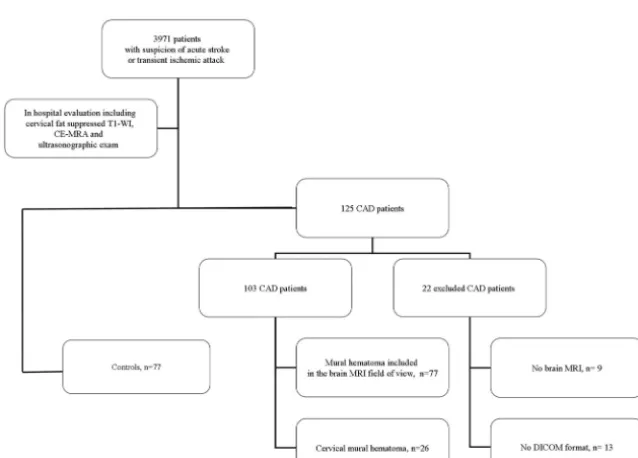
122) or conventional DSA (n⫽3). One hundred three patients with a total of 130 CADs met the following inclusion criteria: 1) brain MR imaging examinations in DICOM format, and 2) cervical axial fat-suppressed T1-weighted and CE-MRA or DSA. Twenty-two patients were thus excluded (contraindication or no MR imaging examina-tion,n⫽9; no DICOM format,n⫽13). In the 103 patients studied, analysis of axial cervical fat-suppressed T1-weighted sequences— from the aortic arch proceeding to the foramen magnum— demon-strated that the mural hematoma was located in the lower part of the FOV of brain MR images (the petrous segment of the ICA, the V3 segment of the vertebral artery) in 77 patients (74.8%; age range, 23– 66 years) with 88 CADs (67.7%; 67 cCADs, 21 vCADs) (Table 1 and Fig 1). The other 26 patients with 42 dissected arteries vertebral segment,n⫽6; V2 vertebral segment,n⫽15; cervical carotid arter-ies,n⫽21) were excluded. To evaluate the validity of CAD detection, we extracted 77 control patients from the same data base until 1 con-trol patient was individually matched to each case patient, within corresponding sex, age, NIHSS score, and stroke on DWI categories. Controls met the following inclusion criteria: 1) brain MR imaging examinations in DICOM format, and 2) identified etiology for neu-rologic deficit other than CAD. The final study group included 154 patients (98 men, 56 women; mean age, 45.4⫾9.4 years; age range, 23– 66 years).
MR Imaging
All brain MR imaging examinations were performed on a 1.5T Signa MR imaging unit (GE Healthcare, Milwaukee, Wisconsin) by using a standardized protocol: 6-mm-thick sagittal T1-weighted imaging (TR/TE, 230/5.1 ms; matrix, 320⫻224; acquisition time, 40 seconds), located from 1 mastoid process to the other; 6-mm-thick bicommis-sural axial FLAIR imaging (TR/TE/TI, 9802/159.1/2300 ms; matrix, 256⫻192; acquisition time, 2 minutes 18 seconds); gradient recalled-echo T2-weighted imaging (TR/TE, 480/13 ms; matrix, 256⫻224; flip angle, 25°; acquisition time, 1 minute 25 seconds); DWI using spin-echo echo-planar imaging (TR/TE, 5000/88.2 ms; matrix, 128⫻ 128; acquisition time, 48 seconds); and 3D time-of-flight angiography of the circle of Willis (TR/TE, 27/3.6 ms; acquisition bandwidth, 25 kHz; flip angle, 20°; FOV, 240⫻240⫻65 mm; matrix, 256⫻256; acquisition time, 2 minutes 56 seconds; single-slab acquisition seg-mented in 93 contiguous axial sections 0.7 mm thick), located from the vertex to the upper part of the second cervical vertebra.
Image Analysis
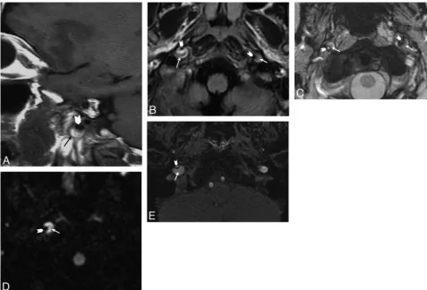
Intracranial MR imaging examinations were anonymized and ran-domly numbered. Two blinded observers independently reviewed the 5 brain MR sequences of each examination on a dedicated worksta-tion (Advantage Windows 4.1, GE Healthcare). Each sequence data-set was read separately and randomly analyzed in 2 different sessions separated by at least 2 months to minimize recall bias. Observers had to search for a crescentic hypersignal of the carotid or vertebral wall and for an increased external diameter (Fig 2). They had to conclude, for each sequence, whether a CAD was present. If by the end of the readings at least 3 sequences were positive, CAD was diagnosed. The standard of reference for CAD detection was represented by the con-sensus reading of the 2 observers, performed 2 months after the initial interpretation.
Statistical Analysis
The Statistical Package for the Social Sciences software (Version 15.0; SPSS, Chicago, Illinois) was used for the analysis. The Mann-Whitney
[image:2.594.52.286.60.133.2]Fig 1.Flow chart showing inclusion of patients. Table 1: Patient characteristics
77 Patients with CAD
77
Controls P Age (yr, mean) 45.7⫾9.2 45.0⫾9.5 0.65 Male (%) 49 (63.6) 49 (63.6) 1 NIHSS score (mean) 3.2⫾4.9 3.4⫾5.4 0.85
Stroke on DWI 41 41 1
[image:2.594.135.454.483.712.2]Utest and the Fisher exact test were used to compare, respectively, continuous parametric variables (age, NIHSS score) and categoric variables (sex, stroke on DWI sequence) between the 2 populations. Simplecoefficients and their 95% confidence intervals were used to assess inter- and intraobserver agreement. We calculated the sensitiv-ity of brain MR imaging in patients with and without CAD and com-pared the detection rate in cCAD and vCAD. We also comcom-pared the detection rate of CAD in occluded and nonoccluded arteries and in MR imaging performed⬎3 hours after the onset of symptoms versus within the first 3 hours after the onset of symptoms. We further com-pared the consensus reading with the radiologic report of each brain MR imaging. A 2-sidedPvalue⬍.05 was considered statistically significant.
Results
Cases and controls did not differ significantly for clinical, de-mographic, and stroke status on DWI data (Table 1). Among the 77 patients and 88 CADs (67 cCADs and 21 vCADs), 58 patients (75.3%) had 64 cCADs (72.7%), 17 (22.1%) patients had 18 vCADs (20.5%), 2 patients (2.3%) had both cCAD and vCAD (6.8%, 3 cCADs and 3 vCADs). Inter- and intraob-server agreement was excellent (⫽ 0.87, 95% confidence intervals: 0.57– 0.94, and⫽0.91, 95% confidence intervals: 0.56 – 0.96, respectively). Fifty-nine of the 77 patients with CAD (76.6%) and 73 of the 77 patients without CAD (94.8%) were correctly classified (Table 2). Only 18 of the 77 patients
with CAD (23.4%) had been correctly diagnosed according to the initial brain MR imaging report. Brain MR imaging dem-onstrated cCAD more frequently than vCAD (54/58, 93.1%, and 5/19, 26.3%, respectively;P⬍.0001). In 6 patients with CAD and 7 without it, MR imaging was performed within 3 hours after onset, with a correct classification in 83.3% and 100% of cases, respectively (not significant) (Table 2). In cases of occlusion, patients with and without CAD were correctly classified in 28/32 (87.5%) and 22/25 (88%) cases, respectively (not significant).
[image:3.594.53.531.42.366.2]Fig 2.Illustration of CAD on brain MR imaging sequences.A, Saggital T1-weighted image in a 42-year-old woman with left ICA dissection.BandC, Axial FLAIR (B) and gradient recalled-echo T2-weighted (C) images in a 45-year-old man with bilateral ICA dissection.DandE, Axial DWI (D) and native sections of 3D time-of-flight angiography of the circle of Willis (E) in a 38-year-old man with right ICA dissection. Note the increased external diameter, crescentic mural thickening (arrows), and eccentric lumen (arrowheads) of the dissected ICA in all 3 patients.
Table 2: Detection rate of CAD in 77 patients and 77 controls
Overall No. (%)
No. of Patients
(%)
No. of Controls
(%) p Correct rating 132/154 (85.7) 59/77 (76.6) 73/77 (94.8) .002
cCAD – 54/58 (93.1) – ⬍.0001 vCAD – 5/19 (26.3) –
Onset to MRI⬍3 hr 12/13 (92.3) 5/6 (83.3) 7/7 (100) .5 cCAD – 5/6 (83.3) –
vCAD – 0 –
Onset to MRI⬎3 hr 120/141 (85.7) 54/71 (76.0) 66/70 (94.3) .004 cCAD – 49/52 (94.2) –
vCAD – 5/19 (26.3) –
Occlusion 50/57 (87.7) 28/32 (87.5) 22/25 (88) 1 Stenosis or
irregularity
[image:3.594.301.532.572.730.2]Discussion
The main findings of this case-control study were as follows: 1) Nearly 75% CADs were included within the FOV of brain MR imaging; and 2) more than three-quarters of such acute CADs could be diagnosed by using brain MR imaging only. These results might have clinical implications for patients who are not initially suspected of having CAD on the basis of available history at the time of protocol and at institutions where cervi-cal CE-MRA is not coupled with MR brain imaging. Despite stroke imaging recommendations, cervical imaging is fre-quently delayed, with only half of patients with stroke having undergone carotid imaging within 12 weeks after the stroke event.6In 2 prospective population-based studies, the median
(interquartile range) times from the presenting event (stroke or TIA) to carotid imaging was 33 (12– 62) days.5
Conse-quently, stroke brain MR imaging can contribute to a better and earlier identification of CAD in patients with stroke.
The fact that the readers performed significantly better than the radiologists in the initial brain MR imaging report (77% versus 23%) supports the contention that the interpre-tation of direct signs of CAD on brain MR imaging is a learn-able skill; the excellent interobserver agreement between a se-nior neuroradiologist and a resident with ⬍3 months’ experience in neuroimaging demonstrates that the interpreta-tion of highly conspicuous imaging findings requires little spe-cialized training and can also be achieved in regular clinical practice.
Although several studies have described the proximal ana-tomic location of spontaneous CAD, albeit with conflicting results,8,9data on the cranial extension of the mural
hema-toma are scarce. In the present study, we report that the mural hematoma extended in the cranial direction, involving the V3 segment or the petrous segment of the ICA in nearly 75% of CADs. This has to be kept in mind in clinical practice, because a possible intracranial extension of CAD should be considered before beginning an anticoagulant treatment. For patients with CAD and severe and sudden headache, many authors consider a lumbar puncture mandatory, to rule out a sub-arachnoid hemorrhage.10
Ischemic stroke in CAD can be preceded by at least 1 TIA in 10%–15% of patients, with latencies between stroke and TIA ofⱕ17 days.10This group is of particular interest because
patients with TIA are increasingly examined with brain MR imaging. The stroke may be prevented by immediate recogni-tion of the CAD signs on brain MR imaging and initiarecogni-tion of antithrombotic or anticoagulation treatment.3
Mural hematoma can be definitely recognized on brain MR imaging even in the case of occlusion or in thrombolysis-eli-gible patients. In the latter case, it is important to avoid any delay in initiating treatment as a result of artery imaging. Some data suggest that an occluded large intracranial vessel is less likely to be recanalized by intravenous fibrinolysis alone if the parent cervical lumen is occluded or severely reduced.11
Sev-eral studies on retrospective nonrandomized series12-15have
reported the outcome in patients with hyperacute stroke due to CAD, with both cervical occlusion or high-grade stenosis and intracranial occlusion treated by stent-assisted endovas-cular thrombolysis or thrombectomy. Such endovasendovas-cular treatments were reportedly safe and effective and compared favorably with intravenous recombinant tissue plasminogen
activator.14Thus, in institutions that have chosen MR imaging
as the prime screening imaging technique in hyperacute stroke, standard brain MR imaging may reveal specific signs of CAD, thereby enabling faster triage of those patients who may derive the greatest benefit from endovascular thrombolysis.
Although the mismatch hypothesis has not yet been tested conclusively,16 penumbral selection, PWI, is being used to
screen patients for acute thrombolytic therapy.17The clinical constellation of a large middle cerebral artery stroke with a substantial mismatch between DWI and PWI in the 3- to 6-hour time window is for many stroke neurologists an indi-cation for the off-label use of intravenous tissue plasminogen activator. Cervical CAD may lead to a misinterpretation of the PWI. A mismatch between a larger PWI abnormality, due to reduced lumen of the dissected artery, and a smaller DWI lesion, due to an embolic small-vessel stroke, may erroneously identify patients as being suitable candidates for recanaliza-tion therapies.
Our study has several limitations. First, it is unlikely to obviate cervical imaging if stroke brain MR imaging does not demonstrate any mural hematoma. Second, this analysis was retrospective and only 1 control was associated with each case, resulting in a population with a prevalence of 50% dissection. Third, the delay from onset to brain MR imaging was hetero-geneous, with ⬍10% of patients imaged within the first 3 hours after onset. Another potential limitation is that some patients with an extracranial arterial dissection were missed, because an intimal tear may occur and extend without mural hematoma.
Conclusions
Stroke brain MR imaging can allow early detection of CAD before dedicated imaging of the cervical arteries is performed. Although the absence of mural hematoma does not com-pletely rule out CAD and does not obviate cervical imaging, stroke brain MR imaging can contribute to a better and earlier identification of patients with stroke who are suitable candi-dates for anticoagulation treatment or revascularization ther-apy. These results serve to remind neuroradiologists and gen-eral radiologists that much information can be derived from stroke brain MR imaging.
References
1. Schievink WI.Spontaneous dissection of the carotid and vertebral arteries.
N Engl J Med2001;344:898 –906
2. Debette S, Leys D.Cervical-artery dissections: predisposing factors, diagnosis, and outcome.Lancet Neurol2009;8:668 –78
3. Engelter ST, Brandt T, Debette S, et al.Antiplatelets versus anticoagulation in cervical artery dissection.Stroke2007;38:2605–11
4. Latchaw RE, Alberts MJ, Lev MH, et al.Recommendations for imaging of acute ischemic stroke: a scientific statement from the American Heart Association.
Stroke2009;40:3646 –78
5. Fairhead JF, Mehta Z, Rothwell PM.Population-based study of delays in ca-rotid imaging and surgery and the risk of recurrent stroke.Neurology
2005;65:371–75
6. Halliday AW, Lees T, Kamugasha D, et al.Waiting times for carotid endarter-ectomy in UK: observational study.BMJ2009;338:b1847
7. Bossuyt PM, Reitsma JB, Bruns DE, et al.Towards complete and accurate re-porting of studies of diagnostic accuracy: the STARD initiative.Radiology
2003;226:24 –28
8. Mokri B, Houser OW, Sandok BA, et al.Spontaneous dissections of the verte-bral arteries.Neurology1988;38:880 – 85
clinical, conventional angiographic, CT, and MR findings.J Comput Assist Tomogr1996;20:185–93
10. Arnold M, Bousser MG, Fahrni G, et al.Vertebral artery dissection: presenting findings and predictors of outcome.Stroke2006;37:2499 –503
11. Zangerle A, Kiechl S, Spiegel M, et al.Recanalization after thrombolysis in stroke patients: predictors and prognostic implications.Neurology2007;68: 39 – 44
12. Georgiadis D, Lanczik O, Schwab S, et al.IV thrombolysis in patients with acute stroke due to spontaneous carotid dissection.Neurology 2005;64: 1612–14
13. Derex L, Nighoghossian N, Turjman F, et al.Intravenous TPA in acute isch-emic stroke related to internal carotid artery dissection.Neurology2000;54: 2159 – 61
14. Lavallee PC, Mazighi M, Saint-Maurice JP, et al.Stent-assisted endovascular thrombolysis versus intravenous thrombolysis in internal carotid artery dis-section with tandem internal carotid and middle cerebral artery occlusion.
Stroke2007;38:2270 –74
15. Baumgartner RW, Georgiadis D, Nedeltchev K, et al.Stent-assisted endovascu-lar thrombolysis versus intravenous thrombolysis in internal carotid artery dissection with tandem internal carotid and middle cerebral artery occlusion.
Stroke2008;39:e27–28
16. Schabitz WR.MR mismatch is useful for patient selection for thrombolysis: No.Stroke2009;40:2908 – 09