organic papers
Acta Cryst.(2005). E61, o4379–o4381 doi:10.1107/S1600536805039164 Saminathanet al. C
5H12N+C6H2N3O7
o4379
Acta Crystallographica Section EStructure Reports
Online
ISSN 1600-5368
Piperidinium picrate
Kolandaivelu Saminathan,a Chellamuthu
Muthamizhchelvanband
Kandasamy Sivakumara*
aDepartment of Physics, Anna University,
Chennai 600 025, India, andbDepartment of
Physics, SRM Engineering College, Kattankulathur 603 203, India
Correspondence e-mail: [email protected]
Key indicators
Single-crystal X-ray study T= 293 K
Mean(C–C) = 0.003 A˚ Rfactor = 0.040 wRfactor = 0.121
Data-to-parameter ratio = 11.5
For details of how these key indicators were automatically derived from the article, see http://journals.iucr.org/e.
#2005 International Union of Crystallography Printed in Great Britain – all rights reserved
In the title compound, C5H12N +
C6H2N3O7
, the protonated N atom of the cation makes one linear and two bifucated hydrogen bonds with two neighbouring picrate anions. Centrosymmetrically related anions and cations form a
hydrogen-bonded network with a graph-set motif R4
4
(12). The picrate ions are parallel to one another and governed by
–interactions; they form columns along thebaxis.
Comment
Picric acid forms salts or charge-transfer complexes with many organic compounds, particularly with aromatic and aliphatic amines. Crystalline picrates have commonly been used in the preparation of amine derivatives in qualitative organic chemistry (Shrineret al., 1980). Crystal structures of a number of picrate complexes with organic compounds and biological
base molecules such as serotonin, guanine andalanine have
been studied in the past (Takayanagiet al., 1996; Thewalt & Bugg, 1972; Anithaet al., 2005). Our aim is to study the nature
and directionality of the specific N—H O hydrogen bonding
between the molecular ions involving the phenolate O and the protonated N atom and the crystal packing mode. As part of our investigations, we have prepared and determined the
crystal structure of piperidinium picrate, (I)
(Mutha-mizhchelvan, Saminathan, Fraanje et al., 2005a,b;
Mutha-mizhchelvan, Saminathan, SethuSankaret al., 2005a,b,c,d,e).
The bond lengths of the anion show characteristic values,
with C1—O1 [1.242 (2) A˚ ] intermediate between single- and
double-bond character; C1—C2 [1.450 (3) A˚ ] and C1—C6
[1.452 (2) A˚ ] deviate from the standard aromatic C—C value
of 1.375 A˚ , as observed in other picrate salts
(Mutha-mizhchelvan, Saminathan, SethuSankar et al., 2005a,b,c,d,e).
These differences are attributed to the loss of a hydroxyl proton at O1, leading to conversion from neutral to the anionic state of the picrate molecule, where the negative charge is constrained to lie in the ring (Fergusonet al., 1984). The twist angles of the three nitro groups of the picrate ions
show that theorthonitro groups O2—N1—O3 and O6—N3—
O7 deviate from the benzene plane by 16.0 (2) and 50.5 (1),
respectively, and theparanitro group (O4/N2/O5) by 8.7 (2). The tilting of the nitro groups facilitates C—H O hydrogen bonding with the neighbouring cations (Table 2).
In the piperidinium cation, bond lengths involving
proto-nated atom N4 are 1.495 (2) (N4—C7) and 1.488 (2) A˚ (N4—
C11), which are longer than those found in other structures and the value 1.469 A˚ given by Allenet al.(1987). Moreover, the average value of the C—C bond, 1.507 A˚ , is also found to be less than the normal C—C distance. The cation exists in its most stable chair conformation.
Protonated atom N4 makes three hydrogen bonds with two of its neighbouring picrate anions. Of the two H atoms of N4, one is involved in bifurcated hydrogen bonds and the other one in a linear bond (Table 2). The bifurcated hydrogen bonds are of different strengths; the one involving the phenolic O atom (O1) is stronger than the other. Such cases have been
observed in other picrate structures and are in line with the discussions of Tayloret al.(1984).
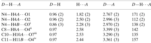
A set of centrosymmetrically related anions and cations
form a graph-set motif of R4
4(12) involving N4—H4A O2
and N4—H4B O3’, as shown in Fig. 2 (Bernstein et al.,
1995). One more hydrogen-bond network is found in this
structure, with N4—H4A O1 and N4—H4A O2
combining together to form a graph-set of motifR21(6).
The picrate ions stack parallel to one another in an offset fashion, with possible–interactions as they are separated by 3.438 A˚ This leads to columnar stacking of the picrate ions,
with the columns running alongaaxis. These anionic columns
are separaed by cationic layers, as shown in Fig. 3.
Experimental
The title compound was prepared by taking equimolar amounts (1:1) of picric acid and piperidine and dissolving them in ethanol. Slow evaporation of the solution resulted in the formation of transparent yellow prism-shaped single crystals.
Crystal data
C5H12N+C6H2N3O7
Mr= 314.26
Triclinic,P1 a= 6.8750 (15) A˚ b= 9.3471 (17) A˚ c= 11.9198 (11) A˚
= 105.393 (12)
= 91.856 (11)
= 111.341 (18)
V= 680.7 (2) A˚3
Z= 2
Dx= 1.533 Mg m
3
MoKradiation Cell parameters from 25
reflections
= 8–15
= 0.13 mm1
T= 293 (2) K Prism, yellow 0.540.520.38 mm
Data collection
Enraf–Nonius CAD-4 diffractometer
!–2scans
Absorption correction: scan (Northet al., 1968) Tmin= 0.873,Tmax= 0.961
2606 measured reflections 2382 independent reflections 1830 reflections withI> 2(I)
Rint= 0.014
max= 25.0
h=2!8 k=11!10 l=14!14 2 standard reflections
every 100 reflections intensity decay: 1%
organic papers
o4380
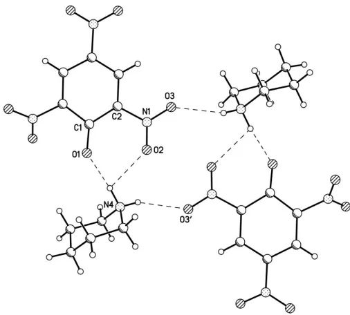
Saminathanet al. C [image:2.610.47.296.72.235.2]5H12N+C6H2N3O7 Acta Cryst.(2005). E61, o4379–o4381 Figure 1
[image:2.610.315.569.73.240.2]The title complex, showing 50% probability displacement ellipsoids and the atom-numbering scheme.
Figure 2
Plot showing the hydrogen-bonded (dashed lines) graph-set motifs:
R4
4(12) involving N4—H4A O2 and N4—H4B O3’, and R21(6)
involving N4—H4A O1 and N4—H4A O2.
Figure 3
Packing of the molecular complex, viewed down the crystallographicb
[image:2.610.43.296.275.503.2]Refinement
Refinement onF2 R[F2> 2(F2)] = 0.040
wR(F2) = 0.121 S= 1.03 2382 reflections 208 parameters
H atoms treated by a mixture of independent and constrained refinement
w= 1/[2
(Fo2) + (0.0615P)2
+ 0.2412P]
whereP= (Fo2+ 2Fc2)/3
(/)max< 0.001
max= 0.43 e A˚
3
min=0.28 e A˚
3
[image:3.610.44.298.209.261.2]Extinction correction:SHELXL97 Extinction coefficient: 0.015 (4)
Table 1
Selected geometric parameters (A˚ ,).
O1—C1 1.242 (2) C1—C2 1.450 (3) C1—C6 1.452 (2)
N4—C11 1.488 (3) N4—C7 1.494 (3)
O1—C1—C2 126.65 (16) O1—C1—C6 122.04 (17)
[image:3.610.43.296.309.381.2]C2—C1—C6 111.30 (15) C11—N4—C7 112.42 (16)
Table 2
Hydrogen-bond geometry (A˚ ,).
D—H A D—H H A D A D—H A
N4—H4A O1 0.96 (2) 1.82 (2) 2.767 (2) 171 (2) N4—H4A O2 0.96 (2) 2.50 (2) 2.996 (3) 112 (2) N4—H4B O3i
0.86 (3) 2.28 (3) 2.970 (2) 138 (2) C8—H8A O4ii
0.97 2.58 3.399 (3) 142 C10—H10A O7iii
0.97 2.53 3.290 (3) 135 C11—H11B O4iv
0.97 2.44 3.361 (3) 157
Symmetry codes: (i)x;yþ1;z; (ii)xþ1;yþ2;z; (iii)x;yþ2;zþ1; (iv)x;yþ2;z.
All H atoms were located in difference Fourier maps. While the H atoms of the protonated N atom were refined isotropically, the C-bound H atoms were refined as riding on their parent atoms, with C— H = 0.93–0.98 A˚ andUiso(H) = 1.2Ueq(C).
Data collection: CAD-4 EXPRESS (Enraf–Nonius, 1994); cell refinement: CAD-4 EXPRESS; data reduction:XCAD4 (Harms & Wocadlo, 1995); program(s) used to solve structure: SHELXS97
(Sheldrick, 1997); program(s) used to refine structure:SHELXL97
(Sheldrick, 1997); molecular graphics:ORTEP3(Farrugia, 1997) and
PLATON (Spek, 2003); software used to prepare material for publication:SHELXL97.
The authors thank Dr Babu Varghese, SAIF, Indian Insti-tute of Technology, Chennai, for collecting the single-crystal data.
References
Allen, F. H., Kennard, O., Watson, D. G., Brammer, L., Orpen, A. G. & Taylor, R. (1987).J. Chem. Soc. Perkin Trans. 2, pp. S1–19.
Anitha, K., Athimoolam, S. & Rajaram, R. K. (2005).Acta Cryst.E61, o1463– o1465.
Bernstein, J., Davis, R. E., Shimoni, L. & Chang, N.-L. (1995).Angew. Chem. Int. Ed. Engl.34, 1555–1573.
Enraf–Nonius (1994). CAD-4 EXPRESS. Version 5.0/1.2. Enraf–Nonius, Delft, The Netherlands.
Farrugia, L. J. (1997).J. Appl. Cryst.30, 565.
Ferguson, G., Ruhl, B. L. & Wieckowski, T. (1984).Acta Cryst.C40, 1740–1742. Harms, K. & Wocadlo, S. (1995).XCAD4. University of Marburg, Germany. Muthamizhchelvan, C., Saminathan, K., Fraanje, J., Peschar, R. & Sivakumar,
K. (2005a).Acta Cryst.E61, o1153–o1155.
Muthamizhchelvan, C., Saminathan, K., Fraanje, J., Peschar, R. & Sivakumar, K. (2005b).Anal. Sci.21, x61–x62.
Muthamizhchelvan, C., Saminathan, K., SethuSankar, K., Fraanje, J., Peschar, R. & Sivakumar, K. (2005a).Acta Cryst.E61, o1377–o1380.
Muthamizhchelvan, C., Saminathan, K., SethuSankar, K., Fraanje, J., Peschar, R. & Sivakumar, K. (2005b).Acta Cryst.E61, o1546–o1548.
Muthamizhchelvan, C., Saminathan, K., SethuSankar, K., Fraanje, J., Peschar, R. & Sivakumar, K. (2005c).Acta Cryst.E61, o2887–o2890.
Muthamizhchelvan, C., Saminathan, K., SethuSankar, K., Fraanje, J., Peschar, R. & Sivakumar, K. (2005d).Acta Cryst.E61, o2910–o2912.
Muthamizhchelvan, C., Saminathan, K., SethuSankar, K., Fraanje, J., Peschar, R. & Sivakumar, K. (2005e).Acta Cryst.E61, o2987–o2989.
North, A. C. T., Phillips, D. C. & Mathews, F. S. (1968).Acta Cryst.A24, 351– 359.
Sheldrick, G. M. (1997). SHELXS97 and SHELXL97. University of Go¨ttingen, Germany.
Shriner, R. L., Fuson, R. C., Curtin, D. Y. & Morrill, T. C. (1980).Qualitative Identification of Organic Compounds, 6th ed., pp. 236–237. New York: Wiley.
Spek, A. L. (2003).J. Appl. Cryst.36, 7–13.
Takayanagi, H., Kai, T., Yamaguchi, S., Takeda, K. & Goto, M. (1996).Chem. Pharm. Bull.44, 2199–2204.
Taylor, R., Kannard, O. & Versichel, W. (1984).J. Am. Chem. Soc.106, 244– 248.
Thewalt, U. & Bugg, C. E. (1972).Acta Cryst.B28, 82–92.
organic papers
Acta Cryst.(2005). E61, o4379–o4381 Saminathanet al. C
supporting information
sup-1 Acta Cryst. (2005). E61, o4379–o4381
supporting information
Acta Cryst. (2005). E61, o4379–o4381 [https://doi.org/10.1107/S1600536805039164]
Piperidinium picrate
Kolandaivelu Saminathan, Chellamuthu Muthamizhchelvan and Kandasamy Sivakumar
Piperidinium picrate
Crystal data
C5H12N+·C6H2N3O7− Mr = 314.26
Triclinic, P1 Hall symbol: -P 1
a = 6.8750 (15) Å
b = 9.3471 (17) Å
c = 11.9198 (11) Å
α = 105.393 (12)°
β = 91.856 (11)°
γ = 111.341 (18)°
V = 680.7 (2) Å3
Z = 2
F(000) = 328
Dx = 1.533 Mg m−3
Mo Kα radiation, λ = 0.71073 Å Cell parameters from 25 reflections
θ = 8–15°
µ = 0.13 mm−1 T = 293 K Prism, yellow
0.54 × 0.52 × 0.38 mm
Data collection
Enraf–Nonius CAD-4 diffractometer
Radiation source: fine-focus sealed tube Graphite monochromator
ω–2θ scans
Absorption correction: ψ scan (North et al., 1968)
Tmin = 0.873, Tmax = 0.961
2606 measured reflections
2382 independent reflections 1830 reflections with I > 2σ(I)
Rint = 0.014
θmax = 25.0°, θmin = 2.5° h = −2→8
k = −11→10
l = −14→14
2 standard reflections every 100 reflections intensity decay: 1%
Refinement
Refinement on F2
Least-squares matrix: full
R[F2 > 2σ(F2)] = 0.040 wR(F2) = 0.121 S = 1.03 2382 reflections 208 parameters 0 restraints
Primary atom site location: structure-invariant direct methods
Secondary atom site location: difference Fourier map
Hydrogen site location: difference Fourier map H atoms treated by a mixture of independent
and constrained refinement
w = 1/[σ2(F
o2) + (0.0615P)2 + 0.2412P]
where P = (Fo2 + 2Fc2)/3
(Δ/σ)max < 0.001
Δρmax = 0.43 e Å−3
Δρmin = −0.28 e Å−3
Extinction correction: SHELXL97, Fc*=kFc[1+0.001xFc2λ3/sin(2θ)]-1/4
supporting information
sup-2 Acta Cryst. (2005). E61, o4379–o4381
Special details
Geometry. All e.s.d.'s (except the e.s.d. in the dihedral angle between two l.s. planes) are estimated using the full covariance matrix. The cell e.s.d.'s are taken into account individually in the estimation of e.s.d.'s in distances, angles and torsion angles; correlations between e.s.d.'s in cell parameters are only
used when they are defined by crystal symmetry. An approximate (isotropic) treatment of cell e.s.d.'s is used for estimating e.s.d.'s involving l.s. planes.
Refinement. Refinement of F2 against ALL reflections. The weighted R-factor wR and goodness of fit S are based on F2,
conventional R-factors R are based on F, with F set to zero for negative F2. The threshold expression of F2 > σ(F2) is used
only for calculating R-factors(gt) etc. and is not relevant to the choice of reflections for refinement. R-factors based on F2
are statistically about twice as large as those based on F, and R- factors based on ALL data will be even larger.
Fractional atomic coordinates and isotropic or equivalent isotropic displacement parameters (Å2)
x y z Uiso*/Ueq
O1 0.2397 (3) 0.94396 (16) 0.22563 (12) 0.0587 (4)
O2 0.1961 (3) 0.67125 (18) 0.05737 (15) 0.0703 (5)
O3 0.1002 (3) 0.65672 (18) −0.11915 (14) 0.0780 (6)
O4 0.2936 (3) 1.1182 (2) −0.24481 (13) 0.0730 (5)
O5 0.3397 (3) 1.34806 (19) −0.12344 (15) 0.0763 (5)
O6 0.5341 (3) 1.39507 (19) 0.30131 (14) 0.0740 (5)
O7 0.2416 (3) 1.2472 (2) 0.34014 (15) 0.0836 (6)
N1 0.1657 (3) 0.73365 (18) −0.01645 (15) 0.0460 (4)
N2 0.3118 (3) 1.2055 (2) −0.14520 (15) 0.0504 (4)
N3 0.3703 (3) 1.27679 (19) 0.27422 (15) 0.0493 (4)
C1 0.2578 (3) 0.9980 (2) 0.14017 (16) 0.0378 (4)
C2 0.2210 (3) 0.9060 (2) 0.01717 (16) 0.0361 (4)
C3 0.2373 (3) 0.9734 (2) −0.07340 (16) 0.0384 (4)
H3 0.2070 0.9083 −0.1510 0.046*
C4 0.2983 (3) 1.1372 (2) −0.04913 (15) 0.0379 (4)
C5 0.3457 (3) 1.2371 (2) 0.06573 (16) 0.0379 (4)
H5 0.3911 1.3480 0.0817 0.045*
C6 0.3241 (3) 1.1688 (2) 0.15443 (15) 0.0365 (4)
N4 0.0594 (3) 0.6685 (2) 0.29316 (15) 0.0474 (4)
H4A 0.129 (3) 0.758 (3) 0.2649 (19) 0.057 (6)*
H4B −0.035 (4) 0.596 (3) 0.237 (2) 0.062 (7)*
C7 0.2150 (4) 0.6041 (3) 0.32664 (19) 0.0566 (6)
H7A 0.2843 0.5754 0.2596 0.068*
H7B 0.1417 0.5077 0.3488 0.068*
C8 0.3775 (4) 0.7270 (3) 0.4274 (2) 0.0594 (6)
H8A 0.4643 0.8164 0.4011 0.071*
H8B 0.4684 0.6791 0.4523 0.071*
C9 0.2784 (3) 0.7893 (3) 0.53097 (18) 0.0526 (5)
H9A 0.3874 0.8743 0.5915 0.063*
H9B 0.2056 0.7032 0.5639 0.063*
C10 0.1237 (3) 0.8534 (2) 0.49162 (18) 0.0493 (5)
H10A 0.0567 0.8894 0.5577 0.059*
H10B 0.1989 0.9451 0.4646 0.059*
supporting information
sup-3 Acta Cryst. (2005). E61, o4379–o4381
H11A −0.1225 0.6371 0.4225 0.062*
H11B −0.1376 0.7703 0.3695 0.062*
Atomic displacement parameters (Å2)
U11 U22 U33 U12 U13 U23
O1 0.0884 (11) 0.0402 (8) 0.0443 (8) 0.0175 (7) 0.0082 (7) 0.0180 (6)
O2 0.0997 (13) 0.0396 (8) 0.0693 (11) 0.0220 (8) 0.0124 (9) 0.0194 (8)
O3 0.1180 (15) 0.0386 (8) 0.0531 (10) 0.0173 (9) −0.0039 (9) −0.0050 (7)
O4 0.1092 (14) 0.0771 (11) 0.0394 (9) 0.0413 (10) 0.0112 (9) 0.0205 (8)
O5 0.1179 (15) 0.0522 (10) 0.0612 (10) 0.0294 (10) −0.0006 (10) 0.0274 (8)
O6 0.0848 (12) 0.0457 (9) 0.0602 (10) 0.0046 (9) −0.0138 (9) −0.0019 (8)
O7 0.1212 (16) 0.0687 (11) 0.0554 (10) 0.0323 (11) 0.0412 (11) 0.0125 (9)
N1 0.0465 (9) 0.0339 (8) 0.0492 (10) 0.0093 (7) 0.0099 (7) 0.0076 (8)
N2 0.0569 (10) 0.0527 (10) 0.0430 (10) 0.0197 (8) 0.0025 (8) 0.0189 (8)
N3 0.0705 (12) 0.0376 (9) 0.0406 (9) 0.0229 (9) 0.0033 (8) 0.0106 (7)
C1 0.0349 (9) 0.0346 (9) 0.0416 (10) 0.0102 (7) 0.0041 (7) 0.0123 (8)
C2 0.0307 (8) 0.0297 (9) 0.0429 (10) 0.0082 (7) 0.0039 (7) 0.0078 (7)
C3 0.0321 (9) 0.0413 (10) 0.0363 (9) 0.0125 (8) 0.0022 (7) 0.0053 (8)
C4 0.0354 (9) 0.0421 (10) 0.0371 (10) 0.0143 (8) 0.0030 (7) 0.0143 (8)
C5 0.0368 (9) 0.0322 (9) 0.0437 (10) 0.0123 (7) 0.0037 (7) 0.0116 (8)
C6 0.0368 (9) 0.0341 (9) 0.0357 (9) 0.0131 (7) 0.0027 (7) 0.0067 (7)
N4 0.0623 (11) 0.0318 (8) 0.0406 (9) 0.0120 (8) −0.0057 (8) 0.0090 (8)
C7 0.0780 (15) 0.0478 (12) 0.0519 (12) 0.0346 (11) 0.0098 (11) 0.0128 (10)
C8 0.0520 (12) 0.0671 (14) 0.0616 (13) 0.0298 (11) 0.0035 (10) 0.0143 (11)
C9 0.0560 (12) 0.0538 (12) 0.0426 (11) 0.0182 (10) −0.0030 (9) 0.0111 (9)
C10 0.0548 (12) 0.0476 (11) 0.0454 (11) 0.0221 (9) 0.0098 (9) 0.0103 (9)
C11 0.0448 (11) 0.0549 (12) 0.0566 (12) 0.0189 (9) 0.0041 (9) 0.0205 (10)
Geometric parameters (Å, º)
O1—C1 1.242 (2) N4—C11 1.488 (3)
O2—N1 1.228 (2) N4—C7 1.494 (3)
O3—N1 1.216 (2) N4—H4A 0.96 (2)
O4—N2 1.222 (2) N4—H4B 0.86 (3)
O5—N2 1.228 (2) C7—C8 1.502 (3)
O6—N3 1.215 (2) C7—H7A 0.9700
O7—N3 1.208 (2) C7—H7B 0.9700
N1—C2 1.449 (2) C8—C9 1.513 (3)
N2—C4 1.442 (2) C8—H8A 0.9700
N3—C6 1.460 (2) C8—H8B 0.9700
C1—C2 1.450 (3) C9—C10 1.513 (3)
C1—C6 1.452 (2) C9—H9A 0.9700
C2—C3 1.376 (3) C9—H9B 0.9700
C3—C4 1.376 (3) C10—C11 1.499 (3)
C3—H3 0.9300 C10—H10A 0.9700
C4—C5 1.387 (3) C10—H10B 0.9700
supporting information
sup-4 Acta Cryst. (2005). E61, o4379–o4381
C5—H5 0.9300 C11—H11B 0.9700
O3—N1—O2 122.07 (17) H4A—N4—H4B 108 (2)
O3—N1—C2 118.30 (17) N4—C7—C8 110.74 (17)
O2—N1—C2 119.52 (16) N4—C7—H7A 109.5
O4—N2—O5 122.99 (17) C8—C7—H7A 109.5
O4—N2—C4 118.35 (17) N4—C7—H7B 109.5
O5—N2—C4 118.65 (17) C8—C7—H7B 109.5
O7—N3—O6 123.29 (19) H7A—C7—H7B 108.1
O7—N3—C6 118.69 (17) C7—C8—C9 112.20 (19)
O6—N3—C6 117.98 (17) C7—C8—H8A 109.2
O1—C1—C2 126.65 (16) C9—C8—H8A 109.2
O1—C1—C6 122.04 (17) C7—C8—H8B 109.2
C2—C1—C6 111.30 (15) C9—C8—H8B 109.2
C3—C2—N1 116.15 (16) H8A—C8—H8B 107.9
C3—C2—C1 123.50 (16) C8—C9—C10 109.83 (17)
N1—C2—C1 120.35 (16) C8—C9—H9A 109.7
C2—C3—C4 119.95 (16) C10—C9—H9A 109.7
C2—C3—H3 120.0 C8—C9—H9B 109.7
C4—C3—H3 120.0 C10—C9—H9B 109.7
C3—C4—C5 121.18 (17) H9A—C9—H9B 108.2
C3—C4—N2 119.14 (16) C11—C10—C9 110.90 (17)
C5—C4—N2 119.68 (16) C11—C10—H10A 109.5
C6—C5—C4 118.38 (16) C9—C10—H10A 109.5
C6—C5—H5 120.8 C11—C10—H10B 109.5
C4—C5—H5 120.8 C9—C10—H10B 109.5
C5—C6—C1 125.61 (16) H10A—C10—H10B 108.0
C5—C6—N3 117.05 (15) N4—C11—C10 109.98 (17)
C1—C6—N3 117.34 (15) N4—C11—H11A 109.7
C11—N4—C7 112.42 (16) C10—C11—H11A 109.7
C11—N4—H4A 107.0 (13) N4—C11—H11B 109.7
C7—N4—H4A 109.8 (13) C10—C11—H11B 109.7
C11—N4—H4B 110.2 (16) H11A—C11—H11B 108.2
C7—N4—H4B 109.8 (16)
O3—N1—C2—C3 −13.3 (3) N2—C4—C5—C6 177.60 (16)
O2—N1—C2—C3 163.00 (17) C4—C5—C6—C1 1.0 (3)
O3—N1—C2—C1 167.30 (18) C4—C5—C6—N3 −178.55 (16)
O2—N1—C2—C1 −16.4 (3) O1—C1—C6—C5 −179.32 (18)
O1—C1—C2—C3 177.71 (18) C2—C1—C6—C5 1.4 (3)
C6—C1—C2—C3 −3.1 (2) O1—C1—C6—N3 0.2 (3)
O1—C1—C2—N1 −2.9 (3) C2—C1—C6—N3 −179.03 (15)
C6—C1—C2—N1 176.32 (15) O7—N3—C6—C5 128.9 (2)
N1—C2—C3—C4 −177.11 (15) O6—N3—C6—C5 −48.8 (2)
C1—C2—C3—C4 2.3 (3) O7—N3—C6—C1 −50.7 (2)
C2—C3—C4—C5 0.4 (3) O6—N3—C6—C1 131.57 (18)
C2—C3—C4—N2 −179.18 (16) C11—N4—C7—C8 −54.9 (2)
supporting information
sup-5 Acta Cryst. (2005). E61, o4379–o4381
O5—N2—C4—C3 170.78 (18) C7—C8—C9—C10 −54.7 (2)
O4—N2—C4—C5 171.52 (18) C8—C9—C10—C11 56.9 (2)
O5—N2—C4—C5 −8.9 (3) C7—N4—C11—C10 57.4 (2)
C3—C4—C5—C6 −2.0 (3) C9—C10—C11—N4 −58.3 (2)
Hydrogen-bond geometry (Å, º)
D—H···A D—H H···A D···A D—H···A
N4—H4A···O1 0.96 (2) 1.82 (2) 2.767 (2) 171 (2)
N4—H4A···O2 0.96 (2) 2.50 (2) 2.996 (3) 112.0 (16)
N4—H4B···O3i 0.86 (3) 2.28 (3) 2.970 (2) 138 (2)
C8—H8A···O4ii 0.97 2.58 3.399 (3) 142
C10—H10A···O7iii 0.97 2.53 3.290 (3) 135
C11—H11B···O4iv 0.97 2.44 3.361 (3) 157