International Journal of COPD
Genetic, host, and environmental interactions
in a 19 year old with severe chronic obstructive
lung disease; observations regarding
the pathophysiology of airflow obstruction
Horiana B Grosu1 Jonathan Killam2 Elvina Khusainova3 James Lozada1
Andrew Needelman4
Edward Eden1
1Division of Pulmonary Critical Care
and Sleep Medicine, 2Department of
Radiology, 3Department of Medicine,
St Luke’s Roosevelt Hospital Center, New York, 4Mid Hudson Medical
Group, Poughkeepsie, New York, USA
Correspondence: Edward Eden Division of Pulmonary/Critical Care and Sleep Medicine, St Luke’s Roosevelt Hospital Center, 1000 10th Avenue, 3A-55, New York, NY, 10019, USA Tel +1 212 523 7341
Fax +1 212 523 8426 Email [email protected]
Abstract: A case of a 19-year-old with severe chronic obstructive pulmonary disease is
presented. This case illustrates genetic (severe alpha-1 antitrypsin deficiency) and host f actors (such as developmental diaphragmatic hernia and the innate response to injury), and e nvironmental (high oxidative stress and lung injury) interactions that lead to severe chronic obstructive lung disease. The development of chronic lung disease was caused by lung injury under high oxidative and inflammatory conditions in the setting of a diaphragmatic hernia. In the absence of normal alpha-1 antitrypsin levels, a pro-elastolytic environment in the early period of lung growth enhanced the development of severe hyperinflation and precocious airflow obstruction.
Keywords: Swyer James Macleod syndrome, alpha-1 antitrypsin deficiency, bronchopulmonary
dysplasia, chronic obstructive pulmonary disease
Introduction
This is a case of a young male who developed severe airflow obstruction as a conse-quence of adverse genetic, host, and environmental factors combining in early life. By the age of 19, this combination of factors led to Swyer James Macleod Syndrome (SJMS), bilateral hyperinflation, and severe airflow obstruction.
SJMS is a radiographic description of localized pulmonary hyperinflation but is considered to result from an acquired postinflammatory bronchiolitis. Its features include demarcated hyperinflation, bronchiectasis, and underdeveloped pulmonary vasculature,1,2 which are demonstrated (Figures 1–4). Although usually unilateral,
abnormalities may occur contralaterally, reflecting the diffuse nature of the inciting injury. In this case, the acute lung injury occurred in the early neonatal period, a con-sequence of congenital diaphragmatic hernia (CDH).
CDH is a life-threatening developmental anomaly with an overall mortality rate of approximately 40%–50%.3,4 Advanced therapeutic approaches including
extracorpo-real membrane oxygenation (ECMO) may lead to increased prevalence of long-term CDH-associated pulmonary impairment in survivors.3–5 The pulmonary impairment
following treatment of CDH would come under the heading of chronic lung disease of youth and includes bronchopulmonary dysplasia (BPD).
BPD is a form of obstructive lung disease that develops in neonates treated with oxygen and mechanical ventilation.6 Prolonged environmental exposure to a high
oxygen concentration leads to oxidative stress with resulting pulmonary epithelial and
Dove
press
C A S E R E P O RT
open access to scientific and medical research
Open Access Full Text Article
International Journal of Chronic Obstructive Pulmonary Disease downloaded from https://www.dovepress.com/ by 118.70.13.36 on 22-Aug-2020
For personal use only.
Number of times this article has been viewed
This article was published in the following Dove Press journal: International Journal of COPD
endothelial damage, ciliary dysfunction, altered surfactant synthesis, and inhibition of normal alveolar development. Young adult survivors of BPD may have structural pulmonary abnormalities, most commonly airflow obstruction.4,6 In this
case the development of chronic airflow obstruction was enhanced by the inheritance of severe deficiency of alpha-1 antitrypsin (AAT).
Severe AAT deficiency is an under-recognized cause of early emphysema. The condition is inherited as an autosomal recessive, usually the result of a single base pair mutation on chromosome 14q, that leads to a conformational change of the protein with polymerization and hepatic retention.7
The resulting decrease in protective anti-neutrophil elastase activity leads to a shift to pulmonary elastolysis in condi-tions of high oxidative stress and neutrophil inflammation. This process leads to the development of early emphysema especially in smokers but, as described in this unique case, also in those subjected to extreme conditions of lung injury in early life. The commonest genotype, PiZ, is estimated to be present in approximately 1 in 200 adult subjects with
chronic obstructive pulmonary disease (COPD) and is usu-ally not diagnosed in young adults. Pi null results in absent synthesis of the protein.
Case report
A 19-year-old Caucasian male presented for the evaluation of lifelong asthma. He was born at 41 weeks’ gestation by normal spontaneous vaginal delivery with an Apgar score of 5 out of 7. Soon after birth he developed cyanosis SpO2 of 50% and respiratory distress due to a large, right-sided diaphragmatic hernia. At operation there was a large lateral diaphragmatic defect and a “nubbin of lung at the hilum.” The course was complicated by persistent respiratory acidosis, severe hypoxemia, bilateral pneumothorax requiring chest tubes, and severe pulmonary hypertension. He also developed
Enterobacter sepsis. On a fraction of inspired oxygen of 100% and high inspiratory pressure ventilation, oxygenation remained tenuous and he was transferred for ECMO on which he remained for 12 days. Staphylococcus aureus was cultured from the blood. Nearly a month later the patient had
Figure 1 Signet ring sign of bronchiectasis (white arrow) and areas of low attenuation in the right lung.
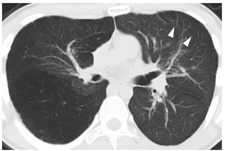
Figure 2 Large demarcated region of low attenuation in the posterior right lung as well as focal area of low attenuation in the left lung (white arrowheads).
Figure 3 The mediastinum is shifted to the left with diffuse low attenuation in the right lung representing air-trapping. Bronchiectasis is demonstrated (white arrow).
Figure 4 Computed tomography scan images showing the right pulmonary artery compared to the left (white arrows).
Dovepress Grosu et al
International Journal of Chronic Obstructive Pulmonary Disease downloaded from https://www.dovepress.com/ by 118.70.13.36 on 22-Aug-2020
improved and was transferred back to the referring hospital for further management. The patient remained on oxygen for greater than 30 days.
During childhood he was treated for asthma with bron-chodilators and inhaled steroids. Wheezing attacks were generally brought on by viral infections. There were no hospitalizations for asthma.
At the age of 19 the patient, who carried the diagnosis of bronchial asthma, was diagnosed with severe AAT deficiency and was referred for evaluation. The patient studied electri-cal mechanics with some exposure to industrial dust but was a nonsmoker. There was no family history of lung or liver disease. He did not have symptoms of reflux. Physical examination revealed a thin male. His vital signs were nor-mal with an oxygen saturation of 98% on room air. He had a right-sided chest deformity with pectus deformity but with normal chest excursion on inspiration. Lung sounds were decreased at the right lung base. There were no signs of right heart failure. The forced vital capacity (FVC) was 71% of predicted, forced expiratory volume in one second (FEV1) was 40%, and the FEV1/FVC ratio of 49% was diagnostic of severe COPD (Global Initiative for Obstructive Lung Disease stage III). After bronchodilator, the FEV1 improved by 200 cc (13%). Lung volumes showed total lung capacity of 118%, functional residual capacity of 148%, and residual volume of 262%. The diffusing capacity was normal. The AAT level was 54 mg/dL (10.4 µM; AAT genotype Z-Null) indicating severe deficiency of AAT. A chest X-ray showed asymmetric right lung hyperinflation.
A computed tomography scan of the chest revealed widespread regions of low attenuation in the right lung, some appearing sharply demarcated (Figures 1–3). There are areas of low attenuation in the left lung as well (Figure 2). The mediastinum is shifted to the left, consistent with air trapping in the right lung (Figure 3). There is diffuse bronchial wall thickening and bronchiectasis in the right lung (Figures 1 and 3) as well as reduction in caliber of the right pulmonary artery compared to the left (Figure 4).
Lung density analysis at threshold of -950 Hounsfield units confirmed the presence of lung attenuation consistent with emphysema involving both lungs. The patient was started on replacement therapy with purified human AAT.
Discussion
This review explores the likely pathophysiologic mecha-nisms involved in this 19-year-old with severe AAT deficiency presenting with severe COPD. The interaction of adverse genetic, host and environmental factors led to
the phenotype of severe airflow obstruction with airspace enlargement. Severe chronic airflow obstruction developed consequent to an interaction of AAT deficiency, neutrophil-mediated inflammation, and oxidative stress-related lung injury sustained during lifesaving treatment for CDH. Features of the presentation point to AAT deficiency as playing a contributing role in the development of the spirometric c riteria for severe COPD. First, the patient has bilateral emphysema; second, the severest emphy-sema is predominantly basal (as is more common in AAT deficiency);7 third, the degree of airflow obstruction as
noted on pulmonary f unction tests is much more severe than is generally reported in the literature where airflow obstruction tends to be milder,8–10 and fourth, the surgeon’s
report suggests that the right lung was hypoplastic and that emphysema of the right lower lobe developed during subsequent lung growth.
AAT, a member of the serine protease inhibitor family, is the major inhibitor of neutrophil elastase. Severe AAT deficiency is found in approximately 1% of cases of COPD and predisposes to the development of premature emphysema in those susceptible.7 Development of emphysema almost
always occurs in middle age so the presence of emphysema in this case is unique. Smoking is by far the most common environmental factor accelerating FEV1 decline in those with the severe deficiency, usually requiring 20–30 years of smoke exposure to cause clinical disease. Cigarette smoke reduces the anti-elastase inhibitory activity of AAT through methionine oxidation and also supports a pro-elastolytic environment through the recruitment of alveolar neutrophils. But in this case it was the severe oxidative stress and neutrophil-mediated inflammation during a period of forma-tive lung development that accelerated the development of emphysema.
BPD, a form of neonatal chronic lung disease, and emphysema share common pathophysiologic features.6 BPD
as a cause of severe airflow obstruction usually develops during lung injury in premature infants yet many of the clinical features of this young man, who was born at term, are consistent with BPD; namely, prolonged oxygen therapy, chronic wheezing, bronchial hyper-responsiveness, dyspnea, and computed tomography scan and lung volumes that show air-trapping.6 The definition of BPD is the requirement for
oxygen 28 days after birth and would be graded in this patient as severe.6 Development of emphysema has also been
described as a consequence of BPD6,11–13 and in this case is
strongly suggested by the focal airspace enlargement and reduced lung attenuation on computed tomography scan.
Dovepress Swyer James MacLeod syndrome with severe alpha-1 antitrypsin deficiency
International Journal of Chronic Obstructive Pulmonary Disease downloaded from https://www.dovepress.com/ by 118.70.13.36 on 22-Aug-2020
In this case, SJMS represents an extreme example of focal emphysema in a patient with severe AAT defi-ciency and diffuse obstructive lung disease. SJMS is usually considered to be the result of post-inflammatory obliterative bronchiolitis, with the characteristic radio-graphic appearance, as in this case, of localized increase in radiographic lucency, bronchiectasis, and decreased vascularity.1,2 As suggested by the operative findings in our
case and in prior work,14,15 SJMS developed over 19 years
from the abnormal development of an immature right lung. It is plausible that genetic, host, and environmental factors have led to airspace enlargement from a paucity of alveolar development with alveolar destruction, favored by protease–anti-protease imbalance during and after oxidative and neutrophil-mediated lung injury.
In the early 1980s ECMO was introduced as salvage therapy for neonates with CDH. Late sequelae include hyper-inflation, airway obstruction, and lower oxygen saturation with exercise.5 ECMO activates neutrophils, resulting in free
circulating elastase concentrations.16 Our patient was exposed
to high-pressure ventilation resulting in barotrauma, very high oxygen concentrations which caused additional oxida-tive stress, inflammation, and acute lung injury with sepsis during the perinatal period. Lung injury occurs with associ-ated local production and systemic release of inflammatory mediators such as interleukin-8, interleukin-6, and tumor necrosis factor. These have been measured in high alveolar concentrations in infants who develop BPD.17,18
Elastin modeling is critical in the development of normal alveolar septation during lung growth19,20 but also
acceler-ated elastin degradation is part of the protease–anti-protease model for emphysema as typified by severe AAT deficiency. The likely consequence of the injury sustained in this case was disruption of normal elastin modeling with destruc-tion of mature elastin and inhibidestruc-tion of normal alveolar development. The latter is a feature more commonly associ-ated with the development of BPD.6 Indeed desmosine, an
elastin breakdown product, is increased in those developing BPD.19 Furthermore AAT is susceptible under high
oxida-tive conditions to methionine oxidation that leads to reduced anti-elastase activity and conversion to a pro-inflammatory molecule.21 Abnormal elastin turnover enhanced by the
extreme lack of anti-protease protection from elastolysis during a critical early period of lung development resulted in impaired alveolar development and the propensity to hyperinflation and structural emphysema.
An association of asthma with AAT deficiency has been suggested. Severe AAT deficiency is likely to increase
s usceptibility to asthma,22 a diagnosis supported in this
patient by the bronchodilator response, the presence of bron-chial hyper-responsiveness, symptom control with standard asthma medications during childhood, and wheezing during respiratory infections. Furthermore, the presence of a bron-chodilator response on spirometry accelerates FEV1 decline in those with AAT deficiency.22 In this young patient the
complex causes of airflow obstruction were not recognized because diagnostic bias evoked asthma as an explanation for the symptoms of wheezing.22
This case illustrates that genetic and host factors and environmental interactions have led to chronic obstructive lung disease in a 19-year-old. The development of chronic lung disease was caused by lung injury under high oxidative and inflammatory conditions in the setting of CDH. In the absence of normal AAT levels and activity, pro-elastolytic lung inflammation in the neonatal period of lung growth enhanced the development of SJMS and precocious o bstructive lung disease.
Disclosure
The authors have no conflict of interest to declare.
References
1. Lucaya J, Gartner S, García-Peña P, Cobos N, Roca I, Liñan S. Spectrum of manifestations of Swyer-James-MacLeod syndrome. J Comput Assist Tomogr. 1998;22(4):592–597.
2. Moore AD, Godwin JD, Dietrich PA, Verschakelen JA, Henderson WR Jr. Swyer-James syndrome: CT findings in eight patients. AJR Am J Roentgenol. 1992;158(6):1211–1215.
3. Colvin J, Bower C, Dickinson JE, Sokol J. Outcomes of congenital diaphragmatic hernia: a population-based study in Western Australia.
Pediatrics. 2005;116(3):e356–e363. Erratum in: Pediatrics. 2006;117(5):1870.
4. Peetsold MG, Heij HA, Kneepkens CMF, Nagelkerke AF, Huisman J, Gemke BJ. The long term follow up of patients with a congenital diaphragmatic hernia: a broad spectrum of morbidity. Pediatr Surg Int. 2009;25:1–17.
5. Hamutcu R, Nield TA, Garg M, Keens TG, Platzker AC. Long-term pulmonary sequelae in children who were treated with extracorporeal membrane oxygenation for neonatal respiratory failure. Pediatrics. 2004;114(5):1292–1296.
6. Baraldi E, Filippone M. Chronic lung disease after premature birth.
N Engl J Med. 2007;8;357(19):1946–1955.
7. Silverman EK, Sandhaus RA. Alpha-1 antitrypsin deficiency. N Engl J Med. 2009;360:2749–2757.
8. Koumbourlis AC, Wung JT, Stolar CJ. Lung function in infants after repair of congenital diaphragmatic hernia. J Pediatric Surg.
2006;41(10):1716–1721.
9. van den Hout L, Sluiter I, Gischler S, et al. Can we improve the outcome of congential diaphragmatic hernia? Pediatr Surg Int. 2009;25(9):733–743.
10. Peetsold MG, Heij HA, Nagelkerke AF, et al. Pulmonary function and exercise capacity in survivors of congenital diaphragmatic hernia. Eur Respir J. 2009;34:1140–1147.
11. Falconer AR, Brown RA, Helms P, Gordon I, Baron JA. Pulmonary sequelae in survivors of congenital diaphragmatic hernia. Thorax. 1990;45(2):126–129.
Dovepress Grosu et al
International Journal of Chronic Obstructive Pulmonary Disease downloaded from https://www.dovepress.com/ by 118.70.13.36 on 22-Aug-2020
12. Howling SJ, Northway WH Jr, Hansell DM, Moss RB, Ward S, Müller NL. Pulmonary sequelae of bronchopulmonary dysplasia survivors: high-resolution CT findings. AJR Am J Roentgenol. 2000;174:1323–1326.
13. Wong PM, Lees AM, Louw J, et al. Emphysema in young adult survivors of mild-moderate bronchopulmonary dysplasia. Eur Respir J. 2008;32:321–328.
14. Beals DA, Schloo BL, Vacanti JP, Reid LM, Wilson JM. Pulmonary growth and remodeling in infants with high-risk congenital diaphrag-matic hernia. J Pediatr Surg. 1992;27(8):997–1001.
15. Hislop A, Reid L. Persistent hypoplasia of the lung after repair of congenital diaphragmatic hernia. Thorax. 1976;31(4):450–455. 16. Fortenberry JD, Bhardwaj V, Niemer P, Cornish JD, Wright JA,
Bland L. Neutrophil and cytokine activation with neonatal extracorporeal membrane oxygenation. J Pediatr. 1996;128(5 Pt 1): 670–678.
17. Jobe AH, Bancalari E. Bronchopulmonary dysplasia. Am J Respir Crit Care Med. 2001;163:1723–1729.
18. Bourbon JR, Boucherat O, Boczkowski J, Crestani B, Delacourt C. Bronchopulmonary dysplasia and emphysema: in search of common therapeutic targets. Trends Mol Med. 2009;15(4):169–179.
19. Bruce MC, Schuyler M, Martin RJ, Starcher BC, Tomashefski JF Jr, Wedig KE. Risk factors for the degradation of lung elastic fibers in the ventilated neonate. Implications for impaired lung develop-ment in bronchopulmonary dysplasia. Am Rev Respir Dis. 1992; 146(1):204–212.
20. Bland RD, Ertsey R, Mokres LM, et al. Mechanical ventilation uncouples synthesis and assembly of elastin and increases apoptosis in lungs of newborn mice. Prelude to defective alveolar septation during lung development? Am J Physiol Lung Cell Mol Physiol. 2008;294(1): L3–L14.
21. Li Z, Alam S, Wang J, Sandstrom CS, Janciauskiene S, Mahadeva R. Oxidized {alpha}1-antitrypsin stimulates the release of monocyte chemot-actic protein-1 from lung epithelial cells: potential role in emphysema.
Am J Physiol Lung Cell Mol Physiol. 2009;297(2):L388–L400. 22. Eden E. Asthma and COPD in alpha-1 antitrypsin deficiency. Evidence
for the Dutch hypothesis. COPD. 2010;7(5):366–374.
Dovepress Swyer James MacLeod syndrome with severe alpha-1 antitrypsin deficiency
International Journal of Chronic Obstructive Pulmonary Disease downloaded from https://www.dovepress.com/ by 118.70.13.36 on 22-Aug-2020
International Journal of COPD
Publish your work in this journal
Submit your manuscript here: http://www.dovepress.com/international-journal-of-copd-journal
The International Journal of COPD is an international, peer-reviewed journal of therapeutics and pharmacology focusing on concise rapid reporting of clinical studies and reviews in COPD. Special focus is given to the pathophysiological processes underlying the disease, intervention programs, patient focused education, and self management protocols.
This journal is indexed on PubMed Central, MedLine and CAS. The manuscript management system is completely online and includes a very quick and fair peer-review system, which is all easy to use. Visit http://www.dovepress.com/testimonials.php to read real quotes from published authors.
Continuing medical education questions
1. Typical features of Swyer James Macleod Syndrome are: a) Bronchiectasis
b) Lower lobe fibrosis c) Vascular attenuation d) Lobar hyperinflation
2. Alpha-1 antitrypsin deficiency: a) Promotes pulmonary elastolysis b) May be misdiagnosed as asthma
c) Is caused by the retention of alpha-1 antitrypsin polymers in the liver d) Is a major inhibitor of neutrophil elastase
3. Bronchopulmonary dyplasia:
a) Is a condition of impaired alveolar development
b) Is diagnosed by a history of prolonged neonatal oxygen therapy c) May present with airflow obstruction in adults
d) Is excluded by a normal chest X-ray 4. In early life, alveolar development:
a) Is dependent on normal elastin modeling b) May be impaired during oxidative stress c) Is dependent on neutrophil elastase activity d) Is dependent on lung injury
5. The host factor(s) that likely caused the phenotype of Swyer James Macleod Syndrome and emphysema in this case was (were)
a) Alpha-1 antitrypsin deficiency
b) The consequences of extracorporeal membrane oxygenation c) Developmental abnormality of the diaphragm
d) Innate response to sepsis
1 a) T b) F c) T d) T 2 a) T b) T c) T d) T 3 a) T b) T c) T d) F 4 a) T b) T c) T d) F 5 a) F b) F c) T d) T
Figure 5 Answers to Continuing medical education questions.
Abbreviations: T,true; F, false.
Dovepress
Dove
press
Grosu et al
International Journal of Chronic Obstructive Pulmonary Disease downloaded from https://www.dovepress.com/ by 118.70.13.36 on 22-Aug-2020