Scholarship@Western
Scholarship@Western
Electronic Thesis and Dissertation Repository
9-20-2013 12:00 AM
A six week modified sprint interval training program incorporating
A six week modified sprint interval training program incorporating
extended exercise bouts does not increase maximal cardiac
extended exercise bouts does not increase maximal cardiac
output
output
Alan R. Smith
The University of Western Ontario
Supervisor
Dr. Peter W.R. Lemon
The University of Western Ontario Graduate Program in Kinesiology
A thesis submitted in partial fulfillment of the requirements for the degree in Master of Science © Alan R. Smith 2013
Follow this and additional works at: https://ir.lib.uwo.ca/etd
Part of the Exercise Science Commons
Recommended Citation Recommended Citation
Smith, Alan R., "A six week modified sprint interval training program incorporating extended exercise bouts does not increase maximal cardiac output" (2013). Electronic Thesis and Dissertation Repository. 1707.
https://ir.lib.uwo.ca/etd/1707
This Dissertation/Thesis is brought to you for free and open access by Scholarship@Western. It has been accepted for inclusion in Electronic Thesis and Dissertation Repository by an authorized administrator of
MAXIMAL CARDIAC OUTPUT
(Thesis format: Monograph)
by
Alan Robert Smith
Graduate Program in Kinesiology
A thesis submitted in partial fulfillment of the requirements for the degree of
Master of Science
The School of Graduate and Postdoctoral Studies The University of Western Ontario
London, Ontario, Canada
ii
Abstract
Sprint interval training (SIT) improves maximal oxygen uptake (V O2max) and exercise
performance but not maximal cardiac output (Q max). The brevity of typical SIT bouts (30
-seconds) might hinder improvements in Q max. The purpose of this study was to determine whether extended duration SIT (up to 45 second bouts) improves Q max. Pre-/Post-SIT (or
control) V O2max, Q max, maximum stroke volume (SVmax), maximum heart rate, arterial-mixed venous oxygen difference, and 5-minute run distance were measured. SIT progressed from 4x30s to 7x45s “all-out” efforts (4 min recovery) over 6 wk (3x/wk) on a manually driven treadmill. Following SIT, V O2max improved (pre-=3.6±0.8 vs post=3.8±0.8 L·min-1;
p=0.012). Increases in Q max (pre-= 26.13±5.09 vs post=27.10±4.82 L·min-1; p=0.166) and SVmax (pre-= 138±27 vs post=144±28 mL·beat-1; p=0.095) were not significant. These data suggest 6 wk of extended SIT bouts (up to 45 s) does not increase Q max significantly and the observed increase in V O2max was due primarily to non-significant improvements in
Q max and SVmax.
Keywords
iii
Acknowledgments
I am very grateful to the many people who have helped me during my time at the University of Western Ontario. In particular, I must thank my supervisor, Dr. Peter Lemon. I have learned a great deal from him; his knowledge, expertise, and approach to research and teaching have greatly influenced me. I would like to thank Drs. Don Paterson and John Kowalchuk for allowing us to use their lab space to collect data. Thank you to the students in the Canadian Centre for Activity and Aging for accommodating this project and providing assistance when needed.
I must also thank my friends, colleagues, and lab mates in kinesiology. I am especially indebted to Stephanie and Dylan; without their suggestions, hard work, and involvement this thesis wouldn’t have been possible.
iv
Table of Contents
Abstract ... ii
Acknowledgments... iii
Table of Contents ... iv
List of Tables ... vi
List of Figures ... vii
List of Abbreviations ... viii
List of Appendices ... x
Chapter 1 ... 1
1 Introduction ... 1
1.1 Exercise performance, maximal oxygen uptake, and exercise training ... 2
1.2 Endurance training ... 3
1.3 Interval training ... 4
1.4 Central adaptations... 5
1.5 Peripheral adaptations ... 7
1.6 Summary, purpose, hypothesis ... 10
Chapter 2 ... 12
2 Methods ... 12
2.1 Participants ... 12
2.2 Study design ... 12
2.3 Tests ... 14
2.4 Measurements ... 15
2.5 Statistical analysis ... 17
2.6 Excluded data points ... 17
v
3 Results ... 18
3.1 Maximal oxygen uptake (V O2max; mL·kg lean mass-1·min-1) ... 18
3.2 Maximal oxygen uptake (V O2max; L·min-1) ... 19
3.3 Maximal cardiac output (Q max; mL·kg lean mass-1·min-1) ... 19
3.4 Maximal cardiac output (Q max; L·min-1) ... 20
3.5 Maximum heart rate (HRmax; beats·min-1) ... 21
3.6 Maximum stroke volume (SVmax; mL·kg lean mass-1·beat-1) ... 21
3.7 Maximum stroke volume (SVmax; mL·beat-1) ... 21
3.8 Arterial-mixed venous oxygen difference (a-v O2diff; mL O2·100mL blood-1·kg lean mass-1) ... 22
3.9 Arterial-mixed venous oxygen difference (a-v O2diff; mL O2·100mL blood-1) ... 22
3.10 Five minute run: total distance (m) ... 23
3.11 Five minute run: change in distance (∆d; m) ... 24
Chapter 4 ... 25
4 Discussion ... 25
4.1 Effect on V O2max ... 25
4.2 Effect on Q max ... 26
4.3 Effect on a-v O2diff ... 27
4.4 Effect on 5-minute run ... 29
4.5 Summary ... 30
4.6 Future directions ... 31
4.7 Conclusion ... 31
References ... 32
Appendices ... 44
vi
List of Tables
Table 1. Participant characteristics. ... 12
vii
List of Figures
Figure 1. Maximal oxygen uptake (V O2max; mL·kg lean mass-1·min-1) ... 18
Figure 2. Maximal cardiac output (Q max; L·min-1) ... 20
Figure 3. Maximum stroke volume (SVmax; mL·beat-1) ... 22
Figure 4. Arterial-mixed venous oxygen difference (a-v O2diff; mL O2·100mL blood-1) ... 23
Figure 5. Total distance run (m) ... 24
viii
List of Abbreviations
∆d – change in distance
a-v O2diff – arterial-mixed venous oxygen difference
bFBF – basic fibroblast growth factor
CON – control
ET – endurance training
FABPpm – plasma membrane fatty acid-binding protein
FAT/CD36 – fatty acid translocase/cluster of differentiation 36
GLUT4 – glucose transporter type 4
HIIT – high intensity interval training
HR – heart rate
HRmax – maximum heart rate
IT – interval training
LV – left ventricle
MCT4 – monocarboxylate transporter 4
MIT – moderate intensity interval training
NHE1 – sodium-hydrogen antiporter 1
NO – nitric oxide
PAR-Q – physical activity readiness questionnaire
ix Q max – maximal cardiac output
SET – speed endurance training
SIT – sprint interval training
SV – stroke volume
SVmax – maximum stroke volume
VEGF – vascular endothelial growth factor
V O2 – oxygen uptake
x
List of Appendices
Appendix A: Ethics approval notice. ... 44
Appendix B: Letter of information. ... 45
Chapter 1
1
Introduction
Sprint interval training (SIT) is a low-volume, time efficient, exercise training method capable of rapidly improving exercise performance (Burgomaster, Hughes,
Heigenhauser, Bradwell, & Gibala, 2005). Although there are a variety of formats, typically, SIT consists of 4 to 6 bouts of 30-second “all-out” exercise bouts separated by 4 minutes of rest/recovery. SIT is vastly different from traditional high-volume
endurance training (ET; constant-load, submaximal exercise maintained for an extended period of time), as SIT requires ~14 to 23 minutes (of which only 2 to 3 is actual
exercise) while ET usually requires 30 to 60 minutes (of which all is exercise). SIT is performed at supramaximal intensity (~150-200% maximal aerobic power) and ET is typically performed between 60-80% of maximal aerobic power. Despite these obvious differences, SIT and ET result in similar V O2max and exercise performance
improvements (Gibala et al., 2006; MacPherson, Hazell, Olver, Paterson, & Lemon, 2011).
Several types of exercise training elicit key central and peripheral physiological adaptations (Blomqvist & Saltin, 1983; Holloszy & Booth, 1976). Central adaptations occur within the cardiovascular system (resulting in increased oxygen delivery) and peripheral adaptations occur at the level of the muscle (resulting in greater oxygen extraction and utilization). The improved delivery, extraction, and utilization of oxygen alter energy substrate utilization and metabolite production resulting in both improved capacities and exercise performance.
interval duration is insufficient (MacPherson et al., 2011). Time efficiency and potency of SIT make it an attractive training method, but unfortunately the adaptations are not all encompassing (only peripheral adaptations are obtained). Thus, in order to obtain both central and peripheral adaptations, SIT may complement, but not replace ET.
Interestingly, other, less intense forms of interval training utilizing longer duration intervals have been shown to improve Q max (Daussin et al., 2007). This suggests that supramaximal intensity is not a requirement for central adaptations but some exercise duration is. However, neither the necessary intensity nor the duration is clear at the present time. Previous studies have not used 45-second intervals. It is possible that extending SIT bouts to 45-seconds while maintaining an “all-out” intensity would provide a time-efficient method of training that might affect Q max.
The purpose of the current study was to determine if 6 weeks of modified SIT (duration of sprint intervals was lengthened from 30 up to 45 seconds) improves exercise
performance and maximal cardiac output (Q max). Maximal oxygen uptake, Q max, and 5-minute run performance were assessed pre- and post-training, in healthy, young, male and female participants. It was hypothesized that the modified SIT protocol would
generate improvements in Q max, resulting in improved maximal oxygen uptake and exercise performance.
1.1 Exercise performance, maximal oxygen uptake, and
exercise training
Exercise performance is determined by the integration of various body systems (respiratory, cardiovascular, and neuromuscular) (Bangsbo, Mohr, Poulsen,
Maximal oxygen uptake is the greatest amount of oxygen that can be taken up, delivered to, and utilized by muscle tissue to perform exercise (measured as an absolute value: L·min-1; or, measured relative to body mass: mL·kg-1·min-1) (Åstrand, 1956; Jones & Carter, 2000). Oxygen uptake (V O2) is the product of cardiac output (Q ; surrogate for total oxygen delivery) and arterio-venous oxygen difference (i.e. oxygen
extraction/utilization) (Bevegård, 1962; González-Alonso, 2008).
Cardiac output is the volume of blood ejected from the heart per minute (measured in L·min-1). It is the product of stroke volume (SV) and heart rate (HR), and is considered to be the “central” component of any training response because the respiratory system is seldom limiting (Bassett & Howley, 2000).
Arterio-venous oxygen difference (a-vO2diff) is a measure of oxygen extraction by the muscle. Oxygen content of blood is measured at the arterial and venous levels (i.e. before and after the capillary bed in the muscle where oxygen extraction takes place) (Clausen, Klausen, Rasmussen, & Trap-Jensen, 1973). The difference between arterial and venous oxygen content provides a measure of oxygen extraction. Changes in a-vO2diff following exercise training reflect adaptations that occur at the muscle (i.e. improved extraction or utilization) (Coyle et al., 1984; Ekblom, Åstrand, Saltin, Stenberg, & Wallström, 1968). These changes are considered to be “peripheral” changes (Daussin et al., 2008).
Exercise training can elicit adaptations that occur centrally and/or peripherally (Coffey & Hawley, 2007; Daussin et al., 2008). These adaptations increase V O2max as well as exercise performance (Ekblom et al., 1968; Gunnarsson & Bangsbo, 2012; Jones & Carter, 2000). Endurance training (ET) and interval training (IT) are training methods used to elicit these changes. The degree of influence exerted on central and peripheral aspects is determined by the frequency, volume, intensity, and duration of the training method used (Baar, 2006; Buchheit & Laursen, 2013; Laursen, 2010).
1.2 Endurance training
training is aerobic and relies mainly on a mix of lipid and carbohydrate oxidation via the oxidative system (relatively little energy is contributed from ATP-PCr and glycolytic systems) (Jeukendrup, 2002; Spriet, 2002). Endurance training needs to be performed several times per week (at least 2-3) in order to adequately stress the body and promote positive adaptations (Laursen & Jenkins, 2002). Typically, high-volume (long duration) training results in both peripheral and central adaptations, such as increased Q max and a-vO2diff, and produces improvements in both V O2max and exercise performance (Coffey & Hawley, 2007; Holloszy & Booth, 1976)
1.3 Interval training
Interval training differs from traditional ET in that it is a discontinuous workload. Interval training is characterized by repeated blocks (bouts) of vigorous exercise separated by periods of rest or lower intensity exercise utilized for recovery in preparation for the next interval of vigorous exercise (Laursen & Jenkins, 2002). The combinations of exercise intensities, work to “rest” ratios, and number of intervals are endless and many IT
heavily on oxidative metabolism. Similarly to ET, SIT results in comparable improvements in exercise performance; however, these improvements in exercise performance are likely caused by peripheral adaptations, as 6 weeks of SIT does not
increase Q max (Gibala et al., 2006; MacPherson et al., 2011). Sprint interval training is a low-volume, time efficient, intense training method that increases exercise performance
and V O2max (via greater a-vO2diff , but not Q max) (MacPherson et al., 2011).
1.4 Central adaptations
According to the Fick principle, an increase in Q max will increase V O2max unless extraction decreases (Stray-Gundersen et al., 1986). Cardiac output is the product of SV
and HR (so Q max is the product of SVmax and HRmax) (Stray-Gundersen et al., 1986). In response to exercise training, HRmax does not increase; instead HRmax decreases or is unchanged (Blomqvist & Saltin, 1983). Therefore, any increase in Q max must be the result of increased SVmax (Ekblom & Hermansen, 1968).
Stroke volume is directly related to end-systolic and end-diastolic myocardial fibre length. In order to increase SV, diastolic fibre length must be greater and/or end-systolic fibre length must be lesser (Kiil, 1978). Stroke volume can be modulated by intrinsic (e.g. cardiac dimensions, response to inotropic agents) and extrinsic (e.g. blood volume, peripheral resistance) elements which impact upon factors affecting myocardial fibre length (Blomqvist & Saltin, 1983; Kiil, 1978). The main factors that affect
myocardial fibre length are: 1) preload; 2) afterload; 3) inotropy; and 4) myocardial dimensions and performance.
end-diastolic volumes and improves SV (Convertino, 1991; Kanstrup & Ekblom, 1982; Robinson, Epstein, Kahler, & Braunwald, 1966). In response to training there is an increase in blood volume so preload also increases (Kjellberg, Rudhe, & Sjöstrand, 1949; Sawka, Convertino, Eichner, Schnieder, & Young, 2000). Increased preload improves SV through the Frank-Starling mechanism whereby increasing left-ventricular end-diastolic volume leads to myocardial stretching which improves contractility, producing an
increased SV (Ferguson, Gledhill, Jamnik, Wiebe, & Payne, 2001; Konhilas, Irving, & de Tombe, 2002).
Afterload refers to the external forces which oppose the shortening of myocardial fibres (Kiil, 1978). If afterload is decreased, SV will increase (because end-systolic fibre length will decrease) (Blomqvist & Saltin, 1983). Afterload is mainly determined by peripheral resistance (Jaski, Fifer, Wright, Braunwald, & Colucci, 1985). Exercise training also leads to increased capillarization and improvements in the ability to vasodilate (Jensen, Bangsbo, & Hellsten, 2004; Rakobowchuk et al., 2008). These adaptations have a
number of beneficial effects, among them, is a decrease in peripheral resistance (Booth & Thomason, 1991; Laughlin & Roseguini, 2008).
Inotropy refers to the response of the myocardium to substances which affect contractility. Inotropic agents include calcium, catecholamines, and glucagon (Kiil, 1978). Increased inotropy leads to increased force and velocity of contraction (Ross, Covell, Sonnenblick, & Braunwald, 1966). Stroke volume will increase in response to increased inotropy (Blomqvist & Saltin, 1983). Exercise training can enhance myocardial inotropy (Ehsani, Ogawa, Miller, Spina, & Jilka, 1991).
Myocardial performance is determined by structural or functional changes within the myocardium itself that affect contractile force (Blomqvist & Saltin, 1983). Intrinsic contractile performance is difficult to measure but studies in rats have shown contractile performance, calcium handling, and myosin ATPase improvements with exercise training (Baldwin, Cooke, & Cheadle, 1977; Penpargkul, Repke, Katz, & Scheuer, 1977).
Kemi, & Ellingsen, 2001). Myocardial hypertrophy is beneficial, and not pathological, as long as left ventricular (LV) volume is not reduced simultaneously (Pluim, Zwinderman, van der Laarse, & van der Wall, 2000; Vinereanu et al., 2001). Endurance and interval training lead to myocardial hypertrophy which does not impede LV volume (Maron & Pelliccia, 2006; Pelliccia et al., 2002). Weightlifters and people suffering from
hypertension also develop myocardial hypertrophy but this occurs with heart wall thickening which reduces LV volume and hampers cardiac performance and function (Haykowsky, Dressendorfer, Taylor, Mandic, & Humen, 2002; Vinereanu et al., 2001).
There are several ways of measuring adaptations to the central system. It is often difficult to determine exactly which factor is responsible for increases in SV (Blomqvist & Saltin, 1983). Most studies have used animals or isolated muscle fibres and attempted to distinguish between the different factors and how they affect SV (Glower et al., 1985; Sagawa, 1978; Sonnenblick & Downing, 1963). Measuring the independent contribution of preload, afterload, inotropy, and myocardial performance is difficult, while measuring
Q is more practical. Cardiac output can be measured invasively (direct Fick method,
dye-dilution, thermodilution) and non-invasively (acetylene breathing methods, carbon dioxide rebreathe, Doppler ultrasound, impedance). Invasive techniques pose greater risks, require medical and technical assistance, and are not always practical during maximal exercise (Warburton, Haykowsky, Quinney, Humen, & Teo, 1999). Less invasive techniques, particularly open-circuit acetylene methods, are reliable, reproducible, correlate well with values produced from invasive methods, and are
applicable to conditions of maximal exercise. Acetylene is a gas which is soluble in blood and inert (does not bind with haemoglobin). Typically, inspired and expired acetylene concentrations are measured by a mass spectrometer over a number of breaths and rate of disappearance is calculated. As cardiac output increases, the rate of acetylene
disappearance increases. An open circuit acetylene method is an effective and reliable method for measuring maximal cardiac output during exercise (Johnson et al., 2000).
1.5 Peripheral adaptations
Peripheral adaptations are those that occur at the muscle and allow oxygen to be
metabolism (glycolytic capacity, oxidative capacity, substrate utilization), glycogen storage, ion transporters, fibre type, haemoglobin/myoglobin concentration, and capillary density reflect peripheral adaptations (Coffey & Hawley, 2007; Holloszy & Coyle, 1984; Iaia & Bangsbo, 2010).
Improvements in muscle metabolism are related to the energy system stressed by the exercise task. Peripherally, endurance training results mainly in improvements to the oxidative capacity of the muscle whereas interval training augments glycolytic and/or oxidative capacity of muscle (depending on which interval training method is used) (Gollnick & Saltin, 1982; Hawley, 2002; Kubukeli, Noakes, & Dennis, 2002; MacDougall et al., 1998).
Glycolysis is an energy liberating process that involves the breakdown of
glucose/glycogen. Glycolysis takes place in the cell cytoplasm and does not require oxygen to be present. The net result of glycolysis alone is the production of 2 mol of ATP per mol of glucose (Alberts et al., 2004). Glycolytic capacity is often assessed by measuring the concentration and activity of various glycolytic enzymes. Most commonly, phosphofructokinase, pyruvate kinase, lactate dehydrogenase,
glyceraldehyde-3-phosphate dehydrogenase, or aldolase are measured (MacDougall et al., 1998; Mujika & Padilla, 2001; Parra, Cadefau, Rodas, Amigó, & Cussó, 2000). Generally, glycolytic capacity is unaffected by ET but increases in response to interval training (Gollnick et al., 1973; MacDougall et al., 1998).
The oxidative energy system includes the citric acid cycle and electron transport chain. This system relies on a special organelle, called the mitochondria, and oxygen to produce energy. Fat, carbohydrate, and protein can be used as energy sources (fat and
oxidative capacity. Specifically, studies often investigate the activity and/or concentration of citrate synthase, 3-hydroxyacyl-CoA dehydrogenase, succinate dehydrogenase,
pyruvate dehydrogenase, malate dehydrogenase, and cytochrome c oxidase (Dudley, Abraham, & Terjung, 1982; Irrcher, Adhihetty, Joseph, Ljubicic, & Hood, 2003; Mujika & Padilla, 2001). Oxidative capacity increases in response to both ET and SIT (Gibala et al., 2006; Ingjer, 1979; MacDougall et al., 1998).
In addition to affecting muscle glycolytic and oxidative capacity, exercise training can influence substrate utilization. Following training, glucose (GLUT4) and fatty acid (FAT/CD36 and FABPpm) transporter contents increase, muscle glycogen content increases, intramuscular triglyceride content increases, and there is a shift in substrate utilization with an increase in the relative contribution of fat and a decrease in the relative contribution of carbohydrate at a given workload (Burgomaster et al., 2005; Hawley, 2002; Holloszy & Booth, 1976; Juel, 2006).
In addition to GLUT4 and fatty acid transporters, there are other sarcolemmal transport proteins that are affected by exercise. Ion transporters, such as Na+/K+-ATPase, NKCCl, NHE1, MCT1, MCT4, La+/H+, and Na+/HCO3-, exist to prevent disruptions in cellular homeostasis (Juel, 2006). The transition from rest to exercise imposes an immediate increased energy demand. The resulting metabolic products and processes cause ionic imbalances which disrupt cellular homeostasis and have a tremendous influence on muscle function (Cairns & Lindinger, 2008). Changes in muscular ionic concentrations, muscle pH, and metabolite concentrations affect muscle membrane potential, muscle excitability, muscle contractility, and contribute to muscular fatigue (Allen, Lamb, & Westerblad, 2008). Exercise training can increase the muscle content of ion transporter proteins which improves exercise performance by delaying or sequestering ion imbalance related fatigue (Iaia et al., 2008; Juel, 2006; Juel et al., 2004). Endurance training and interval training promote increased protein content of most ion transporters (Juel & Halestrap, 1999; Juel, 2006; McKenna, 1995).
the remainder is dissolved. Haemoglobin binds oxygen at the lung and transports it to the muscle where it is off-loaded to muscle (Wilmore & Costill, 2004a). Conditions during exercise leads to the Bohr effect where the affinity of haemoglobin for oxygen is decreased in the presence lower pH, increased temperature, and the increase in certain exercise-related metabolites (Krustrup, Hellsten, & Bangsbo, 2004). The Bohr effect promotes oxygen off-loading at the muscle without having much effect on loading in the lung due to the shape of the oxygen-haemoglobin dissociation curve (Thomson,
Dempsey, Chosy, Shahidi, & Reddan, 1974). Increases in production of red blood cells and haemoglobin are induced by the hormone erythropoietin (whether endogenous or exogenous) (Silverthorn, 2004). Increases in haemoglobin, irrespective of changes in blood volume, improve exercise performance and oxygen uptake (Kanstrup & Ekblom, 1984). A molecule similar to haemoglobin, myoglobin, is found in muscle tissue. Oxygen binds to myoglobin (oxymyoglobin) facilitating oxygen transport to the mitochondria (myoglobin also acts as an oxygen store) (Wilmore & Costill, 2004b). Theoretically, an increase in myoglobin could improve V O2max but studies have produced equivocal results thus far (Coyle et al., 1984; Jürgens, Papadopoulos, Peters, & Gros, 2000).
The formation of new blood vessels is called angiogenesis. Angiogenesis occurs in response to mechanical (e.g. shear stress, muscle stretch) and chemical (e.g. VEGF, bFBF, NO, MMPs, integrins) stimuli that are produced during exercise (Egginton, 2009). Of particular importance to the exercise response is the proliferation of new muscle capillaries. Increased muscle capillary density improves gas and metabolite exchange between blood and muscle by 1) increasing surface area of capillary in muscle, 2) increasing blood transit time in capillary beds, and 3) reducing the diffusion distance between blood and muscle cells (Iaia et al., 2011; Saltin, 2009).
1.6 Summary, purpose, hypothesis
Recently, SIT has received much attention because it is a time-efficient method of
training and, despite its low volume, results in similar oxidative and exercise performance improvements as ET (Gibala et al., 2006; MacPherson et al., 2011). However, SIT does
Chapter 2
2
Methods
2.1 Participants
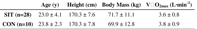
Twenty men and 18 women volunteered for this study. Twenty-eight (14 men; 14 women) performed sprint interval training and 10 (6 men; 4 women) acted as sedentary controls (Table 1). Participants were recreationally active and had not performed interval training for at least 3 months prior to the start of the study. All were given a letter of information regarding this study. Several control subjects were not able to complete the post testing due to noise complaints during the treadmill testing. As a result, the control group is small than planned. Experimental procedures and potential risks were outlined before participants each gave his/her written consent to participate. In addition,
participants completed a PAR-Q to screen for any potential contraindications to exercise. This study was approved by the University of Western Ontario ethics board.
Table 1. Participant characteristics.
Age (y) Height (cm) Body Mass (kg) V O2max (L·min-1)
SIT (n=28) 23.0 ± 4.1 170.3 ± 7.6 71.7 ± 11.1 3.6 ± 0.8 CON (n=10) 23.8 ± 2.3 170.3 ± 7.8 69.9 ± 12.8 3.8 ± 0.9
*P<0.05; SIT = sprint interval training; CON = control.
2.2 Study design
Participants: Participants were placed randomly into either the sprint interval training (SIT) or the control (CON) group. All were familiarized with the testing procedures prior to the study. Pre- and post-testing consisted of a maximal incremental treadmill test, a constant load verification test, a 5-minute manual treadmill performance run, and body composition analyses. These exercise tests were used to assess V O2max, Q max, a
-v O2diff, SVmax, HRmax and exercise performance. Body composition was measured via air displacement plethysmography (BodPod®) and used to normalize V O2max, Q max,
72 hours after the final training session. Prior to testing, participants refrained from alcohol, nicotine, and caffeine for 12 hours and fasted for 3 hours.
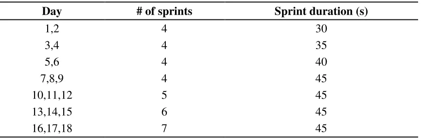
Training: Participants assigned to the training group performed 18 sessions of modified SIT over 6 weeks (Table 2). Specifically, training was performed 3 times per week and sessions were separated by at least 48 hours. Training sessions consisted of “all out” sprints on a manual treadmill (Desmo Pro; Woodway, Waukesha, WI), progressing from 4 x 30-second sprints to 7 x 45-second sprints. Sprints were separated by 4 minutes of active rest/recovery. Training took place on a treadmill set in manual mode. Manual mode is an un-motorized mode in which the tread is propelled solely by the muscular actions of the exerciser.
Table 2. Sprint interval training program.
Day # of sprints Sprint duration (s)
1,2 4 30
3,4 4 35
5,6 4 40
7,8,9 4 45
10,11,12 5 45
13,14,15 6 45
16,17,18 7 45
s = seconds
Familiarization: Prior to the initial testing, participants visited the lab to complete a PAR-Q questionnaire (Thomas, Reading, & Shephard, 1992). During this visit participants were introduced to the lab, the testing equipment, and all procedures to be used.
2.3 Tests
Maximal Incremental Exercise Test: This test was performed on a motorized treadmill
(Model TM310, Trackmaster; Jas Fitness System, Newton, KS). Following 5 minutes of seated rest, participants were familiarized with our open-circuit acetylene cardiac output measurement and 4 resting cardiac output measurements were taken (2 seated followed by 2 standing measurements). Resting measurements provided submaximal reference values and served as practice for participants. Next, participants performed a 5-minute warm-up jog at 9.7 (for men) or 8.0 km·h-1 (for women). Immediately following the warm-up, participants began the incremental exercise test. The test started at a speed of 10.5 (for men) or 8.9 km·h-1 (for women) and increased 0.8 km·h-1 every minute until
participants reached volitional exhaustion. V O2 (mass spectrometry) and heart rate (ECG) were measured continuously throughout the test. A cardiac output measurement was taken when participants indicated they were near exhaustion (approximately 30 seconds remaining).
Constant Load Verification Test: Following the maximal incremental exercise test,
participants were given 5 minutes of active rest/recovery. Then, they began running at a speed equal to 90% of the final speed achieved during the incremental test. Speed remained constant throughout the test and participants were instructed to run as long as possible. Another cardiac output measurement was taken 2 minute into the verification test and used as a submaximal reference value and a third cardiac output measurement was taken when participants indicated they were nearing exhaustion (approximately 30
seconds remaining). V O2 (mass spectrometry) and HR (ECG) were measured continuously throughout the test. This test was used to confirm that the V O2and
Q max values from the incremental test were in fact maximal.
Five Minute Performance Run: Participants completed a 5-minute run for distance on a
mode was given. Two 5-minute runs were completed (separated by at least 24 hours) and the greatest distance covered was recorded to minimize any learning effect.
Body Composition: Air displacement plethysmography (BodPod®) was used to assess
body density. Participants were tested following a 3 hour fast. They wore approved clothing (compression shorts for men, bathing suit or compression shorts plus sports bra for women), placed a lycra cap on their head to minimize hair volume, and removed all jewelry. Thoracic air volume was predicted using an equation from the BodPod® software. Body density was used in the Siri equation to calculate body composition (% fat mass and % lean mass) (Siri, 1961).
2.4 Measurements
Oxygen uptake, heart rate, and cardiac output were measured. Stroke volume and arterial-mixed venous oxygen difference were calculated.
Oxygen Uptake (V O2): V O2 was measured continuously during the maximal
incremental test and verification test. A low dead space (90 mL) bidirectional turbine (VMM-110; Alpha Technologies, Laguna Beach, CA), calibrated prior to testing with a 3.003 L syringe, was used to measure inspired and expired flow rates. The mass
spectrometer (1100 Spectrometer; Perkin-Elmer, Ontario, Canada) used was calibrated with precision-analyzed gas mixtures. Inspired and expired gases were sampled
continuously (50 Hz) and analyzed for concentrations of O2, CO2, and N2. As reported previously, changes in gas concentrations were aligned with gas volumes by measuring the time delay for a square-wave bolus of gas passing the turbine to the resulting changes in fractional gas concentrations as measured by the mass spectrometer. Breath-by-breath alveolar gas exchange was calculated by using algorithms of Beaver et al. (Beaver, Lamarra, & Wasserman, 1981). V O2 values were averaged over 20 seconds. The
greatest averaged value was taken as V O2max. During pre-testing, 5 out of 38
participants showed a plateau in V O2 (oxygen uptake increased <50% of the estimated oxygen requirement), 20 out of 38 reached an RER greater than 1.15, and 31 out of 38 achieved 95% of their age-predicted HRmax. During post-testing, 12 out of 38 participants
out of 38 reached an RER greater than 1.15, and 28 out of 38 achieved 95% of their age-predicted HRmax.
Heart Rate (HR): HR was monitored continuously by electrocardiogram (three-lead
arrangement) using PowerLab L132/ML880 (ADInstruments, Colorado Springs, CO, USA) and recorded using LabChart v7.1 (ADInstruments, Colorado Springs, CO, USA). HR was calculated using a human ECG macro that was included with the software.
Cardiac Output (Q ): An acetylene (C2H2) non-rebreathing technique (breathing a C2H2 gas mixture in an open-circuit system) was used to measure Q at rest and during
exercise.
For acetylene non-rebreathing, participants were connected to a one-way valve assembly with a two-way valve on the inspired side. One input was room air and the other a gas mixture containing known concentrations of acetylene ((C2H2) (0.698%), helium (He) (8.99%), O2 (21%), and N2 (BAL)). The gas mixture was released from the gas tank into a plastic bag (Douglas bag; Hans Rudolph, Inc.). This plastic bag was subsequently attached to the valve leading to the mouth piece. At the end of expiration the valve was turned and inspired air changed from room air to the acetylene mixture. Concentrations of C2H2 and He were then measured continuously for 10 breaths by mass spectrometry. Digitized signals were interpreted by a commercially available software program (BIPS). After 10 breaths, the valve was switched from acetylene mixture back to room air at the end of expiration. Q was calculated based on computer analysis of the rate of
disappearance of acetylene (Johnson et al., 2000).
Stroke Volume (SV): Using HR and Q values, SV was calculated as
SV = Q ÷ HR
Arterial-mixed venous oxygen difference (a-v O2diff): Using V O2 and Q values, a
-v O2diff was calculated as:
2.5 Statistical analysis
Statistical analyses were performed using SigmaPlot for Windows (Version 12.0). All data, except 5-minute run ∆d, were analyzed using a two-way repeated measures
ANOVA. Tukey’s HSD test was used for post-hoc analysis of any significant interactions or main effects. A two-tailed, paired t-test was used to analyze 5-minute run ∆d.
Significance was set at a P < 0.05. Data are presented as means ± standard deviation (SD). All results are presented as group data, but when applicable (findings diverge from group results), sex differences are reported.
2.6 Excluded data points
During post-testing, 2 subjects (a male SIT participant and a male CON participant) had non-physiological Q max values. During post-testing for another subject (a female SIT
participant), we had technical issues while measuring Q max. An error occurred with the
computer calculations and a Q value was never produced. Maximal oxygen uptake and
HRmax data had an n of 38 but, due to the issues mentioned above, Qmax, SVmax, and a
Chapter 3
3
Results
3.1
Maximal oxygen uptake (V
O
2max; mL·kg lean mass
-1·min
-1)
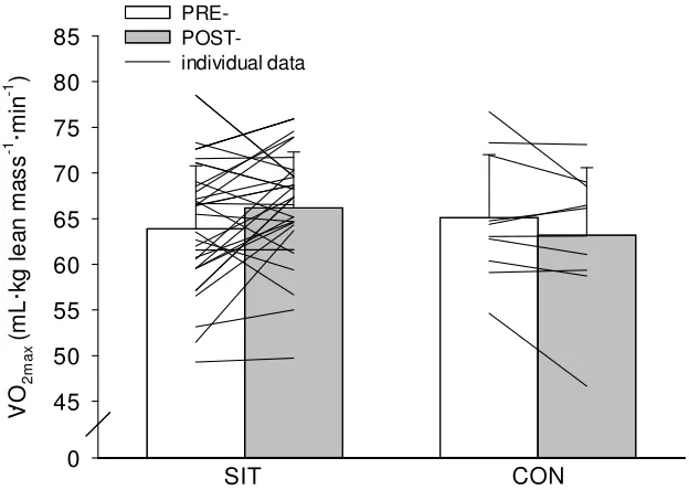
Group Data V O2max: There was a significant group by time interaction (p=0.032). There was a 2.3 mL·kg lean mass-1·min-1 increase in V O2max following SIT (pre-=64±6.9 post=66±6.1 mL·kg lean mass-1·min-1; p=0.024) and V O2max did not change following control (pre-=65±6.9 post=63±7.4 mL·kg lean mass-1·min-1; p=0.244) conditions.
V O2 m a x ( m L ·k g l e a n m a s s -1 ·m in -1 ) 0 45 50 55 60 65 70 75 80 85 PRE- POST-individual data SIT CON .
Figure 1. Maximal oxygen uptake (V O2max; mL·kg lean mass-1·min-1) before (PRE)
and after (POST) 6 weeks of SIT and CON conditions. Group data. Values are
means ± SD. *P < 0.05 (POST significantly greater than PRE). SIT = sprint interval
training; CON = control; lines are individual data.
Sex Differences V O2max: There was a significant group by time interaction for V O2max (p=0.013). There was a 3.3 mL·kg lean mass-1·min-1 increase in V O
2max following SIT (pre-=64±6.3 post=67±7.2 mL·kg lean mass-1·min-1; p=0.015) and V O
change following control (pre-=67±8.7 post=64±9.6 mL·kg lean mass-1·min-1; p=0.150) conditions in men.
There were no main or interaction effects for V O2max in women (p≥0.427). There was
no change in V O2max following SIT (pre-=64±7.6 post=66±4.9 mL·kg lean mass-1·min -1; p=0.604) or control (pre-=63±1.8 post=62±3.2 mL·kg lean mass-1·min-1; p=0.604) conditions in women.
3.2
Maximal oxygen uptake (V
O
2max; L·min
-1)
Group Data V O2max: There was a significant group by time interaction (p=0.014). There was a 0.14 L·min-1 increase in V O2max following SIT (pre-=3.62±0.78 vs
post=3.76±0.80 L·min-1; p=0.012) and V O2max did not change following control (pre-=3.82±0.92 vs post=3.69±0.84 L·min-1; p=0.160) conditions.
Sex Differences V O2max: There was a significant group by time interaction for
V O2max in men (p=0.004). There was a 0.18 L·min-1 improvement in V O2max
following SIT (pre-=4.22±0.59 vs post=4.40±0.61 L·min-1; p=0.018) in men. There was a 0.24 L·min-1 decline in V O2max following control (pre-= 4.38±0.71 vs post=4.13±0.76 L·min-1; p=0.035) conditions in men.
There were no main or interaction effects for V O2max in women (p≥0.363). There was no significant change in V O2max following SIT (pre-=3.03±0.40 vs post=3.13±0.30 L·min-1; p=0.788) or control (pre-= 2.98±0.35 vs post=3.04±0.44 L·min-1; p=0.788) conditions in women.
3.3
Maximal cardiac output (Q
max; mL·kg lean mass
-1·min
-1)
Group Data Q max: There were no main or interaction effects for Q max (p≥0.136).
Sex Differences Q max: There were no main or interaction effects for Q max (p≥0.066).
There was no change in Q max following SIT (pre-=467±54 vs post=477±49 mL·kg lean mass-1·min-1; p=0.245) or control (pre-=426±71 vs post=409±66 mL·kg lean mass-1·min-1; p=0.245) conditions in men.
There were no main or interaction effects for Q max (p≥0.436). There was no change in
Q max following SIT (pre-=464±61 vs post=490±68 mL·kg lean mass-1·min-1; p=0.484) or control (pre-=474±54 vs post=475±37 mL·kg lean mass-1·min-1; p=0.484) conditions in women.
3.4
Maximal cardiac output (Q
max; L·min
-1)
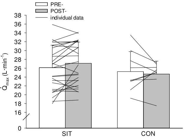
Group Data Q max: There were no main or interaction effects for Q max (p≥0.166). There was no significant change in Q max following SIT (pre-= 26.13±5.09 vs
post=27.10±4.82 L·min-1; p=0.166) or control (pre-= 25.23±4.53 vs post= 24.68±2.92 L·min-1; p=0.166) conditions.
SIT Qm a x ( L ·m in -1 ) 0 16 18 20 22 24 26 28 30 32 34 36 38 PRE- POST-individual data CON ....
Figure 2. Maximal cardiac output (Q max; L·min-1) before (PRE) and after (POST)
6 weeks of SIT and CON conditions. Group data. Values are means ± SD. SIT =
Sex Differences Q max: There was a main effect of group on Q max (SIT=31±2.6 vs CON=27±3.2 L·min-1; p=0.010) in men. There was no change in Q max following SIT (pre-= 30±2.7 vs post=31±2.6 L·min-1; p=0.145) or control (pre-= 28±4.5 vs post=26±1.1 L·min-1; p=0.145) conditions in men.
There were no main or interaction effects for Q max (p≥0.200). There was no change in
Q max following SIT (pre-= 22±2.7 vs post=23±3.4 L·min-1; p=0.671) or control (pre-= 22±3.0 vs post= 23±4.0 L·min-1; p=0.671) conditions in women.
3.5 Maximum heart rate (HR
max; beats·min
-1)
Group Data HRmax: There were no main or interaction effects for HRmax (p≥0.135). There was no change in HRmax following SIT (pre-= 190±8 vs post= 189±8 beats·min-1;
p=0.243) or control (pre-= 185±7 vs post= 186±6 beats·min-1; p= 0.243) conditions.
3.6 Maximum stroke volume (SV
max; mL·kg lean mass
-1
·beat
-1)
Group Data SVmax: There were no main or interaction effects for SVmax (p≥0.109). There was no significant change in SVmax following SIT (pre-=2.5±0.3 vs post=2.6±0.3 mL·kg lean mass-1·beat-1; p=0.109) or control (pre-=2.4±0.4 vs post=2.4±0.3 mL·kg lean mass -1·beat-1; p=0.109) conditions.
3.7 Maximum stroke volume (SV
max; mL·beat
-1)
Group Data SVmax: There were no main or interaction effects for SVmax (p≥0.095). There was no significant change in SVmax following SIT (pre-= 138±27 vs post=144±28
SIT S Vm a x ( m L ·b e a t -1 ) 0 100 120 140 160 180 200 PRE- POST-individual data CON
Figure 3. Maximum stroke volume (SVmax; mL·beat-1) before (PRE) and after
(POST) 6 weeks of SIT and CON conditions. Group data. Values are means ± SD.
SIT = sprint interval training; CON = control; lines are individual data.
3.8 Arterial-mixed venous oxygen difference (a-
v
O
2diff; mL
O
2·100mL blood
-1·kg lean mass
-1)
Group Data a-v O2diff: There were no main or interaction effects for a-v O2diff (p≥0.683). There was no change in a-v O2diff following SIT (pre-=0.25±0.05 vs post=0.25±0.05 mL O2·100mL blood-1·kg lean mass-1; p=0.843) or control (pre-=0.26±0.04 vs post=0.25±0.04 mL O2·100mL blood-1·kg lean mass-1; p=0.843) conditions.
3.9 Arterial-mixed venous oxygen difference (a-
v
O
2diff; mL
O
2·100mL blood
-1)
SIT a -v O2 d if f ( m L O 2 ·1 0 0 m L b lo o d -1 ) 0 8 10 12 14 16 18 20 PRE- POST-individual data CON
Figure 4. Arterial-mixed venous oxygen difference (a-v O2diff; mL O2·100mL blood
-1
) before (PRE) and after (POST) 6 weeks of SIT and CON conditions. Group data.
Values are means ± SD. SIT = sprint interval training; CON = control; lines are
individual data.
3.10 Five minute run: total distance (m)
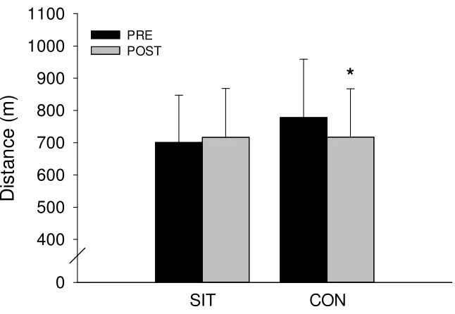
Group Data Total Distance Run: There was a significant group by time interaction (p<0.001) for total distance run (m). There was no improvement in total distance run following SIT (pre-=701.1±146.2 vs post=717.0±151.5 m; p=0.087) and there was a 60.3 m decrease in total distance run following control (pre-=777.8±181.3 vs
post=717.5±149.8 m; p<0.001) conditions.
Sex Differences Total Distance Run: There were no main or interaction effects for total distance run in women (p≥0.370). There was no change in total distance run following
SIT CON
D
is
ta
n
c
e
(
m
)
0 400 500 600 700 800 900 1000 1100
PRE POST
*
Figure 5. Total distance run (m) during 5-minute run before (PRE) and after
(POST) 6 weeks of SIT and CON conditions. Group data. Values are means ± SD.
*P < 0.05 (POST significantly less than PRE). SIT = sprint interval training; CON =
control.
3.11 Five minute run: change in distance (
∆
d; m)
Group Data ∆d: There was a significant ∆d achieved during the 5 min run following SIT
compared with control conditions (SIT=15.94±42.39 vs CON=-60.27±62.76 m; p=0.0002).
Sex Differences ∆d: There was no change in ∆d achieved during the 5 min run following
Chapter 4
4
Discussion
The major findings from the present study were that increasing sprint interval exercise
bout duration from 30 to 45 seconds resulted in an improved V O2max. Also, there was a
4% non-significant increase in Q max whereas a-v O2diff was essentially unchanged following training. Distance completed in the 5-minute run was not improved but there was likely a real improvement in exercise performance with SIT that was masked by the nature of that particular test (see below for details).
4.1 Effect on
V
O
2maxAs in other SIT studies, V O2max was increased following training. However, in the
present study, V O2max was only increased by 3% which is smaller than the 7 to 12% increases seen by others (Burgomaster et al., 2008; MacDougall et al., 1998; MacPherson et al., 2011). Participants’ baseline V O2max in this study was 3.6 L·min-1, compared to 3.6, 3.7, and 2.8 L·min-1 found in similar studies by MacPherson, MacDougall, and Burgomaster, respectively. The sex of participants affects the absolute V O2max measured; due to a smaller blood volume and greater proportion of fat mass, typically
females have lower V O2max values than males. While the baseline values between studies appear similar, the values may actually represent different training statuses based on the composition of each population. The present study included 14 men and 14 women trainees. Burgomaster studied men and women but baseline V O2max was only 2.8 L·min-1, MacPherson included more men than women, and MacDougall studied men exclusively. Our baseline values appear similar to these related studies despite the greater
proportion of female participants, which may be expected to reduce the mean V O2max. The fact that baseline V O2max was not lower than similar studies involving more males might indicate that our participants were actually more trained (for example, men in the
SIT group had a baseline V O2max of 4.2 L·min-1) before the SIT intervention. Changes
In young, trained, healthy humans, breathing normoxic air, cardiac output is often cited as the factor which imposes the greatest limit to V O2max (Bergh, Ekblom, & Åstrand, 2000). Increasing cardiac output (via blood volume expansion, pericardectomy) leads to
an instantaneous increase in V O2max (Kanstrup & Ekblom, 1982; Stray-Gundersen et al., 1986). If subjects in the present study had a relatively high training status, it is possible that the training stimulus to peripheral systems is diminished, and that training
improvements are driven mainly by changes in Q max. This would explain the small
observed increase in V O2max, and agrees with the higher than expected baseline
V O2max.
4.2 Effect on
Q
maxFollowing SIT, participants showed a trend toward increased Q max (4%) which did not reach statistical significance (p=0.166). In agreement with previous research, HRmax did not change following training (Blomqvist & Saltin, 1983). As Q max is the product of HRmax and SVmax, the unchanged HRmax indicates that the trend for Q max to increase was likely related to increased SVmax. In support of this, there was a 5% non-significant increase in SVmax following SIT (p=0.095). While neither change reached significance,
there was considerable variability about each mean (SIT Q max: pre-= 26.13±5.09 vs post=27.10±4.82 L·min-1; SIT SV
max: pre-= 138±27 vs post=144±28 mL·beat-1). It is possible that a real change occurred but it is undetectable due to the variability. Additionally, the other components that might contribute to an increase in V O2max (HRmax and a-v O2diff) were not even approaching significant p values (SIT HRmax
p=0.243; SIT a-v O2diff p=0.803). Thus, our conclusion is that Q max and SVmax likely
increased in response to SIT and were the driving forces in producing a greater V O2max. More subjects and/or a more homogeneous response would be needed to determine this definitively.
for the entirety of the training session including recovery periods(Hazell, Olver,
Hamilton, & Lemon, 2012). So apparently SIT drives heart rate to near maximal values during the exercise bout and remains elevated during the “rest/recovery” periods between bouts. By extending the exercise bout by 15 seconds, there would be a great increase in volume load on the heart because HR would be near maximal during the entirety of the added 15 seconds. Additionally, the exercise was performed at supramaximal intensities so metabolic products as well as increased neural outflow could result in a further
elevated HR during “rest/recovery” in comparison to 30-second SIT. We did not alter the length of the “rest” period (4 minutes) which means the ratio of work:rest was greater compared to 30-second bouts. A greater work:rest ratio will be another factor
contributing to an increased average HR (and cardiac volume load) during each training session.
Sprint interval training is known to be a potent stimulus for a number of other
adaptations. For example, after 1 training session there is elevated PGC-1α and evidence
of mitochondrial biogenesis occurring (Little, Safdar, Bishop, Tarnopolsky, & Gibala, 2011). However as mentioned, SIT, using 30-second bouts does not appear to result in
improvements in Q max. The current study suggests that using 45-second bouts could provide a minimal stimulus because there is a trend for an increased Q max. It is possible that a longer program (e.g. 8 weeks) would result in significant changes to Q max because the stimulus, if real, appears to be weak. Perhaps slightly longer exercise bouts (e.g. 60-80 seconds) could provide a sufficient stimulus.
4.3 Effect on
a
-
v
O
2diffIn contrast to the findings of MacPherson et al. (2011), who showed that 30-second SIT bouts caused an increase in a-v O2diff, there was no change in a-v O2diff when 45-second
exercise bouts were used. Our baseline a-v O2diff values were similar to those of
Improvements in a-v O2diff reflect increased extraction/utilization at the muscle. Sprint interval training using 30-second bouts increase oxidative and glycolytic enzyme concentrations and as a result increases a-v O2diff (Burgomaster, Heigenhauser, & Gibala, 2006; MacPherson et al., 2011). If our subjects were indeed more trained as suggested they would have already experienced increases in enzyme
concentrations/activity prior to the study and therefore would be expected to experience a reduced training adaptation. Importantly, it is also possible that 45-second bouts provide a stimulus that differs from 30-second bouts. Sprint interval training is unique in that the effort is constant (“all-out” or supramaximal) but power output (or running speed in our case) varies throughout the exercise bout. The first 10 seconds are characterized by a large power output and then power output is considerably lower for the remaining 20 to 35 seconds. Hazell et al. (2010) showed that 10-second SIT bouts produced
improvements in aerobic and anaerobic exercise performance that were similar to improvements seen with 30-second bouts. Perhaps SIT is a potent and effective training method largely due to the great power outputs produced during the initial 10 seconds of each bout while the remaining 20 seconds have a negligible effect on training
adaptations. Helgerud et al. (2007) had subjects perform 1 of 4 training programs
(Submaximal endurance training, endurance training at maximal lactate steady state, 1:1 short interval training bouts at 90-95% V O2max, and 1:1 long interval training bouts at
90-95% V O2max). Training frequency and total work were matched, the greatest
improvements in V O2max were associated with higher exercise intensities (interval training was greater than endurance training). SIT is performed at “all-out” intensities and that could be the key to its effectiveness. Because there is an inverse relationship between exercise volume and intensity, by increasing the duration of SIT bouts, we increased the volume and decreased intensity. It is possible that this minimized
improvements in V O2max via a decreased effect on peripheral adaptations. Interestingly, the greater exercise volume vs 30-second bouts may stimulate Q max but, based on our data, insufficiently so to observe a significant increase. Apparently, longer bouts are necessary. Consequently, our choice of 45-second sprint bouts and their effect on both exercise intensity and duration may have contributed to the smaller than expected
4.4 Effect on 5-minute run
The 5-minute run was used to assess the effect of training on exercise performance. Total distance run during the 5 minutes did not change with training and there was a decrease following control conditions.
However, following completion of the study, a number of participants reported that they had modified their pacing strategy and/or effort for the 5-minute run because of the knowledge that it was extremely challenging gained during the initial testing. The tread poses greater than normal resistance so running in manual mode is more difficult than running freely on a road or track. Also, participants must use their upper body to push on the treadmill’s handles in order to properly propel the tread. Running in manual mode is more similar to pushing a sled than running freely. This combination of resistance on the tread and upper body effort made the test quite difficult as indicated. Consistent with this possibility is the observed decreased performance in the control group on the post test. This might explain why the 5-minute run total distance did not improve significantly in the SIT group. As mentioned, there was a main effect of time (pre>post) on total distance run which supports participants’ reports of changing their pacing strategy due to the extreme demands/effort required for the test. During their first run they did not know how hard the test would be and gave a maximal effort. After having completed the test once, participants may have attempted to minimize the associated discomfort by altering their strategy (i.e. pacing themselves), resulting in a poorer performance.
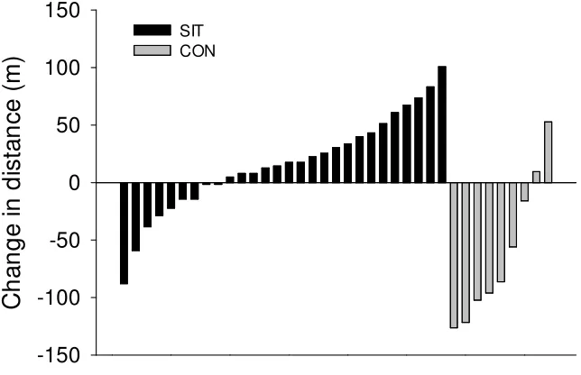
To clarify any coexisting effects of training improvements and voluntary pacing, the post study distance run was compared to the pre-training performance (post-training distance – pre-training distance; ∆d in metres run) to obtain a better representation of performance
changes following SIT (Figure 6). Consistent with pacing affecting the outcome, the majority of control participants actually decreased their performance (∆d was decreased)
while SIT participants exhibited a greater range of responses (with most participants showing improvement or little to no change). If altered pacing strategy (which
participants in both groups reported) is responsible for the control group’s reduction in
∆d, then the SIT group was likely exposed to this effect as well. With this in mind, the
negative effect of pacing, may represent a significant improvement in training status without a significant improvement in absolute distance run.
Individual Data
C
h
a
n
g
e
i
n
d
is
ta
n
c
e
(
m
)
-150 -100 -50 0 50 100 150 SIT CONFigure 6. Individual changes in distance run (∆d) during 5-minute run. SIT = sprint
interval training; CON = control. Each bar represents one participant.
In order to assess exercise performance, a 5-minute run on this type of manually driven treadmill is likely a poor test due to the effort it requires and the discomfort it produces, which may obscure real physiological changes. Tests which have successfully been used to assess exercise performance, such as the Yo-Yo intermittent recovery test, cycling time trials, and running time trials, should be used in future studies to measure exercise
performance (Burgomaster et al., 2006; Iaia et al., 2008; MacPherson et al., 2011) .
4.5 Summary
uptake increased significantly as a result of insignificant increases in Q max and SVmax with no effect on a-v O2diff. The five minute run ∆d following SIT suggests a positive effect from training, despite an overall non-significant difference in absolute distance. Limitations of the treadmill running task may have obscured any underlying training effect, and a future studies should employ a different task.
4.6 Future directions
Based on the findings from this study, longer duration sprint interval training (up to 45-seconds) should not be a replacement for ET. Rather, athletes should use a combination of SIT and ET in order to obtain the gamut of adaptations. Future studies should use
untrained individuals in hopes of uncovering real changes in V O2max, Q max, and a
-v O2diff that may have been obscured by the training status of participants in the current study. Studying the effects of an SIT program which uses a combination of various exercise bout lengths (e.g. 10-second, 30-second, and 60-second bouts) would be of interest. This would help determine if sprint interval exercise could effectively elicit central and peripheral adaptations concurrently. Additionally, due to the unique power profile of SIT exercise bouts, it would be of interest to compare time- and work-matched exercise training using constant-load exercise bouts to SIT exercise bouts.
4.7 Conclusion
References
Abergel, E., Chatellier, G., Hagege, A. A., Oblak, A., Linhart, A., Ducardonnet, A., & Menard, J. (2004). Serial left ventricular adaptations in world-class professional cyclists: implications for disease screening and follow-up. Journal of the American College of Cardiology, 44(1), 144–149.
Alberts, B., Bray, D., Hopkin, K., Johnson, A., Lewis, J., Raff, M., … Walter, P. (2004). How cells obtain energy from food. In Essential Cell Biology (Second., pp. 427–
452). New York: Garland Science.
Allen, D. G., Lamb, G. D., & Westerblad, H. (2008). Skeletal Muscle Fatigue: Cellular Mechanisms. Physiological Reviews, 88, 287–332.
Åstrand, P. O. (1956). Human physical fitness with special reference to sex and age.
Physiological Reviews, 36(3), 307–335.
Baar, K. (2006). To perform your best: work hard not long. The Journal of Physiology, 575(3), 690.
Baldwin, K. M., Cooke, D. A., & Cheadle, W. G. (1977). Time course adaptations in cardiac and skeletal muscle to different running programs. Journal of Applied Physiology: Respiratory, Environmental and Exercise Physiology, 42(2), 267–272.
Bangsbo, J., Mohr, M., Poulsen, A., Perez-Gomez, J., & Krustrup, P. (2006). Training and Testing the Elite Athlete. Journal of Exercise Science and Fitness, 4(1), 1–14.
Bassett, D. R., & Howley, E. T. (2000). Limiting factors for maximum oxygen uptake and determinants of endurance performance. Medicine and Science in Sports and Exercise, 32(1), 70–84.
Beaver, W. L., Lamarra, N., & Wasserman, K. (1981). Breath-by-breath measurement of true alveolar gas exchange. Journal of Applied Physiology: Respiratory,
Bergh, U., Ekblom, B., & Åstrand, P. O. (2000). Maximal oxygen uptake “classical” versus “contemporary” viewpoints. Medicine and Science in Sports and Exercise, 32(1), 85–88.
Bevegård, S. (1962). Studies on the regulation of the circulation in man. Acta Physiologica Scandinavica, 57(S200), 1–36.
Blomqvist, C. G., & Saltin, B. (1983). Cardiovascular adaptations to physical training.
Annual Review of Physiology, 45(46), 169–189.
Bogdanis, G. C., Nevill, M. E., Boobis, L. H., & Lakomy, H. K. (1996). Contribution of phosphocreatine and aerobic metabolism to energy supply during repeated sprint exercise. Journal of Applied Physiology, 80(3), 876–884.
Booth, F. W., & Thomason, D. B. (1991). Molecular and cellular adaptation of muscle in response to exercise: perspectives of various models. Physiological Reviews, 71(2),
541–85.
Brooks, G. A., & Mercier, J. (1994). Balance of carbohydrate and lipid utilization during exercise: the “crossover” concept. Journal of Applied Physiology, 76(6), 2253–2261.
Buchheit, M., & Laursen, P. B. (2013). High-intensity interval training, solutions to the programming puzzle. Part I: cardiopulmonary emphasis. Sports Medicine, 43(5),
313–338.
Burgomaster, K. A., Heigenhauser, G. J. F., & Gibala, M. J. (2006). Effect of short-term sprint interval training on human skeletal muscle carbohydrate metabolism during exercise and time-trial performance. Journal of Applied Physiology, 100, 2041–
2047.
Burgomaster, K. A., Hughes, S. C., Heigenhauser, G. J. F., Bradwell, S. N., & Gibala, M. J. (2005). Six sessions of sprint interval training increases muscle oxidative potential and cycle endurance capacity in humans. Journal of Applied Physiology, 98, 1985–
1990.
Cairns, S. P., & Lindinger, M. I. (2008). Do multiple ionic interactions contribute to skeletal muscle fatigue? The Journal of Physiology, 586(17), 4039–4054.
Clausen, J. P., Klausen, K., Rasmussen, B., & Trap-Jensen, J. (1973). Central training and peripheral circulatory changes after training of the arms or legs. American Journal of Physiology, 225(3), 675–682.
Coffey, V. G., & Hawley, J. A. (2007). The molecular bases of training adaptation.
Sports Medicine, 37(9), 737–763.
Convertino, V. A. (1991). Blood volume: its adaptation to endurance training. Medicine and Science in Sports and Exercise, 23(12), 1338–1348.
Coyle, E. F., Martin, W. H., Sinacore, D. R., Joyner, M. J., Hagberg, J. M., & Holloszy, J. O. (1984). Time course of loss of adaptations after stopping prolonged intense endurance training. Journal of Applied Physiology: Respiratory, Environmental and Exercise Physiology, 57(6), 1857–1864.
Daussin, F. N., Ponsot, E., Dufour, S. P., Lonsdorfer-Wolf, E., Doutreleau, S., Geny, B., … Richard, R. (2007). Improvement of VO2max by cardiac output and oxygen extraction adaptation during intermittent versus continuous endurance training.
European Journal of Applied Physiology, 101, 377–383.
Dudley, G. A., Abraham, W. M., & Terjung, R. L. (1982). Influence of exercise intensity and duration on biochemical adaptations in skeletal muscle. Journal of Applied Physiology: Respiratory, Environmental and Exercise Physiology, 53(4), 844–850.
Egginton, S. (2009). Invited review: activity-induced angiogenesis. European Journal of Physiology, 457, 963–977.
Ehsani, A. A., Ogawa, T., Miller, T. R., Spina, R. J., & Jilka, S. M. (1991). Exercise training improves left ventricular systolic function in older men. Circulation, 83,
96–103.
Ekblom, B., Åstrand, P. O., Saltin, B., Stenberg, J., & Wallström, B. (1968). Effect of training on circulatory response to exercise. Journal of Applied Physiology, 24, 518–
528.
Ekblom, B., & Hermansen, L. (1968). Cardiac output in athletes. Journal of Applied Physiology, 25(5), 619–625.
Ferguson, S., Gledhill, N., Jamnik, V. K., Wiebe, C., & Payne, N. (2001). Cardiac performance in endurance-trained and moderately active young women. Medicine and Science in Sports and Exercise, 33(7), 1114–1119.
Flück, M., & Hoppeler, H. (2003). Molecular basis of skeletal muscle plasticity--from gene to form and function. Reviews of Physiology, Biochemistry and Pharmacology, 146, 159–216.
Gibala, M. J., Little, J. P., van Essen, M., Wilkin, G. P., Burgomaster, K. A., Safdar, A., … Tarnopolsky, M. A. (2006). Short-term sprint interval versus traditional
endurance training: similar initial adaptations in human skeletal muscle and exercise performance. The Journal of Physiology, 575(3), 901–911.
Gollnick, P. D., Armstrong, R. B., Saltin, B., Saubert IV, C. W., Sembrowich, W. L., & Shepherd, R. E. (1973). Effect of training composition on enzyme activity and fiber of human skeletal muscle. Journal of Applied Physiology, 34(1), 107–111.
Gollnick, P. D., & Saltin, B. (1982). Significance of skeletal muscle oxidative enzyme enhancement with endurance training. Clinical Physiology, 2, 1–12.
González-Alonso, J. (2008). Point: Stroke volume does/does not decline during exercise at maximal effort in healthy individuals. Journal of Applied Physiology, 104(1),
275–280.
Gunnarsson, T. P., & Bangsbo, J. (2012). The 10-20-30 training concept improves performance and health profile in moderately trained runners. Journal of Applied Physiology, 113(1), 16–24.
Hawley, J. A. (2002). Adaptations of skeletal muscle to prolonged, intense endurance training. Clinical and Experimental Pharmacology and Physiology, 29, 218–222.
Haykowsky, M. J., Dressendorfer, R., Taylor, D., Mandic, S., & Humen, D. (2002). Resistance Training and Cardiac Hypertrophy: Unravelling the Training Effect.
Sports Medicine, 32(13), 837–849.
Hazell, T. J., MacPherson, R. E. K., Gravelle, B. M. R., & Lemon, P. W. R. (2010). 10 or 30-S Sprint Interval Training Bouts Enhance Both Aerobic and Anaerobic
Performance. European Journal of Applied Physiology, 110, 153–160.
Hazell, T. J., Olver, T. D., Hamilton, C. D., & Lemon, P. W. R. (2012). Two Minutes of Sprint-Interval Exercise Elicits 24-hr Oxygen Consumption Similar to That of 30 min of Continuous Endurance Exercise. International Journal of Sport Nutrition and Exercise Metabolism, 22(4), 276–283.
Holloszy, J. O., & Booth, F. W. (1976). Biochemical Adaptations to Endurance Exercise in Muscle. Annual Review of Physiology, 38, 273–291.
Holloszy, J. O., & Coyle, E. F. (1984). Adaptations of skeletal muscle to endurance exercise and their metabolic consequences. Journal of Applied Physiology: Respiratory, Environmental and Exercise Physiology, 56(4), 831–838.
Hopper, M. K., Coggan, A. R., & Coyle, E. F. (1988). Exercise stroke volume relative to plasma-volume expansion. Journal of Applied Physiology, 64, 404–408.
Iaia, F. M., & Bangsbo, J. (2010). Speed endurance training is a powerful stimulus for physiological adaptations and performance improvements of athletes. Scandinavian Journal of Medicine and Science in Sports, 20(S2), 11–23.
Iaia, F. M., Perez-Gomez, J., Thomassen, M., Nordsborg, N. B., Hellsten, Y., & Bangsbo, J. (2011). Relationship between performance at different exercise intensities and skeletal muscle characteristics. Journal of Applied Physiology, 110, 1555–1563.
Iaia, F. M., Thomassen, M., Kolding, H., Gunnarsson, T., Wendell, J., Rostgaard, T., … Bangsbo, J. (2008). Reduced volume but increased training intensity elevates muscle Na+-K+ pump alpha1-subunit and NHE1 expression as well as short-term work capacity in humans. American Journal of Physiology: Regulatory, Integrative and Comparative Physiology, 294, R966–R974.
Ingjer, F. (1979). Effects of endurance training on muscle fibre ATP-ase activity,
capillary supply and mitochondrial content in man. The Journal of Physiology, 294,
419–432.
Irrcher, I., Adhihetty, P. J., Joseph, A., Ljubicic, V., & Hood, D. A. (2003). Regulation of mitochondrial biogenesis in muscle by endurance exercise. Sports Medicine, 33(11),
783–793.