Clinical, Cosmetic and Investigational Dentistry
Dove
press
O R I G I N A L R E S E A R C H
open access to scientific and medical research
Open Access Full Text Article
Shear bond strength of a bracket-bonding
system cured with a light-emitting diode or
halogen-based light-curing unit at various
polymerization times
Sanjay Prasad Gupta Basanta Kumar Shrestha
Orthodontics and Dentofacial Orthopedics Unit, Department of Dentistry, Tribhuvan University Teaching Hospital, Institute of Medicine, Kathmandu, Nepal
Purpose: To determine and compare the shear bond strength (SBS) of bracket-bonding system cured with light-emitting diode (LED) and halogen-based light-curing unit at various polym-erization times.
Materials and methods: Ninety six human maxillary premolar teeth extracted for orthodontic purpose were divided into four groups, according to the light-curing unit and exposure times used. In the halogen group, the specimens were light cured for 20 and 40 seconds. In the LED group, the specimens were light cured for 5 and 10 seconds. Stainless steel brackets were bonded with Enlight bonding system, stored in distilled water at 37°C for 24 hours and then submitted to SBS testing in a universal testing machine at a crosshead speed of 0.5 mm/minute. Adhesive remnant index (ARI) was used to evaluate the amount of adhesive remaining on the teeth determined by stereomicroscope at 10× magnification.
Results: The highest mean SBS was obtained with the halogen 40 seconds (18.27 MPa) fol-lowed by halogen 20 seconds (15.36 MPa), LED 10 seconds (14.60 MPa) and least with LED 5 seconds (12.49 MPa) group. According to analysis of variance (ANOVA) and Tukey’s multiple-comparison test, SBS of halogen 20 seconds group was not significantly different from halogen 40 seconds group, LED 5 seconds group and LED 10 seconds group, whereas halogen 40 seconds group was significantly different from LED 5 seconds and LED 10 seconds group. The method of light curing did not influence the ARI, with score 2 being predominant.
Conclusion: Polymerization with both halogen and LED resulted in SBS values that were clinically acceptable for orthodontic treatment in all groups. Hence, for bonding orthodontic brackets, photoactivation with halogen for 20 seconds and LED for 5 seconds is suggested. Keywords: shear bond strength, halogen-curing light, LED-curing light, polymerization time
Introduction
Orthodontic appliances may be attached by cementing bands or by bonding brack-ets directly to the enamel surface using a retentive base. The development of firm attachment of brackets directly to the teeth is one of the prominent milestones in the orthodontic field as it enables the efficient movement of the teeth. Firm attachment was initially achieved by cementing bands on all the teeth.
Direct bonding of brackets using acid etching has become contemporary in ortho-dontics fields.1 This was a revolution in the practice of clinical orthodontics, and since
then, there has been a rapid rate of product development in terms of adhesives, brackets and their technical properties.
Correspondence: Sanjay Prasad Gupta Orthodontics and Dentofacial Orthopedics Unit, Department of Dentistry, Tribhuvan University Teaching Hospital, Institute of Medicine, Maharajgunj, PO Box 1524, Kathmandu, Nepal
Tel +977 1 984 358 5923
Email [email protected]
Journal name: Clinical, Cosmetic and Investigational Dentistry Article Designation: ORIGINAL RESEARCH
Year: 2018 Volume: 10
Running head verso: Gupta and Shrestha
Running head recto: LED- or halogen-based curing unit for bracket-bonding system DOI: http://dx.doi.org/10.2147/CCIDE.S155829
Clinical, Cosmetic and Investigational Dentistry downloaded from https://www.dovepress.com/ by 118.70.13.36 on 21-Aug-2020
For personal use only.
Dovepress
Gupta and Shrestha
Orthodontic bonding to the enamel surface leads to sig-nificant improvement in treatment by increasing patient com-fort, better esthetics and decreasing periodontal problems.2–4
The advent of light cure adhesives in 1979 by Tavas and Watts5 became popular because of their various advantages
compared with self-cured adhesive materials.
These advantages are application of a single paste, control of working time, easy removal of excess bonding material, reduced risk of contamination and immediate insertion of the arch wire,6,7 as they provide increased working time for
pre-cise bracket placement and ease of manipulation. Currently, four different technologies are available for curing of dental composites by light that are halogen lamps, lasers, plasma arc lamps and light-emitting diode (LED).
Halogen lamps were the first to be introduced as the source of light. Though it was a boon for the clinical achieve-ments in the dental practice in the initial stages it had many demerits. Quick overheating of the filament of the halogen bulbs made its use restricted for lengthy procedures, espe-cially in orthodontics.
Mills et al8 in 1995 presented the solid-state LED. Since
then, LED has attracted increasing attention as a new source for light-activated polymerization. Many light-curing devices have become popular and it is important to determine the light-curing unit that is most efficient as well as give the desired bond strength. Various studies have been carried out to evaluate the bond strength of bracket-bonding systems using shear testing.9–17
The objective of this study was to determine and compare the shear bond strength (SBS) of the bracket-bonding system cured with LED and halogen-based light-curing unit at vari-ous polymerization times.
Materials and methods
In this study, 96 human bicuspid teeth extracted for orthodon-tic purpose were obtained. The sampling method used was convenient sampling. Extracted teeth were collected from Oral and Maxillofacial Surgery Unit, Tribhuvan University Teaching Hospital, Kathmandu, and stored in distilled water at room temperature to eliminate dehydration and bacterial growth.18,19 All patients had provided written informed
con-sent at the time of treatment that their extracted teeth may be used for future research purposes.
Inclusion criteria were extracted teeth with intact buccal enamel and teeth extracted not more than 3 months until testing, while teeth subjected to caries and any pretreat-ment chemical agent, and that had cracks caused by the pressure of extraction forceps, which is visualized by naked
eye, and developmental defects such as hypoplastic enamel were excluded. This study was approved by the Institutional Review Board, Institute of Medicine, Kathmandu.
The teeth were then mounted on an acrylic block in such a way that the roots were embedded completely into the acrylic up to cementoenamel junction and leaving the crown exposed. The labial surface of the tooth was kept perpendicular to the bottom surface of mold.20 Cuts were
made on both sides of the acrylic block with acrylic trimmer for the purpose of securing it in universal testing machine at the time of debonding.
The mounted specimens were randomly divided into four groups (two halogen and two LED groups) of 24 in each according to the exposure time and the type of light unit and named accordingly as halogen 20 seconds (Ha), halogen 40 seconds (Hb), LED 5 seconds (La) and LED 10 seconds (Lb) groups.21 All specimens were immersed in distilled water
except at the time of bonding and bebonding procedures. Before bonding, the buccal surfaces of all the teeth were cleaned with fluoride-free fine pumice powder in water using a brush at slow-speed micromotor hand piece for 10 seconds for removal of any dirt\calculus\deposits or stains. The teeth were then rinsed thoroughly with water for 10 seconds23,24 and
dried with oil- and moisture-free compressed air. The buccal surface of each tooth was etched for 30 seconds with 37% phosphoric acid in gel form.25 Each tooth was then rinsed with
a distilled water spray for 5 seconds and dried with oil-free air till the etched tooth appeared chalky white.
A thin coat of light-cured adhesive primer Orthosol (Enlight, Ormco Corp., USA) was applied to acid-etched enamel. Enlight composite resin was applied on the 0.022″ slot roth stainless steel double mesh premolar bracket base (Minidiagonale, Leone Co., Sesto, Florentine, Florence, Italy) having a surface area of 8.8 mm2, which was then placed on
the teeth with a holding pincer near the center of the buccal surfaces with sufficient manual pressure that lead the excess material to flow at the margins of the bracket, which was then removed with an exploratory probe before polymerization.
Conventional halogen light-curing system and LED light-curing system for curing orthodontic bracket adhesive were used (Table 1). The light intensity was monitored with radiometers (CM300-2000, APOZA, Taiwan; Table 1).
In the Ha group, light was placed only in an occlusal direc-tion for 20 seconds, while in the Hb group, light was placed both in mesial and distal directions for 20 seconds each.
In the La group, light was placed only in an occlusal direction for 5 seconds, while in the Lb group, light was placed both in mesial and distal directions for 5 seconds each.
Clinical, Cosmetic and Investigational Dentistry downloaded from https://www.dovepress.com/ by 118.70.13.36 on 21-Aug-2020
Dovepress LED- or halogen-based curing unit for bracket-bonding system
Light curing was performed by maintaining the curing tip as close to the bracket as possible with the formation of an angle of 45° with the buccal surface of the tooth.
The specimens (Figure 1) were then stored in distilled water at 37°C for 24 hours26 and submitted to the SBS testing
in a universal testing machine (AG-IC/100 KN, Shimadzu, Japan).
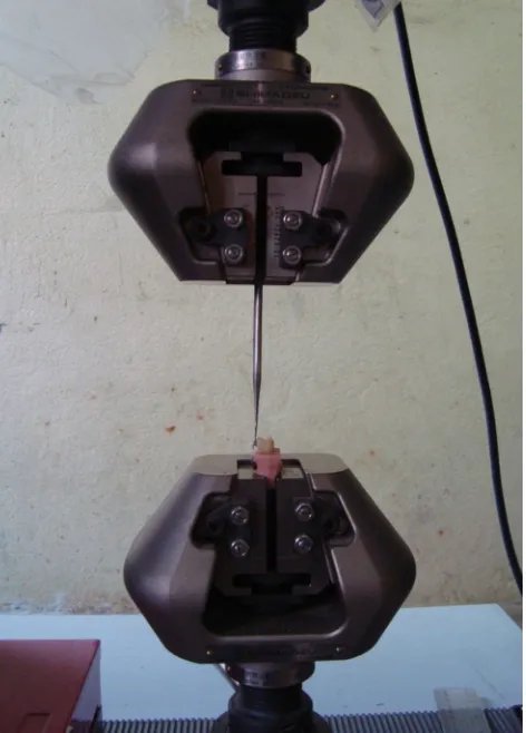
Before debonding, the specimen mounted in its acrylic block with cuts on both side was secured to the lower grip of the machine such that the buccal surface of the tooth with bonded bracket was parallel to the plunger that was attached to the moving crosshead of the universal testing machine (Figure 2).
A chisel-edge-shaped plunger was attached in the mov-able crosshead of the universal testing machine and placed in such a way that the leading edge aimed the enamel–adhesive interface that moves at a crosshead speed of 0.5 mm/min. Load was applied occlusogingivally to the bracket, which produced a shear force at the bracket–tooth interface. A computer connected with the testing machine showed the results of each test that were recorded.
The force required to dislodge the brackets was measured in Newton, and the SBS (MPa) was determined by dividing the force by the bracket base area (8.8 mm2).
SBS MPa Peak load at failure N Specimen surface area m
( ) ( )
(
=
m m2)
After bond strength testing, all specimens were collected and visually examined with a stereomicroscope (Olympus SZX12, Olympus Corp., Japan) at 10× magnification to assess the adhesive remnant index (ARI)21 available at Nepal
Agricultural Research Council, Khumaltar, Kathmandu. The ARI was used to evaluate the amount of resin remaining on the tooth after debonding. The criteria for ARI scoring given by Artun and Bergland28 were used in this study (Table 2).
The data were processed and analyzed by using the SPSS software version 16.0 (SPSS Inc., Chicago, IL, USA). Data were then subjected to analysis of variance (ANOVA) to identify differences in various groups. Tukey’s post hoc Table 1 Characteristics of the halogen and LED units
Appliance Type Wave length (nm) Light intensity (mW/cm2)
QHL 75 (Dentsply, Milford, USA) Halogen 400–500a 620
Galaxy (Shanghai Co., Shanghai, China) LED 430–485a 800
Note:aAccording to the manufacturer’s specifications.
Abbreviation: LED, light-emitting diode.
Figure 1 All samples of four experimental groups.
Figure 2 Close view of crosshead of universal testing machine with sample in situ.
Clinical, Cosmetic and Investigational Dentistry downloaded from https://www.dovepress.com/ by 118.70.13.36 on 21-Aug-2020
Dovepress
Gupta and Shrestha
multiple-comparison test was used to identify where differ-ences occurred.
Kappa test was performed for method error calculation. Chi-square test was performed to determine the differences in ARI scores of all four groups. A p-value of <0.05 was considered to be significant with a 95% confidence limit.
Results
SBS values including mean and SD obtained for the four groups are shown in Table 3.
The highest mean SBS was obtained with the Hb group (18.27 MPa) followed by Ha (15.36 MPa), Lb (14.60 MPa) and least with La (12.49 MPa) groups.
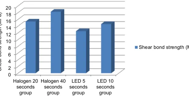
The graphical representation of mean SBS values is shown in Figure 3.
ANOVA was used to compare the mean values of SBS obtained in each group. The test showed that the difference
in the mean values of SBS was statistically significant (p=0.001).
Tukey’s post hoc multiple-comparison test was used for intergroup comparisons. The test showed that the SBS of Ha group was not significantly different from Hb, La and Lb groups. La group was not significantly different from Lb group, whereas Hb group was significantly different from La and Lb groups.
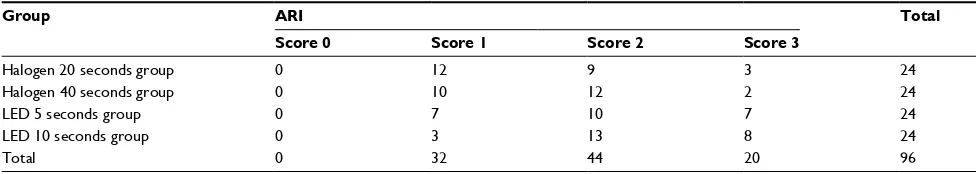
Adhesive index scores based on the amount of resin left on the tooth after debonding of the four groups are shown in Table 4.
The standard error of method, as calculated using kappa test, was 0.933, which shows the good intraobserver reliability.
The chi-square test detected no statistically significant difference in the ARI scores of all the four groups (p=0.065), which means that the method of light curing did not influence the ARI, with the score 2 being predominant.
Discussion
Among different technologies for light curing, halogen lamps and LED are the most commonly used ones. Light power is the important factor that contributes to the level of polymerization. According to Rueggeberg,29 higher the light
power, greater the number of photons reaching the resin and in turn higher number of free radicals available for polym-erization. Halogen unit of mean power of 620 mW/cm2 and
the LED unit of mean power of 800 mW/cm2 were used in
this study.
As during tooth formation there is rapid development of bovine enamel and dentin, bovine enamel has larger crystal grains and more lattice defects compared to human enamel.30
It was reported that bond strength values of permanent bovine enamel are ~35% and 44% below that of the human enamel.31 Table 2 Grading of ARI
ARI
Score 0 No adhesive left on the tooth
Score 1 Less than half of the adhesive left on the tooth Score 2 More than half of the adhesive left on the tooth Score 3 All adhesive left on the tooth, with a distinct impression
of the bracket mesh
Abbreviation: ARI, adhesive remnant index.
Figure 3 Shear bond strength mean values (MPa) in the experimental groups. 0
2 4 6 8 10 12 14 16 18 20
Halogen 20 seconds
group
Halogen 40 seconds
group
LED 5 seconds
group
LED 10 seconds
group
Shear bond strength (MPa)
Shear bond strength (MPa
)
Table 3 Shear bond strength mean values (MPa) in the experimental groups
Group n Mean (MPa) SD
Halogen 20 seconds group 24 15.36 4.10
Halogen 40 seconds group 24 18.27 5.73
LED 5 seconds group 24 12.49 4.23
LED 10 seconds group 24 14.6 5.12
Abbreviation: LED, light-emitting diode.
Clinical, Cosmetic and Investigational Dentistry downloaded from https://www.dovepress.com/ by 118.70.13.36 on 21-Aug-2020
Dovepress LED- or halogen-based curing unit for bracket-bonding system
Though bovine teeth can be used as a substitute for human teeth, the latter is the best substrate for bonding.32
Bond strength studies mostly used distilled water for storing the testing specimens.18,19,33 To standardize the shear
bond test protocols, Fox et al34 recommended distilled water
as a prebonding storage media in the shear bond studies. Studies regarding the effect of the storage media on den-tin bond strength have found that distilled water storage did not adversely affect the bond strength of the teeth stored for up to 6 months.35,36 In vitro bond strength testing comprise
shear, tensile and torsion tests. As failure of the bracket-tooth adhesion is assumed not to be a result of pure tensile and torsional stress, these tests are less often performed, while shear testing is most popular as there is similarity with the clinical debonding situation.37
A methodologic review of SBS studies revealed that crosshead speeds of 0.5 and 1 mm/min are often used in SBS tests.38
Previous studies have shown that LED-curing units are as effective as halogen-curing units. According to Dunn and Taloumis,24 there was no significant difference in bond
strength with LED or halogen-based light-curing units. In this study, the highest mean SBS was obtained with the Hb (18.27 MPa) followed by Ha (15.36 MPa), Lb (14.60 MPa) and least with La (12.49 MPa) groups.
All LED groups presented lower mean SBS value com-pared with the halogen groups. Statistically, mean SBS of Ha group was not significantly different from Hb, La and Lb groups. La group was not significantly different from Lb group, whereas Hb group was significantly different from La and Lb groups.
SBS of Hb group of this study was higher than that in the study done by Dunn and Taloumis,24 Abtahi and Khamverdy,39
Banerjee S and Banerjee R25 and Balasubramanian et al20 while
the study of Turkkahraman and Kucukesmen22 and Cerekja
and Cakirer18 showed similar results.
SBS of Ha group of this study was higher than that in the study by Hui-Ping et al.19
SBS of Hb and Lb groups of this study were higher than those in the study done by Usumez et al15 whereas study by
Dall’Igna et al21 showed the similar results with Lb while
higher with La group when compared with this study. In this study, La group, with a shorter exposure time, showed the lowest mean SBS value, which was adequate bond strength for majority of the clinical situations, as values of 5.9–7.8 MPa proposed by Reynolds and von Fraunhofer3.
According to Lopez,40 the SBS recommended for successful
clinical bonding was estimated to be 7 MPa.
Results of this study are in accordance with the study of Mavropoulos et al41 and Gronberg et al,16 that is, a minimum
time of 5 seconds resulted in SBS values that are sufficient to resist the forces exerted in a clinical situation, while study by Yu et al17 showed divergent results as light curing with an
LED for 4 and 6 seconds showed lower SBS values; when cured for 8 seconds, then only the values were considered to be satisfactory.
The results of our study showed that as light-curing time increased for both light units, there was a gradual increase in mean SBS. Peutzfeldt and Asmussen42 study also is in
accordance with this study. Various authors have also reported the similar findings.15–17,41,43
There was no significant difference in ARI scores among the groups, which is supported by the literature,41,44,45 with the
predominant score being 2. It means that failures occurred mostly at the bracket–adhesive interface after debonding, with the material remaining adhered to the tooth surface that allows removal of composite resin and preservation of the enamel from possible trauma.17,46,47
The results of our study showed that the light unit does not affect the location of orthodontic bond failures because the majority of these occurred at the bracket–composite resin interface (scores 2 and 3) with both light units. According to Gronberg et al16 and Yu et al,17 this failure location could
indicate incomplete resin polymerization at the base of bracket as a result of the short period of light exposure. This diminishes the probability of damage to the enamel during Table 4 ARI scores in the experimental groups
Group ARI Total
Score 0 Score 1 Score 2 Score 3
Halogen 20 seconds group 0 12 9 3 24
Halogen 40 seconds group 0 10 12 2 24
LED 5 seconds group 0 7 10 7 24
LED 10 seconds group 0 3 13 8 24
Total 0 32 44 20 96
Abbreviations: ARI, adhesive remnant index LED, light-emitting diode.
Clinical, Cosmetic and Investigational Dentistry downloaded from https://www.dovepress.com/ by 118.70.13.36 on 21-Aug-2020
Dovepress
Gupta and Shrestha
bracket debonding, which is advantageous in the orthodontic treatment.
One limitation of this study was that being an ex vivo study, it cannot completely reproduce the complex interac-tion processes that occur in the oral cavity.48,49 In a contest of
SBS measurement, several factors can influence the results. This in vitro study fails to simulate factors such as intraoral aging of resin composites, PH and temperature fluctua-tion based on individual’s dietary intake and oral hygiene, complex cyclic loading, microbial attack and enzymatic degradation. Pickett et al50 and Murray and Hobson51 found
that as the biodegradation that occurs in the oral cavity, bond strength values in vivo tend to be lower than those found ex vivo.
Another limitation was the use of a constant crosshead speed of 0.5 mm/minute during debonding in a universal test-ing machine. Accordtest-ing to Eliades and Brantley,52 debonding
in vivo occurs at a higher speed though this load speed is generally used.
Despite the above mentioned limitations, the results of our study suggest that both halogen- and LED-curing unit can be used in bracket bonding with reduced light-curing time without affecting the SBS. This reduction in curing time leads to various advantages like shorter chair time and lower risk of contamination of saliva.
The LED light units are smaller, cordless and lighter with estimated lifetimes of over 10,000 hours, and that do not require a noisy cooling fan.15 Therefore, it seems that they
are a better choice as compared to halogen sources.
Conclusion
Bond strength values between the La and Lb groups with the Ha group were statistically equivalent.
Bonding of orthodontic brackets by photoactivation with halogen for 20 seconds and LED for 5 seconds resulted in SBS values that are clinically acceptable for orthodontic treatment and hence suggested as it requires a reduced clini-cal chair time.
The method of light curing did not influence the ARI, with predominant score 2 among all groups.
Acknowledgment
This study was conducted as a part of a thesis under the guidance of Professor Dr Shrestha.
Disclosure
The authors report no conflicts of interest in this work.
References
1. De Munck J, Van Landuyt K, Peumans M, et al. A critical review of the durability of adhesion to tooth tissue: methods and results. J Dent Res. 2005;84(2):118–132.
2. Buonocore MG. A simple method of increasing the adhesion of acrylic filling materials to enamel surfaces. J Dent Res. 1955;34(6):849–853. 3. Reynolds IR, von Fraunhofer JA. Direct bonding of orthodontic attach-ments to teeth: the relation of adhesive bond strength to gauze mesh size. Br J Orthod. 1976;3(2):91–95.
4. Ozturk B, Malkoc S, Koyuturk AE, Catalbas B, Ozer F. Influence of different tooth types on the bond strength of two orthodontic adhesive systems. Eur J Orthod. 2008;30(4):407–412.
5. Tavas MA, Watts DC. A visible light-activated direct bonding material: an in vitro comparative study. Br J Orthod. 1984;11(1):33–37. 6. Sfondrini MF, Cacciafesta V, Klersy C. Halogen versus high-intensity
light-curing of uncoated and pre-coated brackets: a shear bond strength study. J Orthod. 2002;29(1):45–50.
7. Sfondrini MF, Cacciafesta V, Scribante A, Klersy C. Plasma arc ver-sus halogen light curing of orthodontic brackets: a 12-month clinical study of bond failures. Am J Orthod Dentofacial Orthop. 2004;125(3): 342–347.
8. Mills RW, Jandt KD, Ashworth SH. Dental composite depth of cure with halogen and blue light emitting diode technology. Br Dent J. 1999;186(8):388–391.
9. Hobson RS, McCabe JF, Rugg-Gunn AJ. The relationship between acid-etch patterns and bond survival in vivo. Am J Orthod Dentofacial Orthop. 2002;121(5):502–509.
10. De Munck J, Van Meerbeek B, Yoshida Y, et al. Four-year water degradation of total-etch adhesives bonded to dentin. J Dent Res. 2003;82(2):136–140.
11. Whittaker DK. Structural variations in the surface zone of human tooth enamel observed by scanning electron microscopy. Arch Oral Biol. 1982;27(5):383–392.
12. Sharma-Sayal SK, Rossouw PE, Kulkarni GV, Titley KC. The influence of orthodontic bracket base design on shear bond strength. Am J Orthod Dentofacial Orthop. 2003;124(1):74–82.
13. Graber TM, Vanarsdall RL, Katherine WL. Orthodontics: Current Principles and Techniques. 4th ed. St Louis, MO: Elsevier Mosby Publisher; 2005.
14. Sfondrini MF, Cacciafesta V, Scribante A, Boehme A, Jost-Brinkmann PG. Effect of light-tip distance on the shear bond strengths of resin-modified glass ionomer cured with high-intensity halogen, light-emitting diode, and plasma arc lights. Am J Orthod Dentofacial Orthop. 2006;129(4):541–546.
15. Usumez S, Buyukyilmaz T, Karaman AI. Effect of light-emitting diode on bond strength of orthodontic brackets. Angle Orthod. 2004;74(2):259–263. 16. Gronberg K, Rossouw PE, Miller BH, Buschang P. Distance and time
effect on shear bond strength of brackets cured with a second-generation light-emitting diode unit. Angle Orthod. 2006;76(4):682–688. 17. Yu HS, Lee KJ, Jin GC, Baik HS. Comparison of the shear bond
strength of brackets using the led curing light and plasma arc curing light: polymerization time. World J Orthod. 2007;8(2):129–135. 18. Cerekja E, Cakirer B. Effect of short curing times with a high-intensity
light-emitting diode or high-power halogen on shear bond strength of metal brackets before and after thermocycling. Angle Orthod. 2011;81(3):510–516.
19. Hui-Ping Chen, Hsiu-Ming Hsu, Jia-Kuang Liu,Tzer-Min Lee. Bond strengths of fluoride-releasing orthodontic resins using plasma ARC and halogen lights. J Taiwan Assoc Orthod. 2011;23(2).14–20 20. Balasubramanian MR, Ravi K, Krishna Raj R, Dilip S, Arul Prakash K.
Comparison of shear bond strength of orthodontic bracket using three different curing lights-in vitro study. RGUHS J DentSci. 2011;3(2):3–8. 21. Dall’Igna CM, Marchioro EM, Spohr AM, Mota EG. Effect of curing
time on the bond strength of a bracket-bonding system cured with a light-emitting diode or plasma arc light. Eur J Orthod. 2011;33(1):55–59.
Clinical, Cosmetic and Investigational Dentistry downloaded from https://www.dovepress.com/ by 118.70.13.36 on 21-Aug-2020
Dovepress
Clinical, Cosmetic and Investigational Dentistry
Publish your work in this journal
Submit your manuscript here: https://www.dovepress.com/clinical-cosmetic-and-investigational-dentistry-journal
Clinical, Cosmetic and Investigational Dentistry is an international, peer-reviewed, open access, online journal focusing on the latest clini-cal and experimental research in dentistry with specific emphasis on cosmetic interventions. Innovative developments in dental materials, techniques and devices that improve outcomes and patient
satisfac-tion and preference will be highlighted. The manuscript management system is completely online and includes a very quick and fair peer- review system, which is all easy to use. Visit http://www.dovepress. com/testimonials.php to read real quotes from published authors.
Dove
press
LED- or halogen-based curing unit for bracket-bonding system
22. Turkkahraman H, Kucukesmen HC. Orthodontic bracket shear bond strengths produced by two high-power light-emitting diode modes and halogen light. Angle Orthod. 2005;75(5):854–857.
23. Swanson T, Dunn WJ, Childers DE, Taloumis LJ. Shear bond strength of orthodontic brackets bonded with light-emitting diode curing units at various polymerization times. Am J Orthod Dentofacial Orthop. 2004;125(3):337–341.
24. Dunn WJ, Taloumis LJ. Polymerization of orthodontic resin cement with light-emitting diode curing units. Am J Orthod Dentofacial Orthop. 2002;122(3):236–241.
25. Banerjee S, Banerjee R. A comparative evaluation of the shear bond strength of five different orthodontic bonding agents polymerized using halogen and light-emitting diode curing lights: an in vitro investigation.
Indian J Dent Res. 2010;22(5):731–732.
26. Marquezan M, Lau T, Rodrigues C, et al. Shear bond strengths of orthodontic brackets with a new LED cluster curing light. J Orthod. 2010;37(1):37–42.
27. Oya B, Hacer DA, Bagdagul HK, Sara S. Effect of different light curing systems on the shear bond strength of resin-modified glass ionomer cement and polyacid-modified composite resin. J Dent Oral Hyg. 2009;1(2):17–21. 28. Artun J, Bergland S. Clinical trials with crystal growth conditioning
as an alternative to acid-etch enamel pretreatment. Am J Orthod. 1984;85(4):333–340.
29. Rueggeberg F. Contemporary issues in photocuring. Compend Contin Educ Dent Suppl. 1999;25:S4–S15; quiz S73.
30. Moriwaki Y, Kani T, Kozatani T, Tsutsumi S, Shimode N, Yamaga R. The crystallinity change of bovine enamel during maturation. J Dent Mat. 1968;9:78–85.
31. Barkmeier WW, Erickson RL. Shear bond strength of composite to enamel and dentin using Scotchbond Multi-Purpose. Am J Dent. 1994;7(3):175–179. 32. Rueggeberg FA. Substrate for adhesion testing to tooth structure – review
of the literature. Dent Mater. 1991;7(1):2–10.
33. Finnema KJ, Ozcan M, Post WJ, Ren Y, Dijkstra PU. In-vitro orthodontic bond strength testing: a systematic review and meta-analysis. Am J Orthod Dentofacial Orthop. 2010;137(5):615.e3–622.e3.
34. Fox NA, McCabe JF, Buckley JG. A critique of bond strength testing in orthodontics. Br J Orthod. 1994;21(1):33–43.
35. Goodis HE, Marshall GW, While JM, Gee L, Hornberger B, Marshall SJ. Storage effects on dentin permeability. Dent Mater. 1993;9(2):79–84. 36. Miranda WG, Placido E, Moura SK, Cardoso PE. Influence of postex-traction substrate aging on the microtensile bond strength of a dental adhesive system. J Adhes Dent. 2005;7(3):193–196.
37. Mitchell DL. Bandless orthodontic bracket. J Am Dent Assoc. 1967;74(1):103–110.
38. Julio Orrico de Aragão Pedra e Cal Neto; José Augusto Mendes Miguel. Uma análise dos testes in vitro de força de adesão em Ortodontia [An analysis of the in vitro adhesion in Orthodontics]. Rev Dent Press Ortodon Ortop Facial. 2004;9(4):44–51. Portuguese.
39. Abtahi SM, Khamverdy Z. A comparison of the shear bond strength of orthodontic brackets bonded with light-emitting diode and halogen light-curing units. J Dent. 2006;3(3):107–111.
40. Lopez JI. Retentive shear strengths of various bonding attachment bases.
Am J Orthod. 1980;77(6):669–678.
41. Mavropoulos A, Staudt CB, Kiliaridis S, Krejci I. Light curing time reduction: in vitro evaluation of new intensive light-emitting diode curing units. Eur J Orthod. 2005;27(4):408–412.
42. Peutzfeldt A, Asmussen E. Resin composite properties and energy density of light cure. J Dent Res. 2005;84(7):659–662.
43. Staudt CB, Mavropoulos A, Bouillaguet S, Kiliaridis S, Krejci I. Light-curing time reduction with a new high-power halogen lamp. Am J Orthod Dentofacial Orthop. 2005;128(6):749–754.
44. Davari AR, Yassaei S, Daneshkazemi AR, Yosefi MH. Effect of differ-ent types of enamel conditioners on the bond strength of orthodontic brackets. J Contemp Dent Pract. 2007;8(1):36–43.
45. Cal-Neto JP, Miguel JA, Zanella E. Effect of a self-etching primer on shear bond strength of adhesive precoated brackets in vivo. Angle Orthod. 2006;76(1):127–131.
46. Thind BS, Stirrups DR, Lloyd CH. A comparison of tungsten-quartz-halogen, plasma arc and light-emitting diode light sources for the polymerization of an orthodontic adhesive. Eur J Orthod. 2006;28(1): 78–82.
47. Prietsch JR, Spohr AM, Lima da Silva IN, Pinheiro Beck JC, Silva Oshima HM. Development of a device to measure bracket debonding force in vivo. Eur J Orthod. 2007;29(6):564–570.
48. Oilo G. Biodegradation of dental composites/glass-ionomer cements.
Adv Dent Res. 1992;6:50–54.
49. Eliades T, Eliades G, Watts DC. Structural conformation of in vitro and in vivo aged orthodontic elastomeric modules. Eur J Orthod. 1999;21(6):649–658.
50. Pickett KL, Sadowsky PL, Jacobson A, Lacefield W. Orthodontic in vivo bond strength: comparison with in vitro results. Angle Orthod. 2001;71(2):141–148.
51. Murray SD, Hobson RS. Comparison of in vivo and in vitro shear bond strength. Am J Orthod Dentofacial Orthop. 2003;123(1):2–9. 52. Eliades T, Brantley WA. The inappropriateness of conventional
orthodontic bond strength assessment protocols. Eur J Orthod. 2000;22(1):13–23.
Clinical, Cosmetic and Investigational Dentistry downloaded from https://www.dovepress.com/ by 118.70.13.36 on 21-Aug-2020