_____________________________________________________________________________________________________
ISSN: 2231-0614
SCIENCEDOMAIN international
www.sciencedomain.org
A Light Microscopic Study of the Effects of Smoking
on Apoptosis Process in Epithelial Cells Lining
The Mouth
Kawkab Saleem Al-Kasiy
1, Loay Hatem Ali
2*, Faik Ebrahim Ali Mahmood
1and Ahmed Jasim Mohmmed
11Department of Biology, College of Education for Pure Sciences, Tikrit University, Iraq.
2
Department of Biology, College of Education for Pure Sciences, Anbar University, Iraq.
Authors’ contributions
This work was carried out in collaboration between all authors. All authors read and approved the final manuscript.
Article Information
DOI: 10.9734/BJMMR/2015/15517 Editor(s): (1) E. Umit Bagriacik, Department of Immunology, Gazi University, Turkey. Reviewers: (1) Anonymous, India. (2)Anonymous, Tunisia. Complete Peer review History:http://www.sciencedomain.org/review-history.php?iid=1115&id=12&aid=8980
Received 1st December 2014 Accepted 19th March 2015 Published 27th April 2015
ABSTRACT
Background: It is well known fact that smoking and chewing of tobacco products have number of well documented detrimental effects on the oral cavity. There is an evident that oral cancer risk is related to both intensity and duration of tobacco smoking.
Aim: This study was designed to revel the effects of smoking on the apoptosis process and degeneration of epithelial cells lining mouth.
Methodology: Specimens were collected from epithelial lining cells of heavy smokers (more than 20 cigarettes a day over more than 10 years) and non-smokers mouth. Smears were stained with Giemsa stain to study the epithelial cells.
Results: The results showed that the percentage of apoptotic and degenerated cells in smoker were higher than that of non-smoker. This percentage showed increment with the increment of smoker years. These changes cause congestion of the tissue lining mouth which becomes a good environment to the microorganism growth, which lead to infiltration of inflammatory cells.
Conclusion: From this study it has been concluded that smoke of Tobacco has direct effects on the DNA of epithelial cells lining mouth, which lead to carcinogenic risk.
Keywords: Apoptosis; smoking; epithelial cells.
1. INTRIDUCTION
Tobacco smoke is the leading cause of preventable illness and premature death and the principle risk factor for oral cancer [1]. The smoking and chewing of tobacco products have number of well documented detrimental effects on the oral cavity [2].
The risk of oral cancer has increased in recent decades in many countries in the world. In those countries, in which epidemiological studies have been conducted, it is clear that oral cancer risk is high among smokers [3]. A recent meta-analysis reported 12 studies that estimated oral cancer risk in USA, Uruguay, Italy, Sweden, India, China, Taiwan [4]. It is evident that oral cancer risk is related to both intensity and duration of tobacco smoking. The differential risk between nonsmokers and heavy smokers and the steady progression of risk with increasing amount smoked both provide sufficient evidence for tobacco as a major risk factor for oral cancer. Furthermore most studies show an inverse relation with age when starting to smoke [5]. Several lines of evidence indicate that oral cancers arise as a result of mutagenic events (arising mainly from tobacco and alcohol) causing multiple molecular genetic events in
many chromosomes and genes. The
consequence of this chromosomal (genetic) damage is the impairment of cell regulatory processes leading to acquired capabilities within cells such as self-sufficiency in growth signals, insensitivity to anti-growth signals, evading apoptosis, limitless replicative potential, sustained angiogenesis and tissue invasion and metastasis [6].
Although several studies concerning the effects of smoking on the oral health were done, its effects on apoptosis in the epithelial cells that lining mouth cavity have not yet been studied. Therefore, this research was carried out to observe the effects of smoking on the apoptosis process and degeneration of epithelial cells lining mouthusing light microscope.
2. MATERIALS AND METHODS
2.1 Specimens Collection and Staining
In this study forty epithelial smears were selected from epithelial lining mouth of two groups as follow:
1- Twenty smears from heavy smokers male (smoking more than 20 cigarettes a day without the filter over more than 10 years). 2- Twenty smears from non-smoker male.
Smears were down by using lancet; the epithelial cells were taken from lining mouth and well spread on clean slide with few drops of distilled water. The smears were left at laboratory temperature until they became dry. Then smears were stained with Gimesa stain for six minutes, well washed with distilled water and left until became ready to examine with light microscope.
Apoptotic and necrotic cells percentage were estimated for 100 cells, the apoptotic cells have the followings characteristic feature:
1- Condensation of chromatin materials. 2- Fragmentation of nuclei.
3- Formation of blabbing in the nuclear envelope.
All patients have any disease were neglected.
2.2 Statistical Analysis
Statistical analysis was performed using the Statistical Package for Social Sciences (SPSS for Windows, version 17, SPSS Inc., Chicago, IL). Statistical significance was determined by Chi square test. A p-value of less than 0.001 (p< 0.001) was considered statistically significant.
3. RESULTS
The results showed that there was a cellular change in the epithelial cells lining oral cavity especially after two years smoking.
Table 1 showed the apoptotic and necrotic cells percentage in both smokers and known smokers, in which an increment in the apoptotic percentage in smokers was markedly observed in comparison with non-smokers, apoptotic processes represented with the following characters:
1- Condensation of chromatin materials. 2- Fragmentation of nuclei.
3- Formation of blabbing in the plasma membrane.
Al-Kasiy et al.; BJMMR, 8(3): 256-260, 2015; Article no.BJMMR.2015.446
over more than 10 years) in comparison with non-smokers.
The light microscopic examination of epithelial cells lining mouth of smokers revealed several changes in the epithelial cells, these changes summarized in the following figuers.
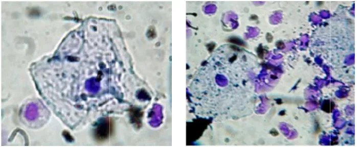
1- Fig. 2 reflects apoptotic changes in epithelial cells of heavy smokers characterized with fragmented nuclei and Formation of blabbing in the nuclear envelope compared with normal cells in non-smokers in Fig. 1.
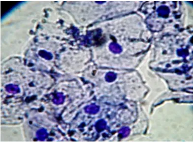
Fig. 1. Normal epithelial cells in cell lining mouth of non-smokers, stained with
Giemsa stain, (400X)
2- Due to these changes, gum easily invaded via micro-organism and finally infiltrated with inflammatory cells (Fig. 3).
4. DISCUSSION
Tobacco smoking is a global problem with the number of tobacco smokers estimated at about 1.3 billion [7]. Tobacco smoking not only increases the risk of system ic disease, but it also contributes to the risk of cancerogenesis [8].
It has long been recognized that there is a strong association between heavy cigarette smoking and gum disease. The gum disease affects gums and bones that support our teeth [9,10]. The immune system has an important role in reducing the harmful foreign bodies that can cause this disease. Smoking cigarettes might affect our immune system, making our body less able to fight against this micro-organism [11]. For this reason smoker gums became more likely to get inflamed, infected and cause problems by attaching to the area where the gum is joined to
the tooth causing deep spaces to form called periodontal pockets. These spaces mean there is less support to hold the tooth in place and more chance of losing smokers their tooth, lead to bleeding gums which are a sign of this problem [12]. However, in smokers, nicotine decreases the blood flow to the gums and this poor blood supply can hide symptoms (like bleeding gums) that would normally give a clue to the start of this disease [13].
Fig. 2. Apoptotic cells in in cell lining mouth of smokers, stained with Giemsa stain, (400X)
Very little is known about the possible impact of tobacco smoke on the apoptosis of epithelial cells and observations vary, from indicating no changes on the level of apoptosis to suggesting increased levels of hypodiploid cells [14,15] in the epithelium of smokers through the inhibition of apoptosis under influence of nicotine [16,17]. However, these observations are based on In vitro cultured keratinocytes and animal models.
Our results showed an increment in apoptotic percent as a result of DNA changes, since DNA is the cell’s “instruction manual.” It controls a cell’s normal growth and function. When DNA is damaged, a cell can begin growing out of control and create a cancer tumor. This happens because toxics in tobacco smoke can destroy or change the cell’s instructions.
Table 1. Represent the mean percentage of normal and apoptotic cells in smokers and non smokers mean percentage of normal cells
Mean percentage of apoptotic cells
* ** *** Total
1.5 4
7 100
11.1 3.1
25.2 100
*No. of apoptotic cells with condense chromatin material.; **No. of apoptotic cells with Formation of blabbing in the
Fig. 3. Accumulation of inflammatory cells in lining stain,
research shows that the Cigarette composed of more than 7000 chemicals. Many of them are toxics in cigarette smoke weaken immunity defense mechanism [18]. When this happens, cells keep growing without being stopped. For this reason, smoking can cause cancer. Sever investigation indicate that oral cancers arise as a result of mutagenic events (arising mainly from tobacco and alcohol) causing multiple molecular genetic events in many chromosomes and genes [19]. The consequence of this chromosomal (genetic) damage is the impairment of cell regulatory processes leading to evading apoptosis.
Our findings indicated that long-term smoking significantly affects oral mucosa. It has been concluded the following:
1- Smoking has marked effects on oral lining mucosa increases apoptotic processes. 2- Oral mucosa became suitable milieu for
micro-organism development due to the increment in degeneration and apoptosis percent.
3- Smoking has marked effects on the DNA of oral mucosa since apoptosis was increased in smoker.
Table 1. Represent the mean percentage of normal and apoptotic cells in smokers and non smokers mean percentage of normal cells
Mean percentage of normal cells
Mean of necrotic cells
Non-smokers 83b
4.5a
Smokers 33b
27.6a
of apoptotic cells with condense chromatin material.; **No. of apoptotic cells with fragmented nuclei.;*** No. of apoptotic cells with Formation of blabbing in the nuclear envelope.; a,bValues are significantly different (
0.001) based on Chi square test
flammatory cells in lining mouth of smokers, stained with Giemsa stain, (400X). Inflammatory cells
shows that the Cigarette composed of more than 7000 chemicals. Many of them are toxics in cigarette smoke weaken immunity defense mechanism [18]. When this happens, cells keep growing without being stopped. For this reason, smoking can cause cancer. Several investigation indicate that oral cancers arise as a result of mutagenic events (arising mainly from tobacco and alcohol) causing multiple molecular any chromosomes and genes The consequence of this chromosomal s the impairment of cell regulatory processes leading to evading
term smoking significantly affects oral mucosa. It has been
Smoking has marked effects on oral lining apoptotic processes. Oral mucosa became suitable milieu for
organism development due to the increment in degeneration and apoptosis
Smoking has marked effects on the DNA of oral mucosa since apoptosis was
5. CONCLUSION
From this study it has been concluded that smoke of Tobacco has direct effects on the DNA of epithelial cells lining mouth, which lead to carcinogenic risk.
CONSENT
All authors declare that ‘written informed consen was obtained from the patient for publication of this case report and accompanying images.
ETHICAL APPROVAL
All authors hereby declare that all experiments have been examined and approved by the appropriate ethics committee and have therefore been performed in accordance with the standards laid down in the 1964 Declaration of Helsinki.
COMPETING INTERESTS
Authors have declared that no competing interests exist.
Table 1. Represent the mean percentage of normal and apoptotic cells in smokers and
non-of normal
smokers Smokers
of apoptotic cells with fragmented nuclei.;*** No. Values are significantly different ( p<
mouth of smokers, stained with Giemsa
From this study it has been concluded that smoke of Tobacco has direct effects on the DNA of epithelial cells lining mouth, which lead to
All authors declare that ‘written informed consent for publication of this case report and accompanying images.
All authors hereby declare that all experiments have been examined and approved by the appropriate ethics committee and have therefore been performed in accordance with the ethical e 1964 Declaration of
Al-Kasiy et al.; BJMMR, 8(3): 256-260, 2015; Article no.BJMMR.2015.446
REFERENCES
1. Gandini S, Botteri E, Iodice S, Boniol M, Lowenfels AB, Maisonneuve P, Boyle P. Tobacco smoking and cancer: A meta-analysis. Int J Cancer. 2008;122(1):155-64.
2. Policy statements: Oral diseases related to tobacco use. The International Association
for Dental Research website.
Available:http://www.iadr.com/i4a/pages/in dex.cfm?pageid=3465#TOBACCO (Accessed 5 September 2011).
3. Warnakulasuriya S. Global epidemiology of oral and oropharyngeal cancer. Oral Oncol. 2009;45:309-316.
4. Gandini S, Botteri E, Iodice S, et al. Tobacco smoking and cancer: A meta-analysis. Int J Cancer. 2008;122(1):155-164.
5. Llewellyn CD, Johnson NW,
Warnakulasuriya KA. Risk factors for oral cancer in newly diagnosed patients aged 45 years and younger: A case-control study in Southern England. J Oral Pathol Med. 2004;33(9):525-532.
6. Hanahan D, Weinberg RA. The hallmarks of cancer. 2000;100(1):57-70.
7. Thun M, Peto R, Boreham J, Lopez AD. Stages of the cigarette epidemic on entering its second century. Tob Control. 2012;21:96–101.
8. Gagat M, Grzanka D, Izdebska M, Maczynska E, Grzanka A. Nornicotine impairs endothelial cell-cell adherens junction complexes in the EA. hy 926 cell line via structural reorganization of F-actin. Folia Histochem Cytobiol. 2013;51(3):179– 192.
9. Warnakulasuriya S. Global epidemiology of oral and oropharyngeal cancer. Oral Oncol. 2009;45:309-316.
10. Zygogianni Anna G, Kyrgias George, Karakitsos Petros, Psyrri Amanta, Kouvaris John, Kelekis Nikolaos, Kouloulias Vassilis. Oral squamous cell cancer: Early detection and the role of
alcohol and smoking. Head & Neck Oncology. 2011;3:2.
11. Palmer RM, Wilson RF, Hasan AS, et al. Mechanisms of action of environmental
factors-tobacco smoking. J Clin
Periodontol. 2005;32(6):180-195.
12. Dietrich T, Maserejian NN, Joshipura KJ, et al. Tobacco use and incidence of tooth loss among US male health professionals. J Dent Res. 2007;86(4):373-377.
13. Maryam Rad, Shahla Kakoie, Fateme Niliye Brojeni, Nasim Pourdamghan. Effect of long-term smoking on whole-mouth salivary flow rate and oral health. J Dent Res Dent Clin Dent Prospect. 2010;4(4): 110-114.
14. Banerjee AG, Gopalakrishan VK,
Vishvanatha JK. Inhibition of nitric oxide-induced apoptosis by nicotine in oral epithelial cells. Mol Cell Biochem, 2007; 305:113–121.
15. Donetti E, Gualerzi A, Bedoni M, et al. Desmoglein 3 and keratin 10 expressions are reduced by chronic exposure to cigarette smoke in human keratinized oral mucosa explants. Arch Oral Biol. 2010;10:815–823.
16. Schwartz J, Muscat J, Baker V, et al. Oral cytology assessment by flow cytometry of DNA adducts, aneuploidy, proliferation and apoptosis shows differences between smokers and non-smokers. Oral on Col. 2003;39:842–854.
17. Assis GF, Ceolin DS, Marques ME, Salvadori DM, Ribeiro DA. Cigarette smoke affects apoptosis in rat tongue mucosa: Role of Bcl-2 gene family. J Mol Hist. 2005;36:483–489.
18. Ryder MI. The influence of smoking on host responses in periodontal infections. Periodontol. 2007;43:267-277.
19. Stone JG, Jones NJ, Mc. Gregor AD, et al. Development of a human biomonitoring assay using buccal mucosa: Comparison of smoking-related DNA adducts in mucosa versus biopsies. Cancer Res. 1995;6:1267-1270.
© 2015 Al-Kasiy et al.; This is an Open Access article distributed under the terms of the Creative Commons Attribution License
(http://creativecommons.org/licenses/by/4.0), which permits unrestricted use, distribution, and reproduction in any medium,
provided the original work is properly cited.
Peer-review history:
The peer review history for this paper can be accessed here: