DEVELOPMENT AND HOMEOSTASIS
Caroline Hutter
UNIVERSITY COLLEGE LONDON
AND
IMPERIAL CANCER RESEARCH FUND
ICRP supervisor: Dr Fiona M. Watt UCL supervisor: Dr Josephine C. Adams
A thesis submitted in partial fulfilment of the requirements for the
degree o f Doctor o f Philosophy, University o f London
All rights reserved
INFORMATION TO ALL USERS
The quality of this reproduction is dependent upon the quality of the copy submitted.
In the unlikely event that the author did not send a complete manuscript and there are missing pages, these will be noted. Also, if material had to be removed,
a note will indicate the deletion.
uest.
ProQuest U643423
Published by ProQuest LLC(2016). Copyright of the Dissertation is held by the Author.
All rights reserved.
This work is protected against unauthorized copying under Title 17, United States Code. Microform Edition © ProQuest LLC.
ProQuest LLC
789 East Eisenhower Parkway P.O. Box 1346
Integrins are a ubiquitously expressed family of cell surface receptors that mediate cell- matrix and in some cases also cell-cell adhesion. In addition to just anchoring the cell to its surroundings, they play a crucial role in tissue morphogenesis and maintenance. In the epidermis p i integrins have been implicated in regulating keratinocyte proliferation and
differentiation.
The aim of the work presented in this thesis is to elucidate the role of p i integrins in the
epidermis by exploiting cells that lack the p i subunit. Since deletion of the p i integrin gene in mice causes embryonic lethality before the development o f the epidermis, I have taken two experimental approaches: Differentiation of pi-null embryonic stem (ES) cells
into kératinocytes in vitro and generation of pi-null kératinocytes via the Cre/Lox system.
ES cells that are homozygous null for the pi integrin subunit fail to differentiate into kératinocytes in vitro but do differentiate in teratomas and wild-type/p 1 -null chimeric mice. I found that the impaired differentiation is due to a reduced sensitivity o f pi-null ES cells to soluble growth factors in comparison to their wild-type counterparts. I showed that pi-null ES cells can be partially rescued by factors that are secreted by dermal
fibroblasts. I could furthermore demonstrate, that TGFa, FGFIO and KGF had an inductive effect on keratinocyte differentiation in vitro.
By isolating kératinocytes from mice with a floxed pi integrin gene that expressed Cre under the keratin 5 promoter and by expressing EGFP-Cre in fl/fl kératinocytes using retoviral infection, I could study the behaviour of pi-null kératinocytes in vitro. I showed that these cells have a reduced ability to adhere to various extracellular matrices and that they are not able to spread and to migrate due to a failure to organize the actin
cytoskeleton and form focal adhesions. In addition, I demonstrated that pi-null
Abstract 3
Table of Contents 4
List of Figures 7
List of Tables and Illustrative Material 9
Abbreviations 10
Acknowledgements 11
CHAPTER 1: INTRODUCTION 12
1.1. STRUCTURE AND FUNCTION OF THE SKIN 12
1.1.1. Epidermis and its appendages 12
1.1.2. The dermo-epidermal j unction/ basement membrane 16
1.1.3. Dermis 17
1.2. EMBRYONIC STEM CELLS 18
1.2.1. What are embryonic stem cells ? 18
1.2.2. In vitro differentiation of ES cells 20
1.3. INTEGRINS AND THEIR LIGANDS 22
1.3.1. Structure of integrins 23
1.3.2. Functional properties of integrins 28
1.3.3. Keratinocyte integrins 3 0
1.4. AIMS OF THESIS 35
CHAPTER 2: MATERIALS AND METHODS 42
2.1. CELL CULTURE 42
2.1.1. General cell culture solutions 42
2.1.2. Cultured cell types 43
2.1.3. Isolation and culture of primary mouse embryo fibroblasts 44
2.1.4. D3 and G201 ES cells 44
2.1.5. De-epidermised dermis (DED) culture 46
2.1.6. Epidermal kératinocytes 46
2.1.7. Retroviral producer cells and retoviral infection of keratinoctes 49 2.1.8. Preparation of adenovirus and adenoviral infection o f kératinocytes 51
2.1.10. Adhesion assays using freshly isolated mouse kératinocytes 53
2.2. IMMUNOLOGICAL METHODS 54
2.2.1. General solutions 54
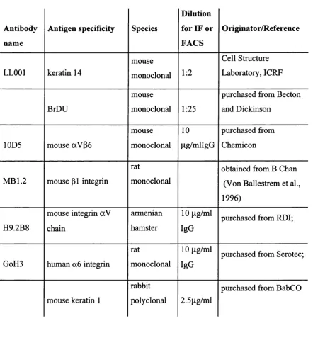
2.2.2. Antibodies 55
2.2.3. Preparation of cells for immunofluorescence staining 58 2.2.4. Preparation of cryosections of DED and mouse epidermis 58
2.2.5. Immunofluorescence staining protocol 59
2.2.6. Flow cytometry 61
2.3. PROTEINS 62
2.3.1. Extraction of total proteins 62
2.3.2. Measuring protein concentration 62
2.3.3. SDS PAGE and immunoblotting 62
2.3.4. Staining for lacZ activity 66
2.4. MOLECULAR BIOLOGY 67
2.4.1. Stocks and general solutions for DNA techniques 67
2.4.2. General DNA techniques 69
2.5. LIST OF SUPPLIERS AND DISTRIBUTORS 74
CHAPTER 3: SOLUBLE GROWTH FACTORS RESTORE THE ABILITY OF
P1 INTEGRIN DEFICIENT ES CELLS TO DIFFERENTIATE INTO
KERATINOCYTES 76
3.1. INTRODUCTION 76
3.2. RESULTS 77
3.2.1. In vitro differentiation of p i null cells 77 3.2.2. Basement membrane does not stimulate ES cell differentiation into
kératinocytes 77
3.2.3. Fibroblasts stimulate ES cells to differentiate into kératinocytes 78 3.2.4. ES cell differentiation into kératinocytes is stimulated by secreted
fibroblast factors 80
3.2.5. Effects o f specific growth factors on ES cell differentiation 81
3.3. DISCUSSION 82
4.1. INTRODUCTION 108
4.2. RESULTS 109
4.2.1. Isolation of kératinocytes from the K5Cre fl/fl p i fl/fl mouse and
genotyping 108
4.2.2. FACS analysis of integrin levels 108
4.2.3. Morphology of p i null kératinocytes in culture: Impaired adhesion
and spreading of p 1 null kératinocytes 110 4.2.4. p i null kératinocytes exhibit reduced proliferation and increased
differentiation in vitro 111
4.2.5. Infection of mouse kératinocytes with an adenovirus vector
expressing Cre recombinase 113
4.2.6. Expression of Cre recombinase using retrovirus. 114
4.3. DISCUSSION 117
CHAPTERS: GENERAL DISCUSSION 162
5.1. DERMAL FIBROBLAST - DERIVED GROWTH FACTORS RESTORE THE ABILITY OF p i INTEGRIN DEFICIENT ES CELLS TO DIFFERENTIATE
INTO KERATINOCYTES 162
5.2. ADHESION, SPREADING, AND MOTILITY OF P 1 NULL
KERATINOCYTES IS SEVERELY COMPROMISED 164
5.3. p i NULL KERATINOCYTES HAVE A LOWER PROLIFERATION RATE THAN WILD TYPE KERATINOCYTES AND UNDERGO TERMINAL
DIFFERENTIATION IN VITRO 165
Figure 1.1 The skin 36
Figure 1.2 Integrin mediated cell adhesion 38
Figure 1.3. In vitro differentiation of ES cells 40
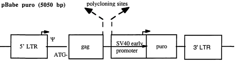
Figure 2.1 Retroviral vector pBabepuro 71
Figure 3.1. Wild type, but not p i null embryoid bodies differentiate into kératinocytes in
vitro. 88
Figure 3.2. Differentiation of embryoid bodies on DED. 90 Figure 3.3. Immunofluorescence staining of cysts formed by wild type ES cells seeded
onto DED. 92
Figure 3.4. Collagen type IV distribution in DED. 94 Figure 3.5. The presence of dermal fibroblasts stimulates ES cells to differentiate into
kératinocytes 96
Figure 3.6. Immunofluorescence staining with anti vimentin of a (31 null ES culture on
DED containing fibroblasts. 98
Figure 3.7. Types of keratin positive ES cell groups. 100 Figure 3.8. Effects of coculture with HDFs on differentiation of ES cells into keratin
positive cells. 102
Figure 3.9. Keratin positive ES cell groups formed by wild type embryoid bodies
cocultured with HDF. 104
Figure 3.10. Effects o f specific growth factors on ES differentiation 106 Figure 4.1. Conditional (31 integrin knockout 121 Figure 4.2. Surface expression of integrins before and after deletion o f p i integrin. 123
Figure 4.3. p i null kératinocytes exhibit impaired adhesion and spreading. 125 Figure 4.4. Loss of focal adhesions and stress fiber formation in p i null kératinocytes. 127
Figure 4.5. Adhesion of wild type vs p i integrin deficient kératinocytes. 129 Figure 4.6. p i integrin deficient kératinocytes incorporate less BrdU, but do not
apoptose. 131
Figure 4.10. Cre recombinase is efficiently introduced into kératinocytes by adenoviral
infection. 139
Figure 4.11. Adenovirus has toxic side effects at the MOI needed for efficient
downregulation of P 1 integrin. 141
Figure 4.12. FACS analysis of p i integrin. 143
Figure 4.13. Immunofluorescence staining o f fl/fl kératinocytes infected with
EGFP Cre. 145
Figure 4.14. Downregulation o f pi integrin after infection with EGFPCre. 147 Figure 4.15. Effects of retroviral infection with EGFPCre on mouse kératinocytes. 149
Figure 4.16. FACS for p i integrin. 151
Figure 4.17. Phase photograph o f fl/fl kératinocytes infected with EGFPCre. 153 Figure 4.18. Phase and fluorescence photographs of different frames. 155 Figure 4.19. Chick p i integrin expression in fl/fl kératinocytes. 157 Figure 4.20. Downregulation of mouse pi integrin after infection with EGFPCre in fl/fl
and fl/fl + chick p i integrin kératinocytes 159 Figure 4.21. Chick p i integrin transduced kératinocytes remain spread after infection
Table 2.1. Primary antibodies 55
Table 2.2. Secondary antibodies 57
Table 3.1. Statistical analysis (p values) of the results of all experiments with specific
growth factors 107
AM 12 gag pol + env AM 12 packaging ceils
bp base pairs
BrdU 5 ’ bromodeoxyuridine
BSA bovine serum albumin
CD cluster o f differentiation antigen
DAB 3,3 -diaminobenzedene tetrahydrochlodride
DCS donor ca lf serum
DED de-epidermised dermis
DEPC diethylpyrocarbonate
DMEM D ulbecco’s modification o f Eagle’s medium
DMSO dimethyl sulphoxide
DNase deoxyribonucleic acid endonuclease
dNTP deoxynucleotide triphosphate
ECM extracellular matrix
EDTA ethyldiaminotetraacetic acid, disodium salt
EOF epidermal growth factor
EGFP enhanced green fluorescent protein
FACS fluorescence activated cell sorter
FAD F I2 + adenine + DMEM
FCS foetal ca lf serum
FITC fluorescein isothiocyanate
OPE gag pol + env86 packaging cells
H&E haematoxylin and eosin staining
HICE hydrocortisone, insulin, cholera enterotoxin and EOF,
HRP horseradish peroxidase
IF keratin intermediate filaments
KIO keratin 10
kDa kilo Dalton
LDH lactate dehydrogenase
LRC label retaining cell
LTR long terminal repeats
MAPK mitogen activated protein kinase
MMTV M olony murine tumour virus
MOI multiplicity o f infection
Mo MuLV M olony murine leukemia virus
N-term amino terminus
O.D. optical density
PBS phosphate buffered saline
PBST PBS/Tween
PCR polymerase chain reaction
pen/strep penicillin/streptomycin
PFU plaque-forming units
PMSF phenylmethanesulphonyl fluoride
PVDF polyvinylidene fluoride
RNase ribonucleic acid endonuclease
rpm revolutions per minute
S.D. standard deviation
SDS sodium dodecyl sulphate
SSC salt sodium citrate buffer
TAE Tris-acetate-EDTA buffer
TBE Tris-borate-EDTA buffer
TCA Trichloroacetic Acid
TEMED N,N,N',N'-tetramethylethylenediamine
TM transmembrane domain
I am grateful to everyone who has helped me during my four years at the ICRP. In particular, I would like to thank Fiona, my supervisor, for giving me the opportunity to work in her laboratory and for her guidance, support and encouragement.
I want to thank all members of the Keratinocyte Laboratory for sharing ideas, bench space, and sometimes even ‘snail cakes’. I am especially grateful to Liz and Simon for looking after me, to my Swiss collaborator Claudia, and to Sally for taking me to Nepal. I also want to thank Teresa for being my bench-neighbour and friend. Thanks also to Catherin, Karen, Richard, Robin, David (O&P), Douglas, James, Sam and Ela.
I also want to thank members from the FACS and the Videomicroscopy laboratories, especially Debbie and Colin, for their assistance.
CHAPTER 1: INTRODUCTION
Integrins are a ubiquitously expressed family of cell surface receptors that mediate cell- matrix and in some cases also cell-cell adhesion. In addition to just anchoring the cell to its surroundings, they play a crucial role in tissue morphogenesis and maintenance. In the epidermis p i integrins have been implicated in regulating keratinocyte proliferation and differentiation. This introduction will begin with a brief overview of the structure and function of the skin, followed by a short summary of ES cell culture. Finally, integrins and their role in the epidermis will be discussed.
1.1. STRUCTURE AND FUNCTION OF THE SKIN
The skin is the largest organ in the body. It consists o f two layers which develop from different germ layers. The outer layer, the epidermis, is a stratified squamous epithelium derived from the ectoderm. The inner layer, the dermis, consists of connective tissue and is of mesodermal origin. Epidermis and dermis are separated by a basement membrane.
1.1.1. Epidermis and its appendages
Interfollicular epidermis
The epidermis is a squamous stratified epithelium. It is made almost entirely of kératinocytes (95%). Other cell types found in the epidermis include melanocytes, Langerhans cells
(dendritic cells) and Merkel cells (sensory receptors).
weeks estimated gestational age (EGA) in humans and between days 13 and 16 days in mice, after the embryonic - fetal transition, the epidermis becomes three layered and an
intermediate layer is formed (Hertle et ah, 1991; Fuchs, 1994; Weiss and Zelickson, 1975). During the early stages of development mitotic activity occurs in all layers (Fuchs, 1994), but as suprabasal cells begin to display morphological signs of differentiation, mitotic activity becomes restricted to the cells in the basal layer. On day 16 of mouse development, the intermedium layer is replaced by the strata spinosum and granulosum, and by day 17 the first comified cells are observed. In human development, it takes about 24 weeks until all the epidermal layers are formed (Hertle et al., 1991).
The same pattern is observed in adult mammalian epidermis. Considering the skin's main function, to form an almost impenetrable barrier, and looking at a histological section might give the impression that the epidermis is a static tissue, but the opposite is the case. The epidermis is a very dynamic epithelium that is constantly being renewed, a process that involves much cell movement and trafficking from different compartments: there is horizontal movement of the undifferentiated cells in the basal layer and vertical movement up to the surface o f the differentiating cells. In hairy skin, the movement is probably even more complex due to a contribution of cells from the hair follicle to the interfollicular epidermis (Jensen et al., 1999).
Morphologically, adult epidermis can be subdivided into four different layers that correspond to different stages o f keratinocyte differentiation (Figure 1 D):
Stratum basale
Markers for the basal layer include the keratins K5 and K14, which form a heterodimer and are expressed in the basal cells of all stratified squamous epithelia (Fuchs, 1994).
Stratum spinosum
Kératinocytes that have committed themselves to terminal differentiation move out of the basal layer and enter the stratum spinosum, which consists of multiple cell layers. The name for this cell layer is derived from the intercellular bridges that are connected by
demsmosomes and look like spines under the light microscope. Cells in this layer (which have lost contact to the basement membrane) become more polygonal and start to flatten parallel to the skin surface. They synthesise differentiation specific keratin filaments and precursors of the comified envelope: The keratins K1 and KIO are expressed instead of K5 and K14 directly above the basal layer and involucrin, a precursor of the comified envelope is synthesised in the upper spinous layers of the epidermis. As the comified envelope is formed, involucrin becomes crosslinked in the comified envelope.
Stratum granulosum
Differentiation continues in the granular layer, as cells and synthesise profilaggrin, precursor of filaggrin, which is important for the aggregation of keratin filaments. Profilaggrin is found in keratohyalin granules that are abundant in these cells and give this layer its name.
Stratum comeum
Epidermal appendages
The epidermal appendages are derived from epithelial germs that are formed during
embryogenesis and lie (with the exception of nails) in the dermis. In mammals they include hair, sebaceous and sweat glands, and nails.
The formation of the appendages is dependent upon epithelial-mesenchymal interaction. The mesenchymal cells of the dermis have an inductive effect on the epidermal cells and
determine the regional character of the skin (Dillingham and Silvers, 1967; Hardy, 1992; Oshima et al., 2001)
The hair follicle contains dermal (dermal papilla and connective tissue sheath) and epidermal (matrix and inner and outer root sheaths) components. It has received much attention in the past years. Mutations and/or epidermal overexpression of developmentally regulated genes have primarily led to severe impairment of hair follicles (Gat et al., 1998; St-Jacques et al., 1998; Zhou et al., 1995; Huelsken et al., 2001). Furthermore, it has been proposed that the hair follicle is a major repository of keratinocyte stem cells in mouse skin (Taylor et al.,
2000).
The development o f the hair follicle can be divided into different stages. First the dermis initiates in the overlying epidermis the formation of an appendage (‘first dermal message’). The epidermis responds by beginning to form hair, feather buds or scale placodes- which appendage is actually formed depends on the origin o f the epidermis, as dermal-epidermal recombination experiments between different classes have shown (Sengel and Mauger,
Once the hair is formed, its lower part undergoes cyclic changes of growth and shedding. The three phases of follicular activity are known as growth (anagen; Figure 1.1 A), regression (catagen) and rest (telogen) (Pans et al., 1999). It is thought that the hair cycle in the adult reproduces some of the embryonic morphogenetic events (Oshima et al., 2001). At every start of the hair cycle, stem cells which reside in a specialised region o f the outer root sheath known as the bulge come in contact with mesenchymal dermal papilla cells and are
stimulated to exit this compartment and migrate down in the outer rooth sheath (ORS) to the hair bulb. While migrating, they contribute to the ORS differentiated layers and once they reach the tip of the hair follicle, they then commit themselves to become inner root sheath (1RS) and hair forming progenitors (Oshima et al., 2001). The catagen is initiated by FGF signalling (Hebert et al., 1994) and results in loss of proliferative activity in the matrix cells, leading to a cessation o f hair elongation. During catagen, the lower (transient) portion of the hair follicle degenerates. In the third phase (telogen) the hair follicle is at rest and no
morphological changes are detectable. In mice the first coat appears at day 4.5 after birth and results from growth of hair formed during embryogenesis. In contrast to humans, hair growth in mice is synchronised. Telogen is reached at day 16 after birth and the first postnatal cycle begins when mice are 4 weeks old.
1.1.2. The dermo-epidermal junction/ basement membrane
The basement membrane separates the epidermis from the underlying connective tissue. It is composed of highly organised extracellular matrix and plays a critical role in organising the skin which goes beyond the provision structural support. In the skin the basement membrane determines cell polarity and has a critical influence on the choice of cell fate. Assembly of a proper basement membrane is dependent on epidermal-mesenchymal interactions (Bohnert et al., 1986).
nidogen. In skin, also type VII collagen is important. It forms the anchoring fibrils that link type IV collagen in the basement membrane to structures called anchoring plaques
(Burgeson, 1993). Fibronectin is abundantly expressed in the dermis and the dermo- epidermal junction, but in normal adult skin there is little fibronectin expressed in the basement membrane itself (Couchman et al., 1990).
Nidogen is a single polypeptide chain that is folded into three globular domains connected by a rod or flexible link. It binds laminin, collagen IV, perlecan and calcium and is essential for connecting the networks of laminins and collagen IV in basement membranes (Dedhar et al.,
1992).
Perlecan is the largest and most common proteoglycan in basement membranes. It serves many functions, including cell attachment, basement membrane assembly and binding of bFGF (Aviezer et al., 1994). Proteoglycans are a diverse family that is characterized by the covalent attachment of one or more glycosaminoglycans (GAG) side chains to a core protein. The highly acidic and hydrophilic GAG (repeating disaccharides) chains have a major
influence on tissue hydration and elasticity, and can show high affinity binding to various ligands.
1.1.3. Dermis
1.2. EMBRYONIC STEM CELLS
1.2.1. What are embryonic stem cells ?
Embryonic stem (ES) cells are pluripotent, non-transformed stem cell lines that are derived from cells of the early epiblast, and can be propagated in culture (Brook and Gardner, 1997). Mouse embryonic stem cells were first isolated from inner cell masses by Evans and
Kaufman (Evans and Kaufman, 1981) and Martin (Martin, 1981). A few years later it was shown that they are able to generate cells o f all lineages when allowed to differentiate and that they are capable o f contributing to many different tissues in chimeric mice, including the germline, when they are transferred back into mouse blastocysts (Bradley et al., 1984).
The concept of a pluripotent cell emerged from work on mouse teratocarcinomas, germ cell tumors that form various somatic tissues. Work in the 1960s and 1970s on cell lines
established from these tumors, embryonal carcinoma (EC) cells, showed that they can serve as models for mammalian development, since they have the potential to differentiate in cells of all three embryonic germ layers in vivo and in vitro (Martin, 1980). EC cell lines that can differentiate derivatives from all three germ layers were also isolated from human
teratocarcinomas (Andrews et al., 1984; Fera et al., 2000)
What defines an embryonic stem cell? One o f the most important features of ES cells is their pluripotency. In mammals it was believed until recently that only the oocyte, zygote, early embryonic cells, primordial germ cells and tumours derived from them have the potential to differentiate into dérivâtes of all three embryonic germ layers (Desbaillets et al., 2000). A simple model would therefore be that a pluripotent cell from the ICM gives rise to committed daughter cells which are restricted to differentiate along specific lineages.
cells) give rise to all three germ layers (Kondo et ah, 2000; Krause et ah, 2001). These experiments suggest two possibilities. One is that tissue specific stem cells might able to 'transdifferentiate' i.e. reprogramme their phenotype when challenged with a new
environment. Another possibility is that there is no intrinsic difference between organ- specific and embryonic stem cells and that the concept of tissue-specific stem cells being restricted in their developmental potential is wrong (Anderson et ah, 2001; Morrison et ah, 2000; Lowell, 2000). One could carry this concept even further and say that being a stem cell is a fimction and not an entity and all cells have a decreasing but recruitable propensity to act as stem cells as they differentiate (Blau et ah, 2001).
However, in contrast to the somatic cells isolated from adult tissue, both ES and embryonic germ (EG; derived from genital ridges of fetuses) (Matsui et ah, 1992) can be cultured in an undifferentiated state for an unlimited period of time without losing their differentiation potential. This characteristic also distinguishes ES from EC cells, which often exhibit karyotype instabilities and variations in the ability to differentiate in multiple tissues (Thomson et ah, 1998).
Taken together, the following criteria can be used to define an ES cell: (i) prolonged undifferentiated proliferation, (ii) clonally derived cultures that have the potential to form derivatives of all three germ layers, (iii) stable karyotype, (iv) derived from pre - or periimplantation embryo.
ES cells can be derived by isolating mouse blastocyts and placing them in culture where, after 5-6 days, the inner cell mass (ICM) has proliferated so much that it is easily recognised. The ICM is then removed from the trophoblast outgrowth and dissociated into smaller aggregates. Two days after the dispersal of the cells colonies o f cells with different morphologies can be observed. The stem-cell-like cells, which can be identified by their appearance and the fact that they proliferate without changing their phenotype, are then selectively removed. The resulting ES cell lines can divide indefinitely without
factor). LIF is a cytokine that acts through a receptor complex composed of a low affinity LIF receptor (LIFRp) and gpl30 (Nakashima and Taga, 1998). One of LIF's downstream
effectors is STAT3, whose activity seems to be sufficient to keep ES cells in the undifferentiated state (Matsuda et al., 1999).
ES cells have revolutionised mammalian genetics because they have provided the basis for random mutagenesis and homologous recombination in mice. In addition to their use as a genetic tool, mouse ES cells are presently the only stem cell population that can be maintained indefinitely in vitro without differentiating. Thus, they provide a resource for investigating the parameters of stem cell growth and the factors that sustain proliferation at the expense of differentiation. Furthermore, with the isolation of human ES cells (Thomson et al., 1998; Reubinoff et al., 2000), translating stem cell research into clinical applications ('therapeutic cloning') has become a widely discussed prospect (Winston, 2001; Antoniou, 2001). In vitro differentiation of ES cells
ES cells can differentiate into multiple lineages in culture. When the factors that keep them in an undifferentiated state are removed, ES cells differentiate spontaneously and - if cultured under appropriate conditions - generate cells of various lineages. The in vitro differentiation of ES cells provides a powerful system to analyse the events that take place during
There exist various protocols for the in vitro differentiation of ES cells. The general principle of all these methods is that the ES cells are removed from their feeder cells or the presence of LIF, and then cultured in a way that allows the formation o f so called embryoid bodies, multicellular structures that contain elements of all three germ layers. This can be achieved by placing the cells in suspension either in methylcellulose containing media or by culturing the ES cells in ’hanging drops’ (Figure 1.3). In some protocols the cells are also cultured directly on stromal cells rather than putting them in suspension first. Once embryoid bodies have formed, they can be transferred to standard culture conditions on tissue culture plastic for studies on adhesive cell development, in suspension cultures or be transplanted into recipient mice.
If embryoid bodies are kept in vitro, a wide range of cell types start to evolve, among them hematopoietic, neuronal, epithelial, muscle and endothelial cells. Interestingly, the cells that differentiate in vitro seem to undergo the same developmental program as their counterparts do to form the embryo in vivo (Keller, 1995).
One major drawback o f the differentiation that takes place in the embryoid bodies is that it is difficult to control and that until now it has not been possible to direct differentiation to only one cell type. However, in the last years immense progress has been made towards this end (Keller, 1995; Lumelsky et al.; 2001; Schuldiner et al., 2000). A range of growth factors has been identified that at least alter the relative proportions of a specific cell type: TGF-p for
example has been shown to induce myogenesis (Rohwedel et al., 1994); retinoic acid directs cells towards neuronal differentiation (Bain et al., 1995), and accelerates cardiac
differentiation (Wobus et al., 1997).
(Oshima et a l, 1983). K14 is first expressed in the single layered ectoderm (which will subsequently differentiate into epidermis) of mouse embryos, from E9.5 onwards in some cells, and from day 14.5 onwards K14 is expressed in all cells of the basal layer (Fuchs,
1994). The regional variation in sites of expression at day 9.5 is consistent with an inductive role of the underlying mesenchyme (Byrne et al., 1994).
K l and KIO are first detected at E l 3.5, as the epidermis begins to differentiate into spinous and granular layers (Byrne et al., 1994). We have previously reported that there is a temporal sequence in the appearance of epithelial markers in wild-type ES cultures: K8 and K18 are expressed first, followed by K5/K14, then Kl/KlO and another epidermal terminal
differentiation marker, involucrin (Bagutti et al., 1996). It thus seems likely that the K14- positive cells that differentiate in ES cell cultures correspond to embryonic ectoderm.
1.3. INTEGRINS AND THEIR LIGANDS
Integrins regulate development and terminal differentiation in many cell types (Hynes, 1992; Hynes et al., 1992; Adams and Watt, 1993; Streuli et al., 1991). Integrins are large cell surface receptors found on almost all cells of multicellular organisms. They were discovered in the early 1980s (Tarone et al., 1982; Neff et al., 1982; Pytela et al., 1985), but not cloned until the mid 1990s (Hemler, 1999).
Integrins seem to be the main receptors by which cells adhere to extracellular matrix. In addition to ECM ligands many integrins have been shown to bind in other cell surface molecules. One distinctive feature of integrins is that cells can regulate their ligand binding activity.
1.1.1. Structure of integrins
many integrin subunits, including a i , 6 ,1 and P l, 3, 5 cytoplasmic domains and a6 ,1 and p3 extracellular domains (de Melker and Sonnenberg, 1999).
Structural knowledge of integrins is still limited. Until recently (with the exception of the I domain, a region in the a subunit which folds independently and can be expressed in
recombinant form) the tertiary structure o f integrins was not known. There are reports that the tertiary structure o f avP3 has been solved (Science 293, 2001), but the original article
was not yet available at the time of writing the thesis. Electron microscopy of integrins suggests that integrins have a globular head region that is connected to the membrane via two stalks (Nermut et al., 1988). The long extracellular domains o f both the a and P subunits are responsible for ligand binding and subunit association, but the cytoplasmic tails also play a role in heterodimer association and the regulation of ligand binding (Takagi et al., 2001 ; Hughes and Pfaff, 1998). It was found that the membrane proximal association of both cytoplasmic tails via complementary negatively and positively charged residues constrains the integrin in an inactive state whereas lack of association in the membrane proximal regions activates the integrin.
The a subunit
The a subunits are composed of 1000-1150 amino acids and their mature size is 140-210 kDa. All integrin a subunits have seven amino terminal repeating sequences that fold into a p-propeller motif that is similar to that found in the p subunits of heterotrimeric G proteins (Springer, 1997).
The a l , a l , a L ,ccm, a x aD and a E subunits (Humphries et al., 2000; Humphries, 2000b; Hemler, 1999) contain an inserted domain (I domain) of about 200 amino acids between P
sheets 2 and 3 o f this P-propeller domain. Three dimensional structures of the I domain show that it has a domain with divalent cation coordination site (Michishita et al., 1993;
complex o f the a2 domain and a triple helical collagen peptide shows that the region of the I
domain that coordinates the ion also engage the collagen (Emsley et al., 2000). There is also growing evidence that the I domains undergo conformational change upon activation and that this regulates cell adhesion (Leitinger and Hogg, 2000; Shimaoka et al., 2000). Those
integrins that lack the I domain seem to use a site within the amino terminal half for ligand recognition, but the exact binding sites have not been determined (Kamata et al., 1995; Plow et al., 2000).
A region that is important for divalent-cation binding has been described in the last three or four C-terminal homologous repeats. The sequence has been described to be similar to the "EF hand" (although it recently has been proposed that these repeats more resemble a (3 hairpin loop than an EF hand (Springer et al., 2000), a motif that is found in many calcium binding proteins including calmodulin and myosin light chain).
Integrins that do not have an I domain often are proteolytically processed in a region close to the carboxyl end o f the extracellular domain, yielding two peptides that are linked by
disulfide bridges. The significance of this proteolytic processing is unclear, but it might play a role in receptor activation (Delwel et al., 1996).
Close to the transmembrane domain in the cytoplasmatic unit, all a subunits have a conserved 'GFFKR' region. Deletion of the membrane proximal GFKKR sequence
constitutively activates LFA-1 (Hibbs et al., 1991), which suggests that this region is required to maintain the integrins in a low affinity state. Calreticulin, a calcium binding protein that seems to involved in transient elevation of calcium upon integrin ligation (Coppolino et al., 1997), directly interacts with the GFFKR motif. Calreticulin null cells have severely
impaired integrin mediated cell adhesion (Coppolino et al., 1997). Another a subunit binding protein is caveolin-1, which is a transmembrane adaptor and has been reported to link Fyn (a tyroine kinase) and recruit She and subsequently Grb2 and Sos and thereby regulate Ras- ERK signalling (Wary et al., 1998).
subunit one can observe a very high degree of cross- species conservation o f the cytoplasmic tails, indicating that cytoplasmic sequences are functionally important.
The P subunit
With the exception of (34 , the (3 subunits have 730-800 amino acids and are 90-130 kDa in
size (Hynes et ah, 1992), They have an I domain-like motif in the amino-terminal half that contains residues essential for ligand and divalent cation binding (Huang et ah, 2000). Mutations in this motif cause loss of adhesive function. Also in the extracellular domain, but nearer to the membrane, is a four repeating motif comprising the 'cysteine-rich', epidermal- growth factor (EOF)- like domains. This region appears to be important in the regulation of integrin function since activating and activation-dependent antibodies bind to this region (Humphries et ah, 2000; Humphries, 2000a) and mutation in this region causes a
constitutively active state (Nolan et ah, 2000). Another domain that is found in the extracellular sequence is a module called PSI
(plexins, semaphorins, integrins) (Bork et ah, 1999).
The cytoplasmic tails of the (3 subunits have been studied extensively. The degree of
sequence conservation between the cytoplasmic domains of the (3 subunit is higher than that of the a subunits (about 60%), and also the cross-species- homology is very high (96-100 %)
(Humphries, 2000b). An exception is the p4 cytoplasmic tail, which is much longer than the other cytoplasmic tails.
T h e p i su bu nit
The p i subunit cytoplasmic tail is sufficient to target proteins to focal adhesions, independent
sequences are also required for the modification of integrin adhesiveness (Hibbs et ah, 1991; O'Toole et al., 1995).
To date, at least 12 proteins have been shown to bind to p i integrin tails. They include talin
(Horwitz et al., 1986), a actinin (Otey and Burridge, 1990), filamin (Pavalko et al., 1989), ILK (Hannigan et al., 1996), FAK (Schaller et al., 1995), Paxillin (Schaller et al., 1995), Rackl (Liliental and Chang, 1998), ICAP-1 (Chang et al., 1997) and CD98 (Zent et al., 2000). Two o f these proteins are kinases: the focal adhesion kinase (FAK) and integrin linked kinase (ILK). It is not clear which, if any, role direct binding of FAK plays (Liu et al., 2000). FAK has been shown to bind isolates of cytoplasmic subunit fragments but it might also interact indirectly with the cytoplasmic domain via the binding o f paxillin or talin (Borowsky and Hynes, 1998; Chen et al., 2000). ILK is a ubiquitously expressed serine- threonine kinase which takes part in focal adhesion formation (Hannigan et al., 1996) and is also involved in various other signalling pathways, intending Wnt signalling (Delcommenne et al., 1998; Novak et al., 1998). Recently a new downstream target of ILK, affixin, has been found which is important for the initial formation of focal adhesions (Yamaji et al., 2001). Talin has a molecular mass of approximately 230 kDa. It links the actin filament bundles to the cytoplasmatic subunit of integrins. The exact talin binding site has not been defined yet, but there exist point and deletion mutants that inhibit talin (Calderwood et al., 1999) and it has been shown that talin is essential for the localisation to focal adhesions (Reszka et al.,
1992). a - actinin colocalizes with integrins in focal adhesions. The a-actinin binding site has
been localised to the membrane - proximal half of the p i tails (Otey and Burridge, 1990). Actin cross links and bundles actin filaments and can also bind various cytoplasmic and membrane bound proteins, including vinculin and a catenin (Nieset et al., 1997). Filamin is
an adaptor molecule (Ohta et al., 1999). It localises to the cortical cytoskeleton and along stress fibres, but also has been detected in focal adhesions (Pavalko et al., 1989). Its recruitment to focal adhesions is stimulated by mechanical stress and leads to actin
pi A. It is phosphorylated in response to cell adhesion to fibronectin. ICAP-1 might regulate cell spreading and migration in CamKII dependent manner (Bouvard and Block, 1998; Bouvard et al., 1998; Zhang and Hemler, 1999). CD98 is a type II transmembrane protein that might be able to regulate integrin activation (Zent et al., 2000).
Five splice variants of the cytoplasmic domains of the p i subunit have been described (de
Melker and Sonnenberg, 1999). The pi A variant is ubiquitously expressed, whereas the expression of the other splice variants is more restricted: p iB is found in the skin, liver, and
in cardiac and skeletal muscle (Balzac et al., 1993). p i C and p i C-2 is found in
haematopoietic cells, liver and kidney (Languino and Ruoslahti, 1992; Svineng et al., 1998), and p i D is expressed in adult cardiac and skeletal muscle (Belkin et al., 1996; Zhidkova et
al., 1995). piB and C which both lack the cyto-2 and cyto-3 motif can not localize to focal contacts and induce tyrosine phosphorylation of cytoplasmic subunits (Meredith et al., 1995; Languino and Ruoslahti, 1992). The plC splice variant has been shown to inhibit the cell cycle in late G l, and p iB can inhibit cell spreading and motility (Balzac et al., 1994; Meredith et al., 1995). In comparison to the PIA, piB expression in kératinocytes is very
low (Kee et al., 2000). p iA and D can both localise to focal contacts, but p iD lacks two consecutive threonine residues between cyto 2 and 3 which appear to be involved in the regulation of ligand binding (de Melker and Sonnenberg, 1999; Wennerberg et al., 1998) Mice with a null mutation of p i D that express pi A instead develop normally but
replacement of the p iA by the p i D isoforms results in embryonic lethal phenotype (Baudoin et al., 1998). p iB is not conserved in the mouse genome (de Melker and Sonnenberg, 1999).
1.3.2 Functional properties of integrins
Integrins have been extensively analysed by mouse genetics. The first report of a targeted mutation was the deletion of the a5 subunit by R. Hynes group in 1993 (Yang et al., 1993b),
p i integrin gene causes peri-implantation lethality, whereas mutations of the respective a
subunits lead to different (mostly lethal) phenotypes at later stages o f development (De Arcangelis and Georges-Labouesse, 2000).
The functions of individual integrins have been elucidated in vitro using various assays: ligand affinity columns, binding of soluble ligands to purified or cellular integrins, use of monoclonal antibodies that block or activate block ligand binding or the overexpression of individual subunits (and mutations therein) in cells.
Integrin binding activity is dependent upon divalent cations, which is explained by the MIDAS and P hairpin motifs in the extracellular subunits. The effects o f cations on integrin binding is cell-type and integrin specific, but some cations, including Mn^^ promote a high affinity state under most conditions (Gailit and Ruoslahti, 1988; Kirchhofer et al., 1991; Elices et al., 1991).
Integrins are capable of bidirectional signalling: they can transduce signals into the cells and activate signal transduction pathways (outside in signalling) and their functional activity (ligand binding affinity) can be modulated by intracellular events (inside out signalling) (Hynes, 1992). Communication between the inner and outer parts of the integrin takes place via conformational change and it recently has been proposed that during activation both subunits unclasp in the integrin C-terminal juxtamembrane region. This conformational change in the integrin is suffient for inside out activation and association with other
molecules; integrin clustering is not required (Takagi et al., 2001). Integrin function is also regulated by diffusion and clustering of integrins in the plasma membrane and lateral association with other membrane components such as TM4SF proteins or syndecans (Hemler, 1998; Woods and Couchman, 2000; Woods et al., 2000).
tyrosine kinases. For example they co-immimoprecipitate with the EGF receptor and PDGF receptor (Giancotti and Ruoslahti, 1999; Miyamoto et al., 1996). They also can bind the same intracellular kinases or adaptors as these growth factor receptors and activate signalling pathways similar to those o f growth factor receptors, including tyrosine phosphorylation of intracellular proteins, elevation of intracellular calcium and activation o f PKC (Clark and Brugge, 1995). A subgroup of pi integrins for example can induce the recruitment of the adaptor molecules She and Grb2 and so activate MAPK (Wary et al., 1996). p i integrins also
have been shown to activate Abl (Lewis, 1996) and suppress ILK (Hannigan et al., 1996). Ligation o f all pi integrins leads to FAK autophosphorylation, thereby creating a binding site
for Src via its SH2 domain. Through the Src family kinases integrin then activates Ras, which leads to the activation of the classical MAPK cascade (Parsons and Parsons, 1997). Other signalling pathways that have been described are the activation of Rac and Rho via the GTPase activating protein Graf (Taylor et al., 1999) or via the recruitment o f Nek
(Schlaepfer et al., 1997).
Many o f these signalling events take place in focal adhesions, which can form very large signalling complexes consisting of integrins and structural and signalling molecules. Focal adhesions are believed to form in a hierachical manner. Experiments using fluorescent- protein tagged molecules showed that paxillin accumulates first, followed by a-actinin (Laukaitis et al., 2001). These complexes then grow in size and complexitiy, as various other components are recruited, including multiple actin binding -proteins and signalling
molecules such as FAK, PKC isoforms, Src and Abl (Yamada et al., 1997). Focal adhesions contain also membrane proteins other than integrins. One example is syndecan-4 , which localizes upon activation by PKC to focal adhesions (Baciu and Goetinck, 1995)
1.3.3. Keratinocyte integrins
Kératinocytes express several integrins. The major integrins in kératinocytes are
further described by immunoprécipitations. Based on their function in the epidermis, they can be divided into two groups: The p i integrins that associate with the actin cytoskeleton, and the a6P4 integrin that mainly associates with the intermediate filament network. The pi
integrins have a pericellular distribution, whereas a6P4 is enriched at the basement
membrane (De Luca et al., 1992; De Luca et al., 1990; Hertle et al., 1991; Hertle et al., 1992).
a 2 p l mediates cell adhesion to collagens I-IV. It also recognises laminin-1 (and possibly
other laminins), but this activity is weaker than interactions with collagen and it is not clear whether it is important in the epidermis (Kirchhofer et al., 1990). The knockout of o2 has not been published yet.
a S p i is primarily a laminin receptor. It interacts more strongly with laminin 5 and 10/11, but
also mediates adhesion to other laminin isoforms and ligands such as collagen and
fibronectin (Hemler, 1999). aS p i associates vsdth tetraspan molecules (Hemler et al., 1996). Insight into the importance of aS p i in the skin came from the knockout of the (x3 gene, which is lethal perinatally mainly due to defects in the kidneys and lungs. Neonatal skin of a3 knockout mice reveals regions of disorganised basement membrane from El 5.5 onwards which is frequently accompanied by blistering at the dermal epidermal junction due to rupture o f the basement membrane (DiPersio et al., 1997). Kératinocytes isolated from these mice can attach to laminin 5 through a6 integrins, but the lack of a3 causes a defect in cell spreading in comparison to wild-type cells. Hodivala-Dilke (Hodivala-Dilke et al., 1998) suggested transdominant inhibition of other integrins in mouse kératinocytes by a 3 p i
integrin: a3 - deficient kératinocytes showed an increase in fibronectin and collagen type IV receptor activities, and she also observed a higher concentration o f actin-associated proteins such as vinculin, talin and alpha-actinin at focal adhesions in oc3 deficient kératinocytes.
assemble a fibronectin matrix due to compensation by a v integrins (Hynes, 1996) a5|3l is
hardly detected in mature epidermis, but gets upregulated during wound healing and in cultured kératinocytes (Nazzaro et al., 1990; Hertle et al., 1991; Hertle et al., 1992). It has been proposed that exposure to dermal fibronectin is one o f the signals that activates keratinocyte migration and proliferation during wound healing (Grinnell, 1992).
a6(34 is primarily a laminin receptor. The distinctive feature of this integrin is the P4 subunit, which has an unusually large cytoplasmic domain containing about 1000 amino acids instead of the about 50 amino acids found in the other P cytoplasmic subunits. In contrast to the pi
integrins, which are involved in the formation of focal contacts, a6P4 forms
hemidesmosomes, which are crucial for an intact dermo-epithelial junction. The knockouts of «6 and P4 essentially have the same phenotype and result in perinatal lethality with severe
blistering of the skin (Georges-Labouesse et al., 1996; van der Neut et al., 1996; Paus et al., 1999). Interestingly, in some a null mice P4 is still expressed in kératinocytes, raising the possibility that P4 is associated with another a subunit (Fassler et al., 1996).
Embryos that lack both aS p i and a6 p s integrins develop epidermal blistering at stage E l 5.5 of development, but they also retain regions where the epidermis adheres to the basement membrane, suggesting alternative adesion mechanisms (DiPersio et al., 2000). Interestingly, epidermal development and differentiation in these regions appears normal which shows that aS p i and a6P4 are not essential for epidermal morphogenesis during skin development
(DiPersio et al., 2000).
a l p l has been reported to be weakly expressed (Hertle et al., 1991; Buck et al., 1990) or absent (De Luca et al., 1990; Nazzaro et al., 1990; Zambruno et al., 1991). In culture, a l is
present only in trace amounts (Adams and Watt, 1991).
aVp5 binds vitronectin. It is readily detected in human keratinocyte cultures (Adams and
Watt, 1991), but in vivo its expression is very low (Hertle et al., 1991) or undetectable (Pellegrini et al., 1992).
al., 1996). In addition it has been described to bind vitronectin (Huang, 1998). The knockout of the a v gene is either embryonic or perinatal lethal due to vascular defects (Bader et al., 1998), whereas the p6 knockout does not have an epidermal phenotype (Huang, 1998)
suggesting, that av|36 is not essential in skin. However, it is upregulated in undifferentiated
squamous cell carcinomas (Jones, 1997), and it recently has been shown that av|36 can regulate matix metalloproteinase - 9 and thereby promote invasion of squamous carcinoma cells (Thomas et al., 2001).
Other integrins reported to be expressed in kératinocytes avp8 and a 9 p l. av^8 has been
described in the eyelid and in adult mouse epidermis (Stepp, 1999) recognises various RGD containing motifs. a 9 p i binds tensacin and is upregulated during wound healing (Stepp and
Zhu, 1997). It also has been described as a limbal marker, but is expressed throughout the basal layer of the epidermis (Stepp et al., 1995).
p i knockout
The embryos die shortly after the initiation o f implantation (Fassler et al., 1995; Stephens et al., 1995).
The trophoblast differentiates without pi and p i is dispensable for the conversion of
trophoectoderm to trophoblast. However the p i null embryos cannot form extraembryonic endoderm and their ICM deteriorates. ES cells isolated from p i null cells are viable, despite of ICM failure in vivo and in vitro. Scanning electron microscopy shows that the surface of pi null ES cell colonies is irregular and that they seem to form fewer cell-cell junctions than
wild type controls, p i null ES cells do not adhere to fibronectin or laminin, but still exhibit a
low level of vitronectin binding comparable to wild type cells. They also do not adhere well to a feeder layer of fibroblasts (Fassler and Meyer, 1995; Fassler et al., 1995; our own observation).
mature endoderm - derived lineages (Fassler and Meyer, 1995). Surprisingly, p i null cells
can participate in normal embryonic development, and chimeric mice are readily obtained if the overall contribution of the |3l null cells is louver than 25%. The percentage of pi null
cells in adult chimeric mice varies from tissue to tissue: Skin and skeletal muscle contain the highest levels of p i integrin, whereas p i null cells never contribute to hematopoietic organs such as bone marrow, thymus, spleen (Fassler and Meyer, 1995).The fact that the percentage of p i null cells is relatively high in the skin of the chimeric mice was surprising, since in vitro data obtained from studies with human kératinocytes clearly indicated a crucial role for p i integrins in contolling keratinocyte fate: Firstly, it has been shovm that human epidermal
stem cells can be distinguished from transit amplifying cells by the expression level of pi integrins which is 2-3 higher in the stem cells. When human kératinocytes are analysed on the basis of whether they are rapidly (within 20 minutes) or slowly adhering to the pi
integrin ligand type IV collagen, the rapidly adhering cells have a high proliferative potential, whereas cells that adhere slowly divide only a few times before all of their progeny undergo terminal differentiation. Thus, rapidly adherent cells resemble stem cells and the slowly adherent cells behave like transit-amplifying cells. This observation holds true whether cells are tested directly after isolating them from epidermis or have been in culture (Jones et al.,
1995; Jones and Watt, 1993). Secondly, p i integrin ligand binding is able to suppress
suspension induced terminal differentiation o f kératinocytes (Adams and Watt, 1989; Watt et al., 1993). The analysis of mutations in the p i integrin subunit for the ability to regulate suspension-induced differentiation indicated that the signal transduced by pi integrins is an
instruction ‘do not differentiate’ (Levy et al., 2000). Interestingly, Levy et a l could also show that the regulation of terminal differentiation is independent of p i mediated adhesion. In addition, the expression of a CDS/ p i integrin chimera, which has earlier been shown to
All these findings argue for a role of p i integrin in keeping kératinocytes in the stem cell
compartment, but there are also arguments against this hypothesis. One is that not all cells
expressing high levels of p i integrin are stem cells. High pi integrin marks 20-45% of the basal cells, which is at least double the proportion that are estimated to be stem cells in vivo
(Jones et al., 1995; Jones and Watt, 1993).
Recently, mice with a targeted deletion of p i integrin have been generated by using the cre- lox system. p i integrin was deleted by crossing mice with a floxed p i integrin to mice that
carried Cre either under the K5 (Brakebusch et al., 2000) or under the K14 (Raghavan et al., 2000) promoter.
The mice die within the first six weeks after birth. Their basement membrane is severely disrupted and the skin shows extensive blisters at the dermal-epidermal junction. There is impaired proliferation of kératinocytes and hair matrix cells. Severe hair follicle
abnormalities lead to dermal inflammation and subsequent fibrosis. In both mice a
proliferation defect is observed, but they do not seem to have a higher rate o f initiation of differentiation, arguing against a role of pi integrin in keeping mouse kératinocytes in the
1.4. AIMS OF THESIS
The aim of the work presented in this thesis is to elucidate the role of p i integrins in the
epidermis by exploiting cells that lack the p i subunit. Firstly, I have tried to investigate the role of p i integrins in epidermal development using vitro differentiation of p i null ES cells.
Figure 1.1. The skin
A) H&E staining of mouse skin in anagen.
B) Stem cell model suggesting that transit amplifying cells have more restricted
differentiation potential than stem cells. There might also be distinct transit cell populations for interfollicular epidermis, hair follicle and sebaceous gland.
C) Schematic representation of the structure o f the hair follicle.
STEM CELL TRANSIT, CELL TRANSIT, CELL TRANSIT CELL
SEBACEOUS GLAND
HAIR FOLLICLE
INTERFOLLICULAR EPIDERMIS
B asal layer
H air shaft P erm a n en t
seg m en t S e b a c e o u s
g la n d
B u lg e
- in n e r ro o t sheath
O u ter root sh e a tit C y clin g >
seg m en t /
C o rtex
B u lb M atrix
D erm a l p ap
il-Terminal differentiation
Î
4P CORNIFIED GRANULAR SPINOUS BASAL LORICRIN PROFILLAG-GRINK1, K10
INVOLUCRIN
Figure 1.2. Integrin mediated cell adhesion
A) Schematic representation of an integrin heterodimer
Putative Nike
domain
Cysteine rich
region
EF-Hand repeats
rrrrrn
lum muu
f f ï ïT f\JUJUiiCytoplasmic domain
Adapted from Newham&Humphries (1996)
B
K ER A TIN IPs
AC TIN
A C T IN IN
PLEC TIN
S r c & _ ^ V IN CU LI
BPAG1 PA X ILLIN
FAK
Figure 1.3. In vitro differentiation of ES cells
formation of embryoid bodies suspension
adherent
(5 ) % ) -► C o ) ( m ) Q i ) H► o o o O H-l
1 1 mu era__
day 1
day 2
day 5/9
day 27
T
InvolucrinI K10. K1
CHAPTER 2: MATERIALS AND METHODS
2.1. CELL CULTURE
2.1.1. General cell culture solutions
The Central Cell Services of Imperial Cancer Research Fund provided sterile distilled deionised water (dHiO) and solutions that are indicated by TCRF’. All reagents used were of tissue culture grade and kept sterile.
Phosphate buffered saline tPBS. ICRFl
8g NaCl, 0.25g KCl, 1.43g Na2HP0 4 and 0.25g KH2PO4 were dissolved in 11 dH20, the pH
was adjusted to 7.2 and the solution was autoclaved. PBS ABC was PBS supplemented with
ImM CaCl2 (B ) and ImM MgCb (C ).
Tris buffered saline tTS)
lOx stock solution was prepared by dissolving 24.2g Trizma base and 80g NaCl in 11 dH20. The pH was adjusted to 7.6 and the solution was autoclaved.
EDTA solution tversene. ICRF)
8g NaCl, 0.2g KCl, 1.15g Na2HP0 4, 0.2g KH2PO4 and 0.2g ethyldiaminotetraacetic acid
(EDTA) and 1.5ml 1% (w/v) phenol red solution were dissolved in 11 dH20, the pH was adjusted to 7.2 and the solution was autoclaved.
2 X HBS for Calcium Phosphate Coprecipitation Transfection
8.0 g NaCl, 6.5 g HEPES (sodium salt) and 10 ml o f Na2HP0 4 stock solution (5.25 g
Na2HP0 4 dissolved in 500 ml H2O) ad 500ml; pH was adjusted to 7.0 and the solution was
Trypsin solution QCRF)
8g NaCl, 0.1 g Na2HP0 4, Ig D-glucose, 3g Trizma Base, 2ml 19% (w/v) KCl solution and
1.5ml o f 1% phenol red solution were dissolved in 200ml dH20, the pH was adjusted to 7.7 and 0.06g penicillin and 0.1 g streptomycin (Gibco BRL) were added. 2.5g pig trypsin (Difco, 1:250) was dissolved in 200ml dH20; air was bubbled through the solution until the trypsin dissolved. The trypsin solution was added to the Tris-buffered saline, made up to 11 with dH20, sterilised by filtration through 0.22pm filter (Millipore) and stored at -20°C.
Mitomvcin C stock solution
Mitomycin C is an inhibitor of DNA synthesis . It is used to metabolically inactivate J2-3T3 cells for the keratinocyte cultures. 4 mg mitomycin C powder (Sigma) was dissolved in 10ml PBS. The stock solution (0.4 mg/ml) was sterilised by filtration through a 0.22pm filter, aliquoted and stored at -20°C. In the treatment of mouse embryonic fibroblasts, mitomycin C solution was added to the cell culture medium at a final concentration of 6 pg/ml.
Puromvcin stock solution
lOOmg puromycin powder (Sigma) was dissolved in 50ml PBS. The stock solution (2 mg/ml) was sterilised by filtration through a 0.22pm filter, aliquoted and stored at
-20°C.
2.1.2. Cultured cell types
The wild type embryonic stem cellline D3 and the p i deficient cellline D3/G201 were
obtained from Reinhard Fassler. Mouse epidermal kératinocytes were isolated from newborn mice, grown and serially passaged as described below. Mouse embryo fibroblasts were used as feeder cells for supporting ES cell growth. Human dermal fibroblasts and human
kératinocytes were isolated from neonatal foreskins and used for coculture with differentiating ES cells. Ecotropic retroviral packaging cells, GPE, and amphotropic
infecting mouse kératinocytes. All cell lines were cultured on plastic dishes or flasks of tissue culture grade (Becton-Dickinson or Nunc) in a humidified incubator at 37°C with 5% CO2
(human cells and ES cells) or at 32°C with 8% CO2 (mouse kératinocytes). Media or any
solutions added to cells were first warmed to 37°C. All cell lines were confirmed by the ICRP Cell Production Unit as being negative for mycoplasma infection.
2.1.3. Isolation and culture of primary mouse embryo fibroblasts
Mouse embryos were dissected at 13.5. days p.c. Head and liver were removed and the carcasses were transferred into a petri dish containing trypsin and minced with sterile razor blades. The minced embryos were then transferred in a 50ml screw cap tube containing trypsin/EDTA 1:3 and put on a shaker at room temperature for 25 mins. Cell suspension was passed through 70pm cell strainer and cells were plated on gelatin coated tissue culture dishes in DMEM (Gibco BRL ) plus 10% PCS.
When cells approached confluence they were harvested by rinsing them 2 times with trypsin and then washing the cells off by rinsing them in serum-containing culture medium.Mouse embryo fibroblasts (MEFs) cells were replated at a dilution of 1:5 to 1:10. MEFs were used as feeder cells for no more than 3 passages.
Preparation o f MEFs as feeder cells
The culture of undifferentiated wild type ES cells is supported by co-cultivation with mitotically inactivated MEFs cells, which are referred to as feeder cells. Feeder cells were incubated with 0.6 pg/ml mitomycin C for 3h at 37°C in order to inhibit mitosis. The treated feeders were washed in PBS, harvested and plated at 1/2 confluent density. ES cells were added within 24 hours o f preparing the feeder layer.
2.1.4. D3 and G201 ES cells
line D3 was cultured on a feeder layer of mitomycin C treated primary mouse embryo fibroblasts. ES cells were passaged every 1-3 days.
Freezing and thawing o f ES cells
Cells were harvested and centrifuged. The cell pellet was resuspended gently in 3ml DCS containing 10% (v/v) sterile dimethyl sulphoxide (Gibco, BRL). 1ml cell suspension was frozen in each cryotube (Nunc) in an insulated box at -70°C overnight and then transferred to liquid nitrogen for long term storage. Cells were thawed by transferring the cryotube of cells from liquid nitrogen directly to a water bath at 37°C. As soon as the cell suspension was thawed, it was added to 10ml medium and centrifuged at lOOOrpm for 4 minutes. The recovered cells were washed one time in PBS ABC and then plated.
In vitro differentiation
A schematic representation of the in vitro differentiation protocol is shown in Figure 1.3. Embryoid body formation was initiated in 20pl hanging drops which contained 600 cells in
DMEM supplemented with 20% (v/v) fetal calf serum (DCS,), 2mM L-glutamine,
nonessential amino acids, 50U/ml penicillin/streptomycin and 50pM (3-mercaptoethanol (all
reagents from Gibco, BRL).Thereafter, the embryoid bodies were cultured in suspension for another 3 (wild type) or 7 (pi null) days. The cells were incubated for different times in suspension in order to achieve equivalent development (Bagutti et al., 1996). The 5 (wild type) and 9 (Pl null) day old embryoid bodies were allowed to attach to tissue culture dishes
of dead, de-epidermised dermis (DED). Cells started to grow out from the embryoid bodies as soon as cells had attached. Outgrowths were examined at various stages o f differentiation.
2.1.5. De-epidermised dermis (DED) culture
Preparation o f DED
remaining dermis was cut into Icm^ pieces, placed into cryovials and snap frozen in liquid nitrogen. The vials were allowed to thaw at room temperature for 20 minutes and then frozen again in liquid nitrogen. This cycle of freezing and thawing was repeated 10 times in order to kill all of the cells in the dermis. After the last cycle the vials were stored at -70°C until use.
Growing ES cells on DEDs
The vials were thawed and the pieces of DED were placed on tissue culture inserts (Becton- Dickinson) with the denuded epithelial surface uppermost and exposed to the air. ES cells were harvested and centrifuged at lOOOrpm for 4 minutes. The cell pellet was resuspended in 50p,l culture medium and then plated onto the denuded epithelial surface of the DEDs.
Cultures were fed every 2-3 days for 2 weeks.
In some experiments DEDs were repopulated with dermal fibroblasts or kératinocytes before ES cells were added. To this end, DEDs were turned upside down and fibroblasts or
kératinocytes were added. After 5 days, DEDs were orientated with the epidermal side up and ES cells were added.
2.1.6. Epidermal kératinocytes
Isolation and Culture o f Primary Mouse Kératinocytes
through a sterile, 70-pm nylon filter (Becton Dickinson, Franklin Lakes, NJ) to remove comified sheets. Kératinocytes were seeded onto tissue culture plates at a density of ~2-4 xlO^ cells/cm^. A keratinocyte cell line was generated by repeated subcloning at high density as described (Romero et al., 1999).
Keratinocyte culture medium (FAD+ FCS + HICE)
Calcium free FAD powder (F12 + adenine + DMEM (Imperial Labs)) was supplemented with 3.07 g/1 NaHCO], 100IU/1 penicillin and 100 pg/l streptomycin. FAD medium (ICRF) was bubbled with CO2 until the pH dropped below 7.0 and then sterilised by filtration
through a 0.22pm filter. Medium was stored at 4°C until use.
Stock solutions of additives were kindly prepared by Simon Broad (Keratinocyte Lab, ICRF). 10'^ M cholera enterotoxin (ICN) was stored at 4°C. Hydrocortisone (Calbiochem) was dissolved in 95% ethanol at 5 mg/ml and stored at -20°C. 100 pg/ml recombinant human epidermal growth factor (Austral Biologicals) was prepared by first dissolving in 1/100 volume O.IM acetic acid (BDH) before adding to FAD medium containing 10% (v/v) batch- tested foetal calf serum (FCS, Imperial Laboratories) and stored at -20°C. The additives were combined into a lOOOx ‘cocktail’ (HCE): 1ml hydrocortisone, lOOpl cholera enterotoxin and 1ml epidermal growth factor stock solutions were added to 7.9ml FAD medium with 10% FCS and stored at -20°C. The final concentrations in the medium were 10’^® M cholera enterotoxin, 0.5 pg/ml hydrocortisone and 10 pg/ml epidermal growth factor. lOOOx insulin stock solution (5 mg/ml in 5mM HCl, Sigma) was stored at -20°C. The final concentration in the medium was 5pg/ml insulin.
Complete keratinocyte medium (FAD + FCS + HICE) was prepared by adding 10% (v/v) FCS, ‘cocktail’ and insulin solutions to the FAD medium prior to use. Complete medium was stored at 4°C for up to 10 days. Mouse kératinocytes were cultured in medium with a reduced concentration of calcium ions in order to prevent differentiation. The FCS used to
described for standard FAD medium, and adding 1% of complete standard keratinocyte medium. The calcium concentration of complete low calcium FAD medium is O.lmM. For kératinocytes infected with retrovirus, puromycin was added to the culture medium at Igg/ml
Preparation o f calcium- free serum
Chelex 100 resin (100-200 mesh, sodium form; BioRad) was swollen at 40g/l in distilled water, titrated to pH 7.4 with NaOH and filtered through Whatman N o.l paper. 24g swollen resin was added to 100ml FCS and the mixture was stirred at room temperature for 2 hours. The Chelex was then removed by filtration through Whatman N o.l paper and the FCS was sterilised through a 0.22gm filter. Sterile chelated FCS was stored in aliquots at -20°C.
Serial culture o f mouse kératinocytes
Mouse kératinocytes were grown on collagen coated dishes. Fresh medium was given to kératinocytes every 2 days. Kératinocytes were passaged just before they reached confluence. The cultures were rinsed once with versene and then incubated in 5ml trypsin/versene
solution (1 part trypsin and 4 parts versene) at 37°C for about 10 minutes, until all
kératinocytes had detached. 5ml medium was added to the suspension and the number of cells was counted using a haemocytometer. The cells were pelleted and resuspended in medium and plated onto collagen coated dishes.
2.1.7. Retroviral producer cells and retoviral infection of keratinoctes
Retroviral producer cell culture
Helper free ecotropic packaging cells, GP + E-86 (abbreviated as GPE), and amphotropic packaging cells, GP + envAM12 (abbreviated as AM 12), were obtained from Dr P. Patel of the Institute of Cancer Research, London. These cells were designed in conjunction with the pBabe puro vector to reduce the risk of generation of wild type Mo MuL Virus via