THE EFFECTS OF EXPERIMENTAL
OUTFLOW OBSTRUCTION ON THE
DEVELOPING BLADDER
by
Nikesh Thiruchelvam
The Nephro-Urology Unit
Institute o f Child Health
University College London
30 Guilford Street
ProQuest Number: U643348
All rights reserved
INFORMATION TO ALL USERS
The quality of this reproduction is dependent upon the quality of the copy submitted. In the unlikely event that the author did not send a complete manuscript and there are missing pages, these will be noted. Also, if material had to be removed,
a note will indicate the deletion.
uest.
ProQuest U643348
Published by ProQuest LLC(2016). Copyright of the Dissertation is held by the Author. All rights reserved.
This work is protected against unauthorized copying under Title 17, United States Code. Microform Edition © ProQuest LLC.
ProQuest LLC
789 East Eisenhower Parkway P.O. Box 1346
1. Abstract
In utero bladder outflow obstruction (BOO) of the human developing bladder, commonly
caused by posterior urethral valves, produces significant bladder dysfunction with
symptoms o f urinary incontinence, poor bladder emptying and urinary tract infection.
Furthermore, many of these boys suffer significant renal impairment. To investigate the
antenatal events that lead to this postnatal dysfunction, 1 have used an experimental fetal
ovine model o f in utero BOO.
1 found that the obstructed fetal bladder became larger, heavier and had grown; this was
accompanied by an increase in proliferation and apoptosis within the detrusor muscle
layer and an increase o f apoptosis with no increase in proliferation within the lamina
propria layer. Also documented was a downregulation o f the anti-death protein Bcl-2 and
an upregulation of the pro-death protein Bax. Moreover, activated caspase-3, an effector
o f apoptotic death, was increased in obstructed bladders.
1 found the obstructed fetal bladder became hypocontractile and denervated and exhibited
greater atropine-resistant contractions. Nitric oxide-mediated relaxations were also
present. In addition, 1 observed increased compliance and reduced elasticity and
depletion of cells. Furthermore, the obstructed fetal bladder became hypocontractile; in
addition to denervation, this may result from a reduction in the elastic modulus that may
prevent any extramuscular components from sustaining force.
These discoveries provide insight into why the fetal bladder functions poorly postnatally
when exposed to bladder outflow obstruction and suggests potential molecular avenues
for fetal bladder manipulation. Future strategies will characterise the in vivo pressure
changes after obstruction, contractile properties of isolated detrusor myocytes and
Table of Contents
page1. Abstract 2
Table o f Contents 4
List o f Figures 11
List o f Tables 14
Abbreviations 15
2. Introduction 19
2.1 Posterior urethral valves 20
2.1.1 Definition 20
2.1.2 Diagnosis and presentation 21
2.1.3 PUV fetal bladder morphology 21
2.1.4 Fetal management 23
2.1.5 Postnatal treatment 24
2.1.6 Bladder dysfunction 24
2.1.7 Renal dysfunction 26
2.2 Prune-belly syndrome 26
2.3 Embryology o f the developing and obstructed urinary tract 27
2.3.1 Bladder development 27
2.3.2 Kidney development 28
2.5 Experimental strategies for investigating in utero bladder outflow obstruction 32
2.5.1 Feta sheep urinary tract embryology 34
2.6 The molecular biology of the developing and obstructed fetal bladder 35
2.6.1 The developing bladder 35
2.6.2 Bladder growth, hypertrophy and hyperplasia in the obstructed fetal bladder 37
2.6.3 Apoptosis 38
2.6.4 Apoptosis in the urinary tract 40
2.7 The physiology o f the developing and obstructed fetal bladder 42
2.7.1 Ultrasound studies o f the developing bladder 42
2.7.2 Ontogeny o f animal fetal bladder physiology 43
2.7.3 Physiology of the obstructed fetal bladder 44
2.7.4 In vivo fetal cystometry 45
2.8 Concluding remarks 46
3. Experimental strategy 61
3.1 Hypotheses 61
3.2 Aims 61
4. Material and Methods 63
4.1 Experimental model 63
4.1.1 Animals 63
4.1.2 Experimental Design 63
Molecular biology 64a
Physiology 65
4.2 Biochemistry and Molecular biology 67
4.2.1 Dry weight measurement 67
4.2.2 Protein quantification 68
4.2.3 DNA extraction and quantification 69
4.2.4 Histology 70
Tissue processing and embedding 70
Tissue sectioning 71
Staining 71
Masson’s Trichrome 71
Elastin van Geison 72
4.2.5 Morphometric analysis 72
4.2.6 Immunohistochemistry 73
Pretreatment 73
Antibodies and signal detection 74
a-smooth muscle actin, PCNA, Bcl-2 and Bax immunohistochemistry 75
Proliferative index 76
4.2.7 Western blot 77
Protein electrophoresis 78
Electroblotting 80
4.2.8 TUNEL in-situ end-labelling 83
Apoptotic index 83
4.2.9 Activated caspase-3 84
4.2.10 Statistics 85
4.3 Physiology 86
4.3.1 In vitro contractility studies 86
Muscarinic neurotransmission 88
Purinergic neurotransmission 88
Nitrergic neurotransmission 89
The effect o f the mucosa 90
Detrusor thickness calculation 90
Neuronal protein immunohistochemistry and western blot 90
4.3.2 Filling cystometry 91
Wall stress 92
4.3.3 Biomechanical stretch studies 93
4.3.4 Radiotelemetered fetal bladder cystometry 95
Radiotelemetry equipment 95
Study design and implantation 96
Validation by simultaneous filling cystometry 97
4.3.5 Statistics 98
5. Results 110
5.1 Experimental model 110
5.1.2 Summary 111
5.2 Molecular biology 111
5.2.1 Histology, aSM A expression and morphometric analyses 111
5.2.2 Dry weight, protein content and DNA content 113
5.2.3 Proliferation 114
5.2.4 Apoptosis 115
5.2.5 Activated caspase-3 quantification 115
5.2.6 Bcl-2 and Bax expression 116
5.2.7 The developing fetal bladder 117
5.2.8 Summary 118
5.3 Physiology 119
5.3.1 Contractility studies 119
Muscle strips 119
Contractile properties o f bladder strips - nerve-mediated contractions and
muscarinic responses. 120
Neuronal protein expression 121
Contractile properties o f bladder strips - the effect o f adenosine, ABMA and
atropine-resistant contractions. 122
Contractile properties o f bladder strips - the nitrergic system. 123
Muscle strips 127
Stress-strain relationships of isolated preparations 127
Hysteresis 128
5.3.4 Radiotelemetered fetal bladder cystometry 129
Patterns o f activity 129
Calculation o f detrusor pressure 130
Definitions o f discriminate bladder activity 130
Summary o f bladder activity (Table 3) 130
5.3.5 Summary 132
6. Discussion 172
6.1 The experimental model o f ovine fetal BOO and human disease 172
6.2 The molecular biology o f the developing and obstructed fetal bladder 175
6.2.1 BOO deregulates proliferation and apoptosis in the developing bladder 175
Cell proliferation in the fetal bladder 175
Apoptosis in the fetal bladder detrusor 176
Apoptosis in the fetal bladder lamina propria 177
6.2.2 Molecular correlates of increased apoptosis in fetal BOO 178
6.2.3 Growth and cell turnover in normal maturation o f the fetal bladder 179
6.2.3 Possible mechanisms by which BOO leads to altered fetal bladder cell turnover
180
6.2.4 Possible therapeutic manipulation o f apoptosis and apoptotic correlates 182
6.3 The physiology o f the developing and obstructed fetal bladder 183
6.3.2 Purinergic neurotransmission 185
6.3.3 Nitregic neurotransmission 186
The effect o f the mucosa on nitrergic neuro transmission 188
6.3.4 Filing cystometry 188
6.3.5 Biomechanical studies 190
6.3.6 Radiotelemetered fetal cystometry 192
6.3.7 Sex differences 193
6.4 Biological causes o f fetal bladder hypocontractility 194
6.5 Ovine bladder maldevelopment and human in utero BOO 195
6.6 Future strategies 196
7. Conclusion 200
8. References 202
9. Acknowledgements 240
10. Appendices 242
10.1 Preliminary experiments 242
10.2 Publications 242
10.2.1 Papers 242
List of Figures
pageFigure 1. Cystocopic view o f posterior urethral valves 47
Figure 2. Posterior urethral valves classification 48
Figure 3. Antenatal ultrasound image o f a fetus with PUV 49
Figure 4. Micturating cystourethrogram o f a boy with PUV 50
Figure 5. Abdominal wall defect o f Prune Belly Syndrome 51
Figure 6. Radiological imaging in Prune Belly Syndrome 52
Figure 7. Human urinary tact embryology 53
Figure 8. Posterior urethral valve embryology 54
Figure 9. Innervation o f the bladder 55
Figure 10. Bladder pharmacology 56
Figure 11. Fetal ovine anatomy 57
Figure 12. Apoptosis versus necrosis 58
Figure 13. Apoptosis in organogenesis 59
Figure 14. The caspase cascade 60
Figure 15. Experimental design o f in utero bladder obstruction study 99
Figure 16. Experimental design o f maturation study 100
Figure 17. Sheep surgery 101
Figure 18. Fetal sheep surgery 102
Figure 19. Fetal ultrasound 103
Figure 20. Ponceau S staining 104
Figure 21. Electrophysiology contractility rig setup 105
Figure 23. Biomechanical stretch rig setup 107
Figure 24. D70PCP radiotelemetry implant 108
Figure 25. Setup o f radiotelemetry recording equipment 109
Figure 26. Ultrasonography o f the developing urinary tract 134
Figure 27. Post-mortem specimens 135
Figure 28. Histology o f nephrogenic cortex of fetal kidney 137
Figure 29. Histology o f experimental fetal bladders - M asson’s Trichrome 138
Figure 30. Histology o f experimental fetal bladders - Elastin van Geison 139
Figure 31. aSM A expression in experimental fetal bladders 140
Figure 32. Agarose gel electrophoresis 141
Figure 33. Cell proliferation in experimental bladders 142
Figure 34. Proliferation of cells around muscle bundles in the obstructed group
143
Figure 35. Apoptosis in experimental fetal bladders 144
Figure 36. Activated caspase-3 in experimental fetal bladders 145
Figure 37. Bcl2 and Bax proteins in experimental fetal bladders
-immunohistochemistry 146
Figure 38. Bcl-2 and Bax proteins in experimental fetal bladders - western blot
147
Figure 39. Histology of the developing fetal bladder 148
Figure 43. Force-frequency relations 152
Figure 44. Contraction characteristics to nerve-mediated and carbachol stimulation
153
Figure 45. Innervation in experimental bladders 154
Figure 46. The effect o f adenosine, ABMA and atropine-resistant contractions
155
Figure 47. Nitrergic neurotransmission 156
Figure 48. Nitrergic neurotransmission was nerve-mediated 157
Figure 49. The effect o f the mucosa 158
Figure 50. Filling cystometry 159
Figure 51. Stress-strain relationships o f the fetal bladder 160
Figure 52. Elastic and viscous work 162
Figure 53. Hysteresis 163
Figure 54. Simultaneous pressure recording by filling cystometry and
radiotelemetered cystometry 164
Figure 55. Synchronous bladder and abdominal activity 165
Figure 56. Calculation o f detrusor pressure 166
Figure 57. Immature void 167
Figure 58. Staccato activity in trains 168
Figure 59. Staccato activity pre- and post-void 169
Figure 60. ‘Unstable’ type activity 170
Figure 61. Cell turnover summary 198
List of Tables
pageTable 1 Table 1. Bladder and fetal dimensions 136
Table 2 Biomechanical properties in experimental bladders 161
Abbreviations
aSM A alpha-smooth muscle actin
ACh acetyl choline
ABMA a -P methylene-ATP
ADP adenosine diphosphate
AM amplitude modulation
Bax bcl-2 associated X protein
BCA bicinchoninic acid
Bcl-2 b-cell leukaemia/lymphoma-2
BSA Bovine serum albumin
DAB diaminobenzidine tetrahydrochloride
BOO bladder outflow obstruction
cGMP cyclic guanosine 3 % 5 '-monopbospbate
d bladder wall thickness
°C degrees Celsius
Ca^"^ calcium
CO2 carbon dioxide
DEM data exchange matrix
DNA deoxyribose nucleic acid
ECso concentration required to achieve TmnJ2
ECL enhanced chemiluminescence
ESRF end-stage renal failure
/ frequency of stimulation
PCS fetal calf serum
FITC fluorescein-isothiocyanate
HEPES N-[2-hydroxyethyl]piperazine-N’-[2-ethanesulphonic acid]
IM immature void
K\!2 frequency required to achieve TrmJl
HRP horseradish peroxidase
MHC myosin heavy chain
NaCl sodium chloride
NO nitric oxide
Ü2 oxygen
ODQ lH-[l,2,4]oxadiazolo[4,3-a]quinoxalin-l-one
P luminal pressure
PAR? poly (ADP-ribose) polymerase
PBS prune belly syndrome
PCNA proliferating cell nuclear antigen
PI propidium iodide
^ECso logarithmic transform o f K\n
PGP 9.5 protein gene product 9.5
RNA ribose nucleic acid
a wall stress
S agonist concentration
SDS sodium dodecyl sulphate
SMC smooth muscle cell
ST stacatto activity in trains
S V stacatto activity pre- and post-void
SR sarcoplasmic reticulum
xi and %2 time constants
T Tension
T ’ visco-elastic components o f the total change o f stress with ii
T ” visco-elastic components o f the total change o f stress with i2
TI steady-state elastic components for the period o f stretch
T2 steady-state elastic components for the period o f relaxation
Tmax the estimated maximum tension at high frequencies or high concentrations
T(t) the visco-elastic changes o f stress
Tv bladder tissue volume
TCC transitional cell carcinoma
TEMED N,N,N-N-tetramethylethylenediamine
T espa 3 -aminopropyltriethoxysilane
TTX Tetrodotoxin
TUNEL deoxynucleotidyltransferase-mediated uridinetriphosphate (UTP) nick
u
unstable type bladder activityUTP uridinetriphosphate
uv
ultra-violetwork in deforming the viscous component
We work in deforming the elastic component
2. Introduction
The urinary bladder, hereforth simply called ‘the bladder’ is a dynamic organ that is able
to collect urine excreted by the kidneys and store it securely within the body. When the
time is appropriate, contraction o f the bladder muscular wall, causing a rapid rise in its
internal pressure, and simultaneous relaxation o f its outlet, allows urine to be expelled via
the urethra. It then empties to completion before outlet closure and then relaxes again to
start the next filling cycle. During fetal development, obstruction to this outlet from
whatever cause, results in significant postnatal bladder dysfunction, despite antenatal and
postnatal intervention. By far the commonest cause o f in utero bladder outflow
obstruction (BOO) is posterior urethral valves (PUV), other causes include the Prune-
Belly syndrome, urethral atresia, syringocoeles and Cowper’s gland cysts. In my thesis,
in order to understand these postnatal events, I aim to determine the prenatal events that
occur in the bladder exposed to urinary outflow obstruction. My studies were based on
the hypotheses that in utero BOO results in a) significant alteration to the cell turnover
with deregulation of proliferation, apoptosis and apoptotic regulatory protein expression
and b) significant perturbation to contractility, neurotransmission and the physical
properties o f the developing fetal bladder. To do this, I aimed to procure fetal bladder
samples from an experimental fetal ovine model o f in utero BOO. This is a useful model
as the fetus is o f considerable size that is able to tolerate in utero surgery. Furthermore,
numerous investigators have described how experimental fetal ovine urinary tract
manipulation is able to mimic human disease. I also aimed to describe the changes to
organ growth and cell turnover in the developing and obstructed fetal bladder, identify
contractility, neurotransmission and the physical characteristics in the obstructed fetal
bladder. In this section, I describe the primary causes o f human in utero BOO, the
embryological development o f the human and ovine urinary tract and PUV, innervation
o f the human bladder, experimental strategies o f determining in utero effects o f BOO on
the fetal bladder and current understanding o f the molecular biology, in particular, cell
proliferation and apoptosis, and physiology, o f the developing and obstructed fetal
bladder.
2.1 Posterior urethral valves
2.1.1 Definition
PUV result from a membranous obstruction (Figure 1) to the developing fetal urethra,
and is unique to males. The incidence o f PUV is approximately 1 in 5,000 male births
(Cuckow, 1998; W oolf and Thiruchelvam, 2001) but this figure may change because i)
increasing numbers are detected prenatally and ii) the most severe o f these may be
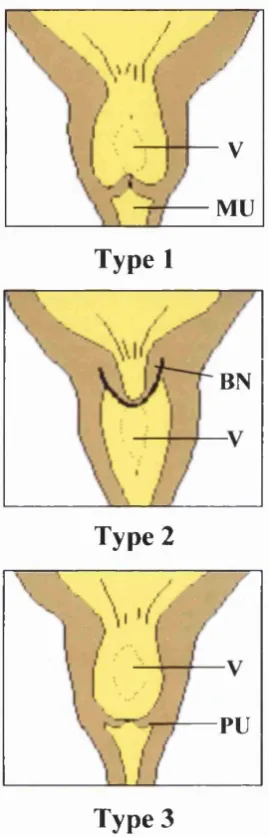
terminated before birth. PUV have been classified as (Figure 2): type I PUV are folds
extending inferiorly from the verumontanum to the membranous urethra; type II PUV are
leaflets or folds radiating from the verumontanum up to the bladder neck; type III PUV
are concentric diaphragms, with a central lumen found within the prostatic urethra
(Young and Frantz, 1919). However, as there is no clear difference in the presentation or
2.1.2 Diagnosis and presentation
With increased use o f antenatal screening, prenatal diagnosis o f PUV is made in 50 % of
cases found to have to have PUV postnatally; fetal PUV ultrasonographic findings
(Figure 3) are hydroureteronephrosis, a distended bladder, and a dilated posterior urethra
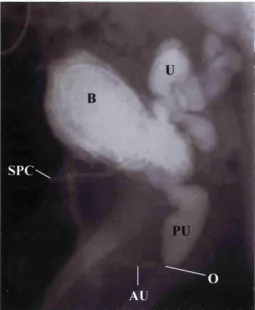
(Hutton et al, 1997). Postnatally, the diagnosis o f PUV must be confirmed by the
micturating cystourethrogram (Figure 4); this contrast study, performed via a urethral or
suprapubic catheter, usually shows the dilated, obstructed posterior urethra as well as
hypertrophy o f the bladder neck, an irregular bladder outline and any associated
vesicoureteric reflux. The ‘classical’ presentation of PUV, however, is within the first
year o f life (Cuckow, 1998). Presentation at birth includes abdominal masses (distended
bladder, hydronephrosis), respiratory distress from pulmonary hypoplasia (secondary to
the oligohydramnios o f obstructive uropathy) and urinary ascites. Others cases present
within the neonatal period with urinary infection, electrolyte disturbance and dehydration.
Later presentation within the first year of life includes failure to thrive, renal failure and
urosepsis.
2.1.3 PUV fetal bladder morphology
Histological and prenatal ultrasound studies o f PUV fetal bladders have documented
varied morphological characteristics. Freedman et al (Freedman et al, 1997) in a study of
human fetuses with BOO (six o f the nine fetuses studied had PUV with a mean gestation
of 29 weeks), documented thickened bladder walls, associated with increased smooth
muscle and collagen, as assessed by computer-assisted image analysis o f stained bladders
et al (1991b) studied seven obstructed fetal bladders with PUV (mean gestation 26
weeks) and using a grid and point-count technique o f stained bladder sections, also
reported increased muscle thickness, but with a decrease in collagen content relative to
muscle. Finally, Workman and Kogan (1990) also found increased muscle thickness in
their study o f four human fetal PUV bladders (mean gestation 27 weeks) as determined
by a grid and point-count technique o f stained bladder sections, but found no increase in
connective tissue deposition.
Studies suggest that the presence o f oligohydramnios and megacystis (an enlarged fetal
bladder) on antenatal ultrasound imaging are highly predictive o f a diagnosis o f fetal
urethral obstruction (Oliveira et al, 2000). However, despite an increasing trend of
antenatal detection o f dilated bladders (Cuckow, 1998), it remains impossible to
distinguish definitely, prenatal posterior urethral valves from other causes o f in utero
bladder distension and oligohydramnios (Callan et al, 1990; Golbus et al, 1982). Abbott
et al (1998) in their study o f antenatal megacystis and hydroureteronephrosis confirmed
PUV postnatally in only 42 % o f cases. Similarly, Holmes et al (2001) in their study o f
fetal intervention for presumed obstructive uropathy found, postnatally, only 39 % of
cases had PUV. Other causes resulting in megacystis include PBS, urethral fibrostenosis,
cloacal anomalies, ureteral duplication, vesico-ureteric reflux and urethral atresia.
Interestingly, large, thinwalled bladders detected on antenatal ultrasound imaging have
detectable dilatation early in the second trimester and those cases detected by the fetal
anomaly scan may represent the severe end of the obstructed spectrum (Thomas, 2001).
This is in keeping with the histological study by Volmar et al (2001) that found that a
kink in the urethra or severe valve obstruction produced thin, dilated bladders whereas
typical mild PUV bladders resulted in thickened dilated bladders. This is also consistent
with antenatal ultrasound investigations by McHugo and Whittle (2001) that suggest a
severe obstructive insult produces a thin-walled dilated bladder whilst mild intermittent
obstruction results in a dilated bladder which is thick walled.
Both histological and radiological investigations suggest that human fetal diseases of
obstructive uropathy are created from a wide spectrum o f severity o f obstructive lesions;
this spectrum appears to result in bladder dilatation with varying degrees o f bladder wall
thickness. These morphological differences influence the range o f clinical symptoms seen
in boys with PUV (Hendren, 1971).
2.1.4 Fetal management
Antenatal intervention exists in the form o f vesicoamniotic shunting and fetal cystoscopy
and endoscopic fulguration (Freedman et al, 1999; Herndon et al, 2000). It is, however,
unclear whether any fetal intervention improves long term renal or bladder function or
prevents lung hypoplasia since prospective studies randomised to control or active
2.1.5 Postnatal treatment
The mainstay o f postnatal management is early bladder drainage by a urethral or
suprapubic catheter; this is accompanied by, if necessary, treatment o f urosepsis, renal
insufficiency, dehydration and electrolyte disturbances. Once the child is stable, valve
ablation by cystoscopy is performed (Smith et al, 1996).
Controversy surrounds the issue o f the management o f PUV in those boys with persistent
renal insufficiency despite adequate catheter drainage o f the bladder. Urinary diversion
with vesicostomy, ureterostomy or pyelostomy has been tried to allow sufficient kidney
drainage in an attempt to permit the obstructed renal parenchyma to recover.
Investigators (Kim et al, 1996; Krueger et al, 1980; Podesta et al, 2002; Reinberg et al,
1992) have found contrary evidence to suggest whether urinary diversion or primary
valve ablation (with expectant follow-up) provides the best renal outcome and which
treatment strategy results in better urodynamic fimction o f the bladder and as such, avoid
later bladder augmentation.
2.1.6 Bladder dysfunction
Various investigations are available to evaluate bladder fimction (and primarily
dysfimction) in the clinical setting and may be collectively referred to as urodynamics.
invasive approach; pressure inside the bladder is measured by a urethral (or suprapubic)
catheter and a second catheter within the abdomen (in most cases placed in the rectum)
allows the abdominal component o f this to be measured separately and subtracted to give
the pressure that is produced by the bladder muscle alone (detrusor pressure). Different
types of bladder activity can be derived from these pressures to characterise bladder
function and dysfunction.
Despite intervention, by various means, bladder dysfunction in PUV boys, often
manifested as incontinence, persists (De Gennaro et al, 1998) (Emir et al, 2002). Three
categories o f bladder dysfunction have been identified in PUV: i) myogenic bladder
failure, characterised by large bladder capacity, large post-micturition residual volumes
and poor contractile function; ii) bladder hyperreftexia with instability and iii) small
capacity bladders with hypertonia or poor compliance (Bauer et al, 1979; Peters et al,
1990). Subsequent studies have revealed not only a large overlap between these
categories but also, that they appear to be time-dependent (De Gennaro et al, 2000;
Holmdahl, 1997). Following valve resection, the bladders o f these boys tend to be
unstable and hypercontractile but later in childhood the bladder capacity increases and the
unstable contractions remain and then slowly diminish. Finally, at or around puberty,
following years o f increased bladder workload, muscle failure sets in leading to large,
floppy bladders. Other factors contributing to myogenic failure include the high fluid
output caused by renal impairment (Cuckow et al, 1997) and the development o f the
2.1.7 Renal dysfunction
The commonest cause o f end-stage renal failure (ESRF) (i.e. chronic renal failure
requiring dialysis or renal transplantation) in children is bilateral renal dysplasia, a term
that describes organs containing undifferentiated and metaplastic tissues such as smooth
muscle and cartilage (Risdon and Woolf, 1998). Just under half the cases o f UK
childhood ESRF caused by renal dysplasia are associated with urinary tract obstruction,
with posterior urethral valves (PUV) being overwhelmingly the commonest specific
diagnosis, accounting for one quarter of all boys with ESRF (Lewis, 1999).
Bladder dysfimction is implicated in the aetiology o f this renal impairment. Investigators
have reported that urinary incontinence after five years o f age (Parkhouse et al, 1988) and
poor bladder compliance and myogenic failure (Lopez et al, 2002) are closely correlated
with poor long term renal outcome. Furthermore, assessments o f outcome o f renal
transplant graft survival into PUV bladders have found variable degrees o f success.
Several studies report that renal transplantation in patients with PUV has less success
than in transplants into patients with normal vesical fimction (Groenewegen et al, 1993;
Reinberg et al, 1988). Others have found no decline in graft survival and long-term renal
outcome between the two groups (Crowe et al, 1998; Ross et al, 1994) although graft
wall muscles (Figure 5); the condition affects 1 in 40,000 births (Wheatley et al, 1996).
Fetal bladder morphology, as assessed by haematoxylin and eosin stained bladder
microspcopy, is variable with thick and thin walled distended bladders observed in
human fetuses with PBS (Shimada et al, 2000). Whilst PBS is considered to be an
‘obstructive’ urinary tract, it is open to debate whether the condition represents either a
case o f functional urinary flow impairment, such as from a bladder neuromuscular
disorder (Volmar et al, 2001), or perhaps results from a transient, fetal physical
obstruction o f the urethra (Jennings, 2000) (Figure 6). In 75 % o f cases, there are
associated malformations o f the cardiopulmonary, gastrointestinal, and orthopaedic
systems (Jennings, 2000) possibly secondary to the oligohydramnios that leads to a
limited intrauterine space, causing fetal compression and deformity. With increased
prenatal imaging, PBS may be detected antenatally, but as for PUV, in utero intervention
(by vesico-amniotic shunting) results in equivocal improvements to renal and bladder
function (Irwin and Vane, 2000; Perez-Brayfield et al, 2001; Shimada et al, 2000).
2.3 Embryology of the developing and obstructed urinary tract
2.3.1 Bladder development
The bladder is derived from the cloaca (Cuckow et al, 2001) which forms from the
hindgut o f the developing human embryo; at around 28 days o f gestation, the cloaca is
separated by an advancing urorectal septum to form an anterior urogenital sinus (that
receives the mesonephric ducts) and a posterior rectum. When this septum reaches the
posterior anal membrane and an anterior urogenital membrane that breaks down in the
seventh week.
The superior urogenital sinus forms the primitive bladder and grows together with the
anterior abdominal wall; the two structures are connected by the allantois (which
connects the apex of the fetal bladder to the umbilical root). The latter forms the urachus
which then obliterates around the third-fourth month o f fetal life (Bourdelat et al, 1998).
By 13 weeks of gestation, the primitive bladder mesenchyme has differentiated
discernible circular and longitudinal strands o f smooth muscle with distinct muscle layers
visible by 16 weeks gestation (Newman and Antonakopoulos, 1989).
During the septation process, the mesonephric ducts become progressively incorporated
into the posterior aspect o f the primitive bladder. At this time, the ureteric orifices also
become incorporated into a more proximal portion o f the posterior wall o f the bladder
with the area between the ureteric orifices and the fused mesonephric ducts forming the
bladder trigone. The distal part o f the urogenital sinus then forms the posterior urethra
with the anterior urethra formed by closure o f the urethral folds (Figure 7).
2.3.2 Kidney development
pronephros then involutes. The mesonephros forms at 24 days o f gestation and produces
small quantities o f urine by the tenth week that drains into the cloaca via the mesonephric
duct. The majority o f the mesonephros involutes with some remnants (with some o f the
mesonephric duct) contributing to the development o f the epididymis, seminal vesicle
and ejaculatory ducts in the male fetus. Prior to this involution, at around 28 days of
gestation, each mesonephric duct gives off a ureteric bud. The bud elongates and
penetrates the mesenchymal metanephros to form the metanephric blastema. The ureteric
bud branches to form the ureter, renal pelvis, calyces and collecting tubules; the
mesenchyme creates the cortex o f the kidney with the first glomeruli forming by 8-9
weeks o f gestation and nephrogenesis continuing until 34-36 weeks gestation (Figure 7).
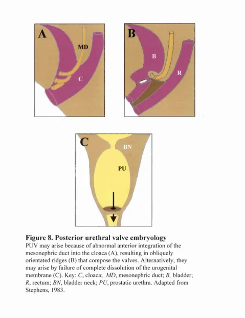
2.3.3 PUV embryology
It has been hypothesised that PUV arise as a result o f the mesonephric ducts entering the
cloaca in a more anterior position than normal with migration o f the ducts impeded by the
enfolding and separation o f the cloaca, thus causing the ducts to fuse in the midline
anteriorly (Stephens, 1983) (Figure 8). It is also possible that valves result by a failure o f
complete dissolution o f the urogenital membrane (Stephens, 1983) (Figure 8).
2.4 Innervation and neurotransm ission in the bladder
2.4.1 Central and peripheral innervation
Micturition or voiding is the process o f emptying the bladder. It is a complex chain of
autonomie nerves in the bladder and somatic and autonomic nerves in the sphincter (the
urethral sphincter has smooth as well as skeletal muscle and therefore an autonomic as
well as a somatic innervation); these events are influenced by the central nervous system
(George, 2001). As the bladder fills with urine, relaxation o f its wall and the maintenance
o f tone in the sphincter allow it to contain the urine at low pressure. When it is distended,
signals indicating a full bladder are transmitted to the spinal cord in sensory (afferent)
nerves and ascend to the brain (Figure 9).
Neurotransmitter release in the brain results in signals being sent back down the spinal
cord, to converge on the two main areas involved in spinal micturition control (George,
2001). These are the thoracolumbar region (thoracic nerves 10,11 and 12 and lumbar
nerves 1 and 2) and the sacral region (sacral nerves 2,3 and 4). The autonomic
parasympathetic and sympathetic nerves (otherwise known as preganglionic fibres) pass
outside the spinal cord to synapse with the cell bodies o f peripheral autonomic nerves in
specialised areas known as ganglia (or plexuses). Neurotransmitter release here results in
impulses leaving the ganglia in postganglionic nerves to reach the bladder muscle,
bladder trigone, or urethra. A further somatic outflow (from O n u f s nucleus) passes
directly to supply the pelvic floor and urethral sphincter via the pudendal nerve.
Additional neurotransmitter release from postganglionic nerve endings adjacent to the
modifies its tone and may effect relaxation and opening o f the bladder outlet (Gosling
and Dixon, 1975).
2.4.2 Overview of bladder detrusor muscle pharmacology
There are several chemicals involved in effecting bladder contraction. These include
neurotransmitters and modulators that allow communication between the central nervous
system, peripheral nervous system and the lower urinary tract in the adult bladder. In
addition, local trophic factors are involved in the maintenance and growth o f nerves
supplying the bladder (Anderson 1993).
Following receptor activation on the bladder, Ca^"^ is released from the sarcoplasmic
reticulum (SR) by a) a chain of membrane-bound reactions that lead to the production of
inositol phosphate or b) calcium influx through the surface membrane (possibly ATP-
mediated). Following SR release o f Ca^"^, the latter binds to calmodulin, and via a
subsequent cascade o f reactions, phosphorylates and activates a portion o f the myosin
molecule. This promotes interaction with actin molecules (with ATP consumption) and
results in an increase in muscle tension. A reduction in the intracellular Ca^^ causes
muscle relaxation probably by SR sequestration and transmembrane calcium flux (Fry
and Wu, 1998) (Figure 10).
For central and peripheral neurotransmission in the adult, acetylcholine (ACh) is released
from preganglionic neurons (from the sacral spinal cord) and activates nicotinic receptors
in excitatory activation o f the bladder via muscarinic receptors (namely M2 and M3
receptors) (Wang et al, 1995) and bladder contraction. Other neurotransmitters postulated
in adult lower urinary tract pharmacology include noradrenaline (Gosling et al, 1999;
Taki et al, 1999), adenosine triphosphate (Elneil et al, 2001), neuropeptide Y,
tachykinins, vasoactive intestinal peptide and calcitonin gene-related peptide (Dixon et al,
1998; Smet et al, 1996) and vasopressin (Holmquist et al, 1991). In addition, sex steroids
may modulate bladder receptors and influence bladder growth (Celayir et al, 2002).
Finally, nitric oxide has a role in neurologically mediated relaxation in the lower urinary
tract, especially in the urethra (Andersson and Persson, 1995). Centrally acting
neurotransmitters include glutamate, dopamine, serotonin, noradrenaline, gamma-amino-
butyric acid, neuropeptides, enkephalins and ACh (de Groat, 1990).
2.5 Experim ental strategies for investigating in utero bladder outflow
obstruction
In attempt to understand fetal bladder structure and function and subsequent changes after
in utero bladder outflow obstruction, investigators have examined the bladders or bladder
myocytes from human fetuses (De La Rosette et al, 2002; Freedman et al, 1997; Kim et
al, 1991a; Kim et al, 1991b; Newman and Antonakopoulos, 1989; Workman and Kogan,
1990) and infants (Cortivo et al, 1981; Deveaud et al, 1998), fetal sheep (Arens et al,
Sutherland et al, 1997), fetal cows (Baskin et al, 1993a; Coplen et al, 1994; Koo et al,
1995), the genetically-manipulated mouse (Bassuk et al, 2000) and the fetal mouse (Park
et al, 1997; Smeulders et al, 2001).
The adult pig bladder has similar anatomical and functional characteristics to the human
bladder (Crowe and Bumstock, 1989; Dalmose et al, 2000; Guan et al, 1994). Although,
the normal fetal porcine bladder has been studied (Olsen et al, 2001), surgical
manipulation o f the fetal porcine urinary tract, to produce an experimental model o f in
utero bladder outflow obstruction, is currently not feasible; this is primarily because of
their multigravid pregnancies necessitating complex technical surgery and probable high
mortalility. As such, investigators have focused principally on the fetal sheep for fetal
urinary tract manipulation, o f the bladder (Cendron et al, 1994; Karim et al, 1993; Levin
et al, 2001; Peters et al, 1992b; Tanagho, 1972), ureters (Santis et al, 2000) and kidneys
(Adzick et al, 1985; Attar et al, 1998; Beck, 1971; Bellinger et al, 1986; Bogaert et al,
1994; Carr et al, 1995; Duncombe et al, 2002; Edouga et al, 2001; Gobet et al, 1999a;
Gobet et al, 1999b; Harrison et al, 1983; Kogan and Iwamoto, 1989; Peters et al, 1992a;
Yang et al, 2001). The fetal sheep is a useful model because mother and fetus tolerate
surgery with little morbidity or mortalility, the fetus is o f considerable size as compared
to small mammals, ewes are relatively easy to handle and they have well established
breeding protocols. Furthermore, studies listed above reveal that experimental bladder
outflow obstruction mimics human disease. Disadvantages include financial expense,
requirement o f a substantial holding facility and extra personnel to perform surgery
2.5.1 Fetal sheep urinary tract embryology
Ovine gestation lasts for 145 days; ovine lower urinary tract development is similar to
human fetal urinary development (Tanagho, 1972). Like the human fetus, the urogenital
sinus in the fetal sheep is formed by division o f the cloaca and later forms the primitive
bladder and urethra. The bladder extends to the umbilicus and opens directly into the
allantois. At mid-gestation, the ovine fetal urethra is well formed but is not used as an
outflow tract from the bladder as the lumen is packed with desquamated epithelial cells
and debris. At this time point, urine from the fetal kidneys pass from the bladder into the
allantoic cavity via the urachus until approximately 90 days o f gestation when the bladder
begins to recede caudally, pulling on the allantoic stalk (Figure 11). Urine progressively
passes directly into the amniotic cavity via the urethra until the urachus fully obliterates
(the exact time this occurs is unknown but it may remain patent up to term). Ovine fetal
allantoic and amniotic sacs remain separate cavities with the former eventually rupturing
into the amniotic cavity to form one sac. This is different to the human fetus where no
equivalent membranes exist; the amniotic cavity expands to obliterate the small chorionic
cavity, between four-eight weeks gestation, to become the sole cavity surrounding the
human fetus (Larson, 2001).
Although three types o f kidney develop during human gestation, the ovine fetus does not
develop a pronephros, but forms a ‘giant glomerulus’, consisting o f approximately 15
regresses from day 40 to 57 (Moritz and Wintour, 1999). As sheep have a more primitive
placenta than the human placenta, the mesonephros is more highly developed than the
human fetus and is able to produce a hypotonic urine from day 18 o f gestation. This urine
passes into the allantoic cavity which increases in volume from 3 ml at day 20 o f
gestation to 20 ml by day 24 (Moritz and Wintour, 1999). This rapid expansion o f the
allantoic membrane is important in developing contact between maternal uterine
calculation and fetal blood vessels, to form placental caruncles. Ovine metanephric
development begins at day 27, with ureteric bud branching. By mid-gestation, urine
production is an impressive 5-6 ml per hour and urine tonicity remains hypotonic
throughout ovine development (Wintour et al, 1996). Also at mid-gestation, the
composition o f the allantoic fluid and fetal urine differ quite significantly because o f
differential rates o f solute removal across the allantoic membranes (Wintour et al, 1986).
Metanephric formation o f new nephrons is complete by day 135 o f gestation and
therefore complete before birth; this is similar to human development and different to
many other mammals (eg mice and rats) where nephrogenesis continues past term
(Matsell and Tarantal, 2002).
2.6 The m olecular biology o f the developing and obstructed fetal
bladder
2.6.1 The developing bladder
Molecular studies in the developing fetal bladder are limited; studies have described the
human (Kim et al, 1991a), mouse (Smeulders et al, 2001), rabbit (Chiavegato et al, 1993;
Lin et al, 2000; Sakurai et al, 1996), cow (Baskin et al, 1994b; Koo et al, 1997; Koo et al,
1998) and sheep (Arens et al, 2000). With interest to my thesis, Smeulders et al (2001)
described how cell proliferation, as assessed by immunohistochemistry and western blot
o f the proliferation marker, proliferating cell nuclear antigen, was highest at inception of
the bladder in the fetal mouse and fell progressively throughout gestation; in addition,
detrusor muscle cell apoptosis, determined using the TUNEL method, was only noted at
embryonic day 14 with no further cell death documented. Also noted was increasing a -
smooth muscle actin and vimentin protein expression throughout gestation representing
increased detrusor smooth muscle differentiation. Study o f the human fetal bladder
revealed increasing muscle thickness and decreasing relative collagen content during
development. Fetal animal studies have shown, that with increasing gestation, as assessed
by western blotting o f myosin heavy chain (MHC) isoforms, there was decreasing
detrusor MHC isoform 1 expression and increasing MHC isoform 2 expression (Arens et
al, 2000; Chiavegato et al, 1993; Lin et al, 2000; Sakurai et al, 1996), suggesting a
prenatal maturation o f detrusor contractile ability. Also noted, by northern and slot blot
analyses and immunohistochemistry of fetal bladders, was decreasing elastin and
collagen III expression and increasing microfibrillar protein and collagen I expression
(Koo et al, 1997; Koo et al, 1998) possibly illustrating the development o f bladder
2.6.2 Bladder growth, hypertrophy and hyperplasia in the obstructed fetal
bladder
BOO in the adult rat bladder has been documented to cause detrusor muscle hypertrophy
and hyperplasia (Uvelius et al, 1984). This appears to occur in a time-dependent manner
that is different to the developing bladder. In the adult rabbit bladder, by nylon taping of
the bladder neck and urethral catheterisation, the reported changes are temporary
submucosal oedema, increased flbrocollagenous connective tissue deposition and then
smooth cell mass increase (in part replacing the connective tissue) by detrusor myocyte
hypertrophy followed by hyperplasia, as assessed by microscopy and point counting
(Brent and Stephens, 1975). By contrast, the growing rabbit bladder exhibited smooth
muscle hypertrophy preceded by smooth muscle hyperplasia (Brent and Stephens, 1975).
Similarly, growth by detrusor hypertrophy and hyperplasia has been documented in the
ovine fetal bladder subjected to BOO by partial urethral obstruction and complete urachal
ligation. Peters et al (1992b) produced ovine BOO at 60 days gestation and sacrificed
fetuses after 35 or 85 days; a marked growth response was found in obstructed bladders
as assessed by increased bladder weight, DNA and protein content, and detrusor smooth
muscle cell size as calculated by computer morphometric analyses. Karim et al (1993)
produced BOO at 9 0 - 1 0 0 days gestation and obtained bladders at term; the authors
reported a significant increase in bladder weight and detrusor DNA content in the
obstructed bladders. Cendron et al (1994) observed a significant increase in bladder
weight at term in ovine fetuses obstructed at 90 days gestation. Finally, Levin et al (2001)
ovine fetuses at 90 days gestation. However, despite these investigations o f bladder
growth, hypertrophy and hyperplasia, little is known of the balance between these
processes and programmed cell death.
2.6.3 Apoptosis
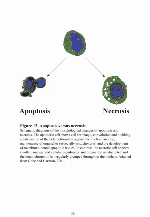
Cell death occurs by necrosis or apoptosis (Kerr et al, 1972) (Figure 12). Necrosis is
uncontrolled cell death that leads to cell swelling, mitochondrial damage and cell
membrane lysis with release o f intracellular contents; this results in an inflammatory
response, with subsequent oedema and damage to the surrounding cells. In contrast,
apoptosis, or programmed cell death, keeps the intracellular content o f the dying cell
sequestered; it is defined by cell shrinkage, nuclear condensation, DNA cleavage and cell
membrane blebbing (containing intracellular contents such as nuclear matter and cellular
organelles). These apoptotic bodies are removed by phagocytes or by neighbouring cells.
Apoptosis has been increasingly implicated in organogenesis during embryo development
(Jacobson et al, 1997) (Figure 13); it is involved in organ sculpting (eg loss o f webs
between digits or loss o f cells in solid structures to form lumina), deleting unwanted
structures (eg loss o f vestigial organs and involution o f the mesonephros), adjusting cell
numbers (eg deleting excess precursors in the metanephric kidney) and eliminating
The key steps in apoptosis is a series o f activations o f proteases called caspases (Hayashi
and Araki, 2002), previously known as interlekin-1 beta-converting enzyme-like
proteases. Caspases have been divided into three groups:
Group I caspase-1, caspase-4, caspase-5, caspase-11
Group II caspase-2, caspase-3, caspase-7
Group III caspase-6, caspase-8, caspase-9, caspase-10
Group I caspases are involved in cytokine processing, Group III caspases activate group
II caspases and the latter group consist o f effector proteins that are able to directly cleave
substrates resulting in apoptosis. The most important o f these, capsase-3, results in cell
death by either activating caspase-activated DNase or directly cleaving cell surface
membranes or cellular enzymes when activated. Furthermore, these caspases are able to
lead to disassembly o f nuclear and cytoskeletal structures, disable cell repair and label
apoptotic cells for phagocytosis (primarily by macrophages) (Zhivotovsky et al, 1997).
Currently, two pathways have been described that activate caspases (Figure 14). Death
factors, such as Fas ligand (Nagata, 1994), bind to death receptors and activate group III
caspases, specifically caspase- 8 and caspase-10. Alternatively, external stimuli, such as
irradiation or chemotherapeutic agents, result in cytochrome C release fi’om mitochondria
which activates the Group III caspase, caspase-9. Subsequently, both pathways converge
with activation o f Group II caspases.
Members o f the protein family, Bcl-2 (B-cell leukaemia/lymphoma-2), have also
molecules, such as Bcl-2 associated X protein (Bax) and anti-death molecules, such as
Bcl-2, play key roles by acting both at the level o f the mitochondria, and at pre- and
postmitochondrial stages, and therefore directly influence cytochrome C release.
2.6.4 Apoptosis in the urinary tract
All studies in this section examined apoptosis by the TUNEL method and apoptosis-
related proteins by immunohistochemistry, unless otherwise stated. Programmed cell
death plays a significant role in renal morphogenesis. During development, a high index
o f proliferation, as assessed by PCNA expression, and a low index o f apoptosis,
accompanied with increased Bcl-2 expression (additionally determined by
cytophotometry) were observed in murine and human renal metanephric cells
(Lichnovsky et al, 1999; Winyard et al, 1996b); apoptosis, as documented by cell
morphology, has also been observed in the nephrogenic region and medullary papilla of
the developing rat kidney (Coles et al, 1993). Furthermore, fetal renal pathology is
associated with aberrations o f apoptosis and apoptosis-related proteins. Increased
apoptosis and decreased Bcl-2 expression has been documented in the stroma o f pre- and
postnatal polycystic and dysplastic human kidneys (Winyard et al, 1996a). In utero
urinary tract obstruction resulted in high rates o f apoptosis in the developing renal pelvis
and tubulointerstitium, accompanied with increased Bax and decreased Bcl-2 expression
Moreover, mice with null mutations of Bcl-2 demonstrate fulminate apoptosis during
metanephric development resulting in renal hypoplasia at birth and multicystic renal
disease later in life (Veis et al, 1993). Similarly adult renal pathology also results in
perturbation o f cell turnover; urinary obstruction in the adult rat resulted in
tubulointerstitial apoptosis with downregulation o f Bcl-2 (Chevalier et al, 2000),
upregulation of caspases (as assessed by ribonuclease protection assays and
immunohistochemistry) (Truong et al, 2001) and increased apoptosis and Bax expression
in smooth muscle cells o f the obstructed ureter (Chuang et al, 2002). Furthermore,
glomerulonephritis, also in the adult rat, resulted in increased apoptosis and caspase-3 (as
assessed by substrate cleavage assays) with a shift in the Bax-Bcl-2 balance (additionally
assessed by western and northern blotting) (Yang et al, 2002).
Less is known o f programmed cell death in the developing fetal bladder. Apoptosis has
been documented in the separation o f the cloaca into the urogenital sinus in embryonic rat
development (as early as embryonic day 12) (Qi et al, 2000) and has also been observed
in the murine fetal bladder (at embryonic day 14 only) (Smeulders et al, 2001). In
addition, immediately prior to birth, the fetal mouse bladder undergoes urothelial cell
desquamation that is accompanied with apoptosis (as assessed by cell morphology and
cytochemistry) (Jezemik et al, 1997) and programmed cell death is also present during
the postnatal restoration o f the urothelium (Erman et al, 2001). In adult pathology,
bladder apoptosis has been documented to a) increase following ischaemia-reperfusion
injury (Saito and Miyagawa, 2002), alpha-1 adrenoceptor antagonist treatment o f benign
(Santarosa et al, 1994) and transitional cell carcinoma (TCC) o f the bladder (King et al,
1996), and b) decrease in diabetes mellitus (Kban et al, 2002) and following stretch of
bladder smooth muscle cells (as assessed by propidium iodide incorporation and flow
cytometry) (Galvin et al, 2002). Finally, Bcl-2 expression in the bladder is associated
with poorly differentiated TCC of the bladder (King et al, 1996) and
immunobistocbemical testing o f activated caspase-3 suggests this molecule is involved in
the progression o f bladder carcinoma from carcinoma-in-situ to invasive bladder cancer
(Burton et al, 2000). However, as there are no studies o f the effects o f fetal urinary
pathology on fetal bladder apoptosis, I have chosen to examine programmed cell death
and apoptosis-related proteins in my experimental model in utero bladder outflow
obstruction.
2.7 The physiology o f the developing and obstructed fetal bladder
Current methods o f studying fetal bladder physiology are human fetal ultrasound and
physiological measurements made in whole organ preparations, muscle strip studies and
in vivo fetal bladders.
2.7.1 Ultrasound studies of the developing bladder
As early as 20 weeks o f gestation, the human fetal bladder is able to store and empty
every 60 minutes by 40 weeks gestation (Hata and Deter, 1992). The maximum fetal
bladder volume increases from 1 ml at 20 weeks gestation to 36 - 54 ml by 40 weeks
gestation (Rabinowitz et al, 1989) with average voiding lasting approximately 9.5
seconds; complete bladder emptying is achieved by 40 weeks gestation.
2.7.2 Ontogeny of animal fetal bladder physiology
Whole bladder studies from the fetal calf reveal that with increasing gestation, the fetal
bladder demonstrates increased compliance (the bladder is able to accommodate
increased urinary volume without an increase in intravesical pressure) (Coplen et al,
1994; Koo et al, 1995). This was confirmed in a study using circularly clamped fetal
bladder strips (Baskin et al, 1994a). It has since been postulated that the change in
compliance during gestation is associated with the documented transformation in
collagen subtype expression (Kim et al, 1991a; Koo et al, 1997). Furthermore, muscle
tone has also been implicated; compliance increases through gestation because the
immature fetal bladder has high active smooth muscle tone that decreases with
maturation allowing for this greater urine accommodation (Coplen et al, 1994; Dean et al,
1997).
Active contractile fimction also develops during gestation. Early gestation whole bladder
preparations from the fetal calf emptied by 50 % when stimulated with the muscarinic
agonist, bethanechol, whilst mid and late gestation preparations emptied to almost
completion (Koo et al, 1995). These age-related differences in contractility may only
By documenting pressure changes within the fetal bladder using indwelling catheters,
ovine fetal bladder contractions were discernable at 1 2 0 days gestation and could be
pharmacologically manipulated (Kogan and Iwamoto, 1989); contractile function
appeared to be under cholinergic and P-adrenergic control. By using a similar method at
the same gestation, nitric oxide also appeared to be active in lower urinary tract function
(Mevorach et al, 1994). Finally, during this period, ovine fetal voiding appeared to
coincide with periods o f electrocortical activity in the fetal brain (Wlodek et al, 1989)
suggesting that descending control o f bladder activity was developed.
Pressure recordings within the fetal pig also show maturation o f bladder function (Olsen
et al, 2001). During the second trimester, fetuses showed no sign o f active storage and
voiding with continuous urine flow with apparent flow increases synchronous with
bladder contractions; this suggests that at this gestation in the fetal pig, the bladder acts
simply as a conduit. By the third trimester, distinct periods o f voiding were observed with
urethral urine flow, bladder contractions (that were o f higher pressure than earlier in
gestation) and the development o f urethral sphincter bursting activity.
2.7.3 Physiology of the obstructed fetal bladder
reported hypocontractile obstructed bladder strips. In contrast, by different methodology,
Peters et al (1992b) reported reduced compliance in the obstructed ovine fetus. Levin et
al (2 0 0 1) also described hypocontractile responses in ovine fetal bladders after short-term
bladder outflow obstruction.
2.7.4 In vivo fetal cystometry
Currently, only three studies have documented pressure characteristics in the fetal
bladder, in the fetal sheep and fetal pig, using externalised catheters. Kogan and Iwamoto
(1989) used intravesical catheters, connected to pressure recorders, in the fetal sheep.
With the ewes ambulatory, fetal bladder pressures were documented following
intravenous ftirosemide administration or by artificially filling the bladder. Similarly,
Mevorach et al (1994) documented bladder contractions in the ovine fetus by artificially
filling the bladder with warm saline and pressures recorded with intravesical catheters.
Finally, under maternal sedation, Olsen et al (2001) reported fetal bladder pressures in the
porcine fetus using intravesical catheters but without any artificial means o f bladder
filling by instillation or forced diuresis. These studies, however, are not without
problems: animals will try to remove catheters on their flanks, animals have imposed
immobility and are stressed, there is an increased risk o f infection and fetal mortality and
studies tend to be o f a short duration. Furthermore, ambulatory urodynamics in humans
suggest that artificial filling and a stressful environment may result in nonphysiological
detrusor activity; compliance remains high, unstable detrusor activity is more fi-equently
observed, voided volumes are lower and maximum voided pressures higher (Robertson et
overcome these difficulties, investigators have used radiotelemetry devices to monitor
bladder pressure. These have a number o f advantages (Mills et al, 2000): reduced stress
to animals (no handling, restraining or tethering to recording machines, no anaesthesia
and no invasive monitoring), monitoring can be over a prolonged period o f time
providing meaningful physiological data, and computer-based monitoring allows accurate
mathematical analysis. Radiotelemetered cystometry had been used successfully to
describe ambulatory pressure recordings in the bladder, by natural filling, in the adult pig
(Mills et al, 2000) and adult monkey (El Ghoneimi et al, 1999) and the obstructed adult
pig (Speakman et al, 1987); such technology has not been evaluated in the fetal bladder.
2.8 C oncluding rem arks
My introduction shows that in utero BOO o f the human fetal bladder results in significant
postnatal urinary tract dysfunction. To examine the fetal consequences o f this
obstruction, investigators have performed limited studies in the human fetal bladder and
developed experimental models o f in utero BOO, primarily using the developing sheep.
These latter studies suggest that obstruction results in a large bladder with impaired
contractility. However, no studies, to date, examine proliferation and apoptosis and
detailed neurotransmission and viscoelasticity in the developing and obstructed fetal
bladder. In the following chapters, I describe the hypotheses and aims o f my thesis and
Figure 1. C ystocop ic view o f posterior ureth ral valves
\ ■ / • / /
y
/
\ (
H - V— MU
T yp e 1
BN
T ype 2
PU
T ype 3
Figure 2. P osterior urethral v alves classification
Figure 3. A n ten atal ultrasound im age o f a fetus w ith PUV
Ultrasound image shows the dilated bladder (B) o f a fetus with
Figure 4. M ictu ratin g cystou reth rogram o f a boy w ith
PUV
h
■
-Figure 5. A b d om in al w all defect o f Prune B elly Syndrom e
Figure 6. R ad iological im aging in Prune B elly Syndrom e
XiT
MD
p d
/
UB
CM
B
A-<MD
CM
CM
AM-Figure 7. H um an urinary tact em b ryology
A-C represents urinary tract embryology during the (A), 5*^ (B) and (C)
o f human fetal gestation. During this period, the mesonephros produces urine and then involutes, the ureteric bud penetrates the metanephros to form the metanephric blastema and subsequently the kidney and the septum divides the
urogenital sinus to form the rectum and the primitive bladder. Key: A, allantois;
M, mesonephros; MD, mesonephric duct; B, metanephric blastema; G, gonad;
UB, ureteric bud; S, septum; CM, cloacal membrane; TF, tail fold; GT, genital
tubercle; UG, urogenital sinus; MT, metanephros; R, rectum; K, kidney; U,
i
1
Figure 8. P osterior urethral valve em b ryology
PUV may arise because o f abnormal anterior integration o f the mesonephric duct into the cloaca (A), resulting in obliquely orientated ridges (B) that compose the valves. Alternatively, they may arise by failure o f complete dissolution o f the urogenital
membrane (C). Key: C, cloaca; MD, mesonephric duct; B, bladder;
R, rectum; BN, bladder neck; PU, prostatic urethra. Adapted from
H ypogastric n e rv e
Levator am + pelvic floo r
ior m esen teric g a n g lio n (sym pathetic)
Pu d endal n erv e (som atic)
Centre
'"'BA
H ypogastric n e rv e (sy m p ath etic)
P ud en dal n e rv e (som atic)
F igure 9. In nervation o f the blad der
To allow for bladder filling, the bladder relaxes it’s tone (by negative feedback o f the sympathetic nervous system) and by maintaining bladder outflow closure by the somatic nerves. With further filling, impulses are carried up the pelvic nerve to enter into the sacral region o f the spinal cord and eventually reach the pontine micturition centre (PMC) in the pons, this area may also be influenced by further higher cerebral control.
y \
ACh
Muscarinic receptor
IP.
Cell membrane
actin
Sarcoplasmic reticulum
calmodulin
Figure 10. B ladd er pharm acology
Following receptor activation on the bladder primarily by acetyl choline (ACh), calcium is released from the sarcoplasmic reticulum by a chain o f
reactions leading to the production o f inositol phosphate (IP3), or by
'KmC /
AC
AM
AmM
U r
Figure 11. Fetal ovine anatom y
Figure shows anatomy o f ovine fetus within maternal uterine cavity; note how bladder urine primarily drains into amniotic cavity and urachal urine
flows into allantoic cavity. Key: F, fetus; K, kidney; B, bladder; U, urethra;
AC, allantoic cavity; AmC, amniotic cavity; Ur, urachus; AmM, amniotic
membrane; AM, allantoic membranes; Ut, uterus. Modified from Wlodek et
i
Apoptosis
Necrosis
Figure 12. A poptosis versus necrosis
Schematic diagrams o f the morphological changes o f apoptosis and
necrosis. The apoptotic cell shows cell shrinkage, convolution and blebbing, condensation o f the heterochromatin against the nuclear envelop,
Organ sculpting
B
Deleting structures
c
Adjusting cell numbers
E
F
Eliminating abnormal cells
♦c
-Figure 13. A p op tosis in organogenesis
D R
extern al stim uli
A
C vtoch rome
Gp II caspases
effector proteins
Figure 14. T he caspase cascade
Apoptosis is activated by two pathways that interact. The binding o f death ligands to the death receptors activate group III and subsequently group II caspases. Alternatively, external stimuli, such as irradiation or chemotherapeutic agents, cause cytochrome C release from mitochondria which activate group II caspases. Either directly, or via effector proteins such as caspase-activated DNase, group II caspases (primarily caspase-3) result in cell death by apoptosis. The BcI-2 family o f apoptotic regulatory proteins act, both at the level o f the mitochondria, and at pre- and
3. Experimental strategy
In utero BOO results in significant postnatal bladder dysfunction. In order to understand
how the fetal bladder responds to this urinary obstruction, the aims o f my thesis were to
examine the following hypotheses.
3.1 H ypotheses
1) In utero bladder outflow obstruction results in significant alteration to the cell turnover
o f the developing fetal bladder with deregulation o f proliferation, apoptosis and apoptotic
regulatory protein expression.
2) In utero bladder outflow obstruction results in significant perturbation to contractility,
neurotransmission and physical properties o f the developing fetal bladder.
To answer these hypotheses, 1 aimed to perform the following experiments.
3.2 Aim s
1) Obtain fetal bladder samples in an experimental fetal ovine model o f in utero bladder
outflow obstruction and sham control animals.
2) Document the changes to organ growth and cell turnover in the fetal bladder exposed
3) Determine the normal maturation of the fetal bladder, with special reference to bladder
growth and cell turnover.
4) Identify molecules involved in neurotransmission in the normal fetal bladder.
5) Ascertain the changes to fetal bladder contractility and neurotransmission after BOO
using electrophysiological bladder strip studies.
6) Discover the changes to the physical characteristics o f obstructed fetal bladders by
examining the pressure-volume relationships in ex vivo whole bladders and
biomechanical visco-elastic properties o f bladder strips, in both experimental groups.
7) Determine the feasibility o f using radiotelemetry implants to document the in vivo