Nijmegen
The following full text is a publisher's version.
For additional information about this publication click this link.
http://hdl.handle.net/2066/27384
Please be advised that this information was generated on 2018-07-07 and may be subject to
change.
Prostate Carcinoma
Een wetenschappelijke proeve op het gebied van de Medische Wetenschappen
Proefschrift
ter verkrijging van de graad van Doctor aan de Radboud Universiteit Nijmegen,
op gezag van de Rector Magnificus prof. dr. C.W.P.M. Blom,
volgens besluit van het College van Decanen in het openbaar te verdedigen
op woensdag 8 februari 2006 des namiddags om 15:30 uur precies
door
Johannes Jacobus Fütterer
geboren op 5 Maart 1975 te Oosterhout
Co-promotor:
dr. G.J. Jager
Manuscriptcommissie: prof. dr. J.W. Leer (voorzitter)
prof. dr. P.H. de Mulder
dr. R.J.A. van Moorselaar (VU)
Printing: PrintPartners Ipskamp Nijmegen
© Jurgen J. Fütterer
ISBN 90-9020328-1
The study presented in this thesis was supported by The Dutch Cancer Society (Koningin
Wilhelmina Fonds: KUN-1999-1969 and KUN 2003-2925), Medrad Europe bv., Guerbet
Nederland bv., Siemens Nederland bv., and Andreas Muehler (CADsciences; www.CAD
Sciences.com).
Chapter 1 General introduction [7]
Chapter 2 MR imaging in local staging of prostate cancer, Prostate Cancer
Imaging, Springer Verlag, in press [17]
Chapter 3 Prostate cancer: Comparison of local staging accuracy of pelvis phased array coil alone versus integrated endorectal pelvic phased array coils,
Submitted [37]
Chapter 4 Combined dynamic contrast-enhanced and proton MR spectroscopic imaging in localizing prostate cancer, Submitted [55]
Chapter 5 Dynamic contrast-enhanced endorectal MR imaging in staging prostate cancer prior to radical prostatectomy – experienced versus less experienced readers, Radiology 2005; 237:541-549 [73]
Chapter 6 Initial experience of 3T endorectal coil MR imaging and 1H-spectro-scopic imaging of the prostate, Invest Radiol 2004; 39:671-680 [93]
Chapter 7 Local staging of prostate cancer using 3T endorectal coil MR imaging – Early experience, Radiology in press [111]
Chapter 8 Precision of integrating functional magnetic resonance in radio-therapy treatment of prostate cancer using fiducial gold marker
registration methods, Radiology 2005; 236:311-317 [127]
Chapter 9 IMRT boost planning on dominant intra-prostatic lesions: gold marker based 3D fusion of CT, dynamic contrast-enhanced and
1H-spectroscopic MR imaging, Int J Radiat Oncol Biol Phys in press [143]
Chapter 10 Summary and conclusions [163] Recommendations and suggestions for further research
Dankwoord [178]
Curriculum vitae [179]
1
Introduction
Prostate cancer is the most common non-cutaneous cancer in the world. In 2005, an estimated 232,090 new cases and 30,350 deaths of prostate cancer are expected in the United States alone. Parallel to these figures, approximately 1 in 10 men will die of prostate cancer [1-2]. In the last 15 years an exponential rise in the number of patients diagnosed with prostate cancer was seen. The increase in the number of older persons and the introduction of prostate-specific antigen (PSA) serum screening programs resulted in an increase in prostate cancer incidence [1, 3-6]. After prostate cancer has been diagnosed, the therapeutic options are determined using nomograms [7-9]. The most frequently used nomogram, the Partin tables, estimates the chance of organ-confined disease, capsular penetration, seminal vesicle invasion and lymph node metastasis, based on the results of the traditional triad of digital rectal examination, biopsy Gleason score and PSA value [10].
Clinical staging to differentiate between localized (surgical candidate) and advanced disease stage (androgen therapy and/or radiotherapy) appear to be unreliable. Curative therapy can be performed only in patients with localized prostate cancer. Accurate staging is therefore especially important for proper disease management. Since 1984 magnetic resonance (MR) imaging have been applied for this purpose. However, the role of MR imaging of the prostate is debated extensively in the literature. Initially MR imaging was performed using a conventional body coil with subsequent limited anatomical detail due to insufficient spatial resolution [11-15]. With the introduction of new MR sequences, new coils and other technical developments numerous studies have attempted to improve local staging. The diagnostic capability of MR imaging in preoperative staging of prostate cancer is currently being established. The results, however, are diverse, with a diagnostic accuracy of 54% - 88% [16-33].
In a meta-analysis of all published studies evaluating the performance of MR imaging in the local staging of prostate cancer, a wide variation in MR imaging performance was demonstrated [34]. A maximum combined sensitivity and specificity of 71% can be obtained, which is in agreement with a previous study by Sonnad et al. (i.e. 74%) [35]. Extrapolating these values to apply in a clinical setting is difficult, given the range of the local staging performance. It is suggested that it is more appropriate to focus on high specificity for extracapsular extension [36-37] in order to reduce overstaging and thus allow a more appropriate selection of patients for prostatectomy.
On T2-weighted MR images, prostate carcinoma represents a low signal intensity area in a bright normal peripheral zone. In addition to carcinoma, the differential diagnosis of an area of low signal intensity includes biopsy haemorrhage, prostatitis, benign prostate hypertrophy, effects of hormonal or radiation treatment, scars, calcifications, smooth muscle hyperplasia, and fibromuscular hyperplasia [38]. Detecting prostate cancer in the central gland is difficult because this area is often involved with benign prostate hyperplasia, which has a signal intensity similar to that of cancer. T2-weighted MR imaging attains a tumor localization accuracy in a range of 67 to 72% [39-40].
Proton MR spectroscopy is a technique that provides metabolic information of the prostate gland that may be used for diagnosing prostate cancer. MR spectroscopic imaging can thus be used for localization of prostate cancer [41-42]. The addition of MR spectroscopic imaging resulted in 90% sextant positive predictive value for tumors in the peripheral zone of the prostate gland [40]. As cancer tissue can occur anywhere in the prostate the use of a three dimensional (3D) method to cover the whole prostate is essential [43]. Only recently the usefulness of MR spectroscopic imaging of the transition zone of the prostate has been studied [44]. The addition of 1H-MR spectroscopy to MR imaging can improve the tumor visualization and determination of tumor extension [45].
Dynamic contrast-enhanced MR imaging is reported to be an effective tool in visualizing prostate pharmacokinetics [46-48]. Prostate cancer demonstrated different enhancement patterns compared to benign tissue [49-51]. The use of four semi-quantitative parameters (i.e. parameters fitted to the time - relative Gadolinium-DTPA concentration curves; start of enhancement, time-to-peak, peak enhancement and washout) [47], and three quantitative parameters (volume transfer constant, permeability surface area and extracellular volume) may be used to increase the localization performance. The number of studies evaluating the use of contrast-enhanced MR imaging in staging of prostate cancer is small with low numbers of patients [23, 28, 33, 52-53]. The role of multi-slice dynamic contrast-enhanced MR imaging in staging prostate cancer has as yet not been evaluated. However, dynamic-contrast enhanced MR imaging demonstrated prostate cancer localization accuracies up to 80% [28, 33]. The combination of high-resolution spatiovascular information from dynamic contrast-enhanced MR imaging and metabolic information from MR spectroscopic imaging has excellent potential to improve localization of prostate cancer in a clinical setting [54].
Current opinion is that localized prostate cancer can be treated successfully by radical prostatectomy in the patient group with life expectancy of 10 to 15 years or more. Nevertheless, benefits of aggressive treatment over watchful waiting in terms of quality-adjusted life expectancy are often small, leading to controversies about the best treatment [55]. Due to a lack of well-controlled comparative trials between surgical and radiotherapeutical interventions, the choice between these treatment modalities is often difficult. New forms of radiation therapy, amongst which intensity-modulated radiation therapy, have been developed in order to improve patient outcome and decrease short- and long-term side effects. The goal of intensity-modulated radiation therapy is to be able to escalate radiation doses in cancerous tissue to up to 90 Gy while keeping doses low (70 Gy) in adjacent prostatic tissue [56].
In the past 20 years, a magnetic field strength of 0.5 to 1.5 tesla (T) has most commonly been used for MR imaging in clinical practice. However, the introduction of MR machines with higher field strengths (> 1.5T) has resulted in improved and more sophisticated applications in neuroradiology [57-58]. MR imaging beyond 1.5T has opened the doors for a variety of improvements in clinical research applications. The signal-to-noise ratio is approximately two times higher at 3T compared to 1.5T. Apart from the increase in signal-to-noise ratio, the spectral resolution at 3T is intrinsically also increased with a factor of two. The improved signal-to-noise ratio can be used for several purposes. First, it is possible to enhance the spatial
resolution in order to achieve improved visualization of subtle morphologic changes. Second, the high signal-to-noise ratio can be used to shorten acquisition time. However, besides these attractive options there are certain factors of concern in body applications of MR imaging at 3T: possibly shorter T2-, and longer T1-relaxation times [59], increased susceptibility differences, the homogeneity of the magnetic field in the organ or tissue of interest, especially for body applications, and radiofrequency coil efficiency. Theoretically an MR technique with an endorectal coil at 3T could produce high spatial, spectral and temporal resolution images. This high spatial resolution images may result in increased staging performance.
The aim of this thesis
When prostate cancer is detected imaging is important to non-invasively show cancer localization, the extent of cancer, and possibly in the near future also aggressiveness of the cancer. Knowledge about localization, extent and aggression of prostate cancer is important as they determine the best therapy. Question which urologists and radiotherapist currently want to be answered are: firstly, is a prostate tumor present and which technique should be used for detection? Secondly, where is the tumor located? With local therapies like modern radiotherapy (intensity-modulated radiation therapy, brachytherapy, or proton beam), the local tumor within the prostate receives an extra dose of irradiation, which may result in better tumor control. Thirdly, is there tumor beyond the prostate capsule? If there is local extension of the cancer outside the prostate, prostatectomy is no longer the appropriate treatment option.
Thus far the localization and local staging performance of MR imaging is disappointing and therefore the implementation of MR imaging into the clinical setting is limited. The performance of MR (spectroscopic) imaging, although well reported, is not sufficiently good to warrant a successful implementation into patient management. The aim of this thesis was to investigate how MR (spectroscopic) imaging may be improved and to determine if the resulting improvements will convince the urologists and radiotherapists to use these advanced MR imaging techniques and if these results will further improve at high field strength (3 tesla). This approach is outlined in the next chapter.
Outline of this thesis
In chapter 2 a general review is presented showing the current possibilities and limitations of staging prostate carcinoma using MR imaging, dynamic contrast-enhanced MR imaging and proton MR spectroscopy. In chapter 3 anatomical detail and local staging performance of MR imaging in prostate cancer using a pelvic-phased array and the integrated endorectal pelvic phased array coils are compared in a prospective study of 82 patients with prostate cancer who underwent radical prostatectomy. The radiological findings were compared with whole mount section histopathology. In chapter 4 the localizing performance of the whole prostate using T2-weighted fast spin echo, 3-dimensional proton MR spectroscopy
and dynamic contrast-enhanced MR imaging are compared in a prospective study of 34 patients, using histopathology as the standard of reference. In chapter 5 the local staging performance of T2-weighted MR imaging and dynamic contrast-enhanced MR imaging was evaluated in a prospective study of 99 patients, using whole mount section histopathology as the standard of reference. Chapter 6 describes the feasibility of prostate MR imaging, dynamic contrast-enhanced MR and MR spectroscopic imaging at 3T using an endorectal surface coil. In chapter 7 the first results of 3T endorectal MR imaging in staging prostate cancer are presented. Chapter 8 compares the accuracy of two image registration methods using gold markers as fiducials. The intensity-modulated radiotherapy treatment of a dominant intraprostatic lesion requires accurate integration of functional MR with treatment CT. Chapter 9 describes a combined approach of dynamic contrast-enhanced MR imaging and 3-dimensional proton MR spectroscopy method in localizing the dominant intraprostatic lesion for intensity-modulated radiation therapy. Chapter 10 consists of the summary of the thesis, the conclusions and recommendations.
References
1. Jemal A, Murray T, Ward E, et al. Cancer statistics 2005. CA Cancer J Clin 2005 55 :10-30 2. Parkin DM, Bray FI, Devesa SS. Cancer burden in the year 2000. The global picture. Eur J
Cancer 2001; 37 Suppl 8:S4-66
3. Barry MJ. Prostate-specific-antigen testing for early diagnosis of prostate cancer. N Engl J Med 2001; 344:1373-1377
4. Quinn M, Babb P. Patterns and trends in prostate cancer incidence, survival, prevalence, and mortality. BJU Int 2002; 90:162-173
5. Quinn M and Babb P. Patterns and Trends in Prostate Cancer Incidence, Survival, Prevalence and Mortality. Part II: Individual Countries. BJU Int 2002; 90:174-184
6. Frankel S, Davey Smith G, Donovan J, Neal D. Screening for prostate cancer. Lancet 2003;
361:1122-1128
7. Ross PL, Scardino PT, Kattan MW. A catalog of prostate cancer nomograms. J Urol 2001; 165:1562-1568
8. Ross PL, Gerigk C, Gonen M, et al. Comparisons of nomograms and urologists’ predictions in
prostate cancer. Semin Urol Oncol 2002; 20:82-88
9. Reckwitz T, Potter SR, Partin AW. Prediction of locoregional extension and metastatic disease in prostate cancer: a review. World J Urol 2000; 18:165-172
10. Khan MA, Partin AW. Partin tables: past and present. BJU Int 2003; 92:7-11
11. Biondetti PR, Lee JK, Ling D, Catalona WJ. Clinical stage B prostate carcinoma: staging with
MR imaging. Radiology 1987; 162:325-329
12. Quint LA, Van Erp JS, Bland PH, et al. Carcinoma of the prostate: MR images obtained with body coils do not accurately reflect tumor volume. AJR Am J Roentgenol 1991; 156:511-516 13. Bezzi M, Kressel HY, Allen KS, et al. Prostatic carcinoma: staging with MR imaging at 1.5 T.
Radiology 1988; 169:339-346
14. Tempany CM, Rahmouni AD, Epstein JI, Walsh PC, Zerhouni EA. Invasion of the neuro- vascular bundle by prostate cancer: evaluation with MR imaging. Radiology 1991; 181:107-112 15. Schiebler ML, Yankaskas BC, Tempany C, et al. MR imaging in adenocarcinoma of the
prostate: interobserver variation and efficacy for determining stage C disease. AJR Am J Roentgenol 1993; 158:559-562; discussion 563-564
16. Rifkin MD, Zerhouni EA, Gatsonis CA, et al. Comparison of magnetic resonance imaging and ultrasonography in staging early prostate cancer. Results of a multi-institutional
cooperative trial. N Engl J Med 1990; 323:621-626
17. Chelsky MJ, Schnall MD, Seidmon EJ, Pollack HM. Use of endorectal surface coil magnetic resonance imaging for local staging of prostate cancer. J Urol 1993; 150:391-395
18. Kier R, Wain S, Troiano R. Fast spin-echo MR images of the pelvis obtained with a phased-array coil: value in localizing and staging prostatic cancer. AJR Am J Roentgenol 1993; 161:601-606
carcinoma: assessment of diagnostic criteria for capsular extension on endorectal coil images. Radiology 199; 193:333-339
20. Quinn SF, Franzini DA, Demlow TA, et al. MR imaging of prostate cancer with an endorectal surface coil technique: correlation with whole-mount specimens. Radiology 1994; 190:323-327 21. Tempany CM, Zhou X, Zerhouni EA, et al. Staging of prostate cancer: results of radiology
diagnostic oncology group project comparison of three MR imaging techniques.
Radiology 1994: 192:47-54
22. Hricak H, White S, Vigneron D, et al. Carcinoma of the prostate gland: MR imaging with pelvic phased-array coils versus integrated endorectal-pelvic phased-array coils. Radiology 1994; 193:703-709
23. Huch Boni RA, Boner JA, Lutolf UM, Trinkler F, Pestalozzi DM, Krestin GP. Contrast-enhanced endorectal coil MRI in local staging of prostate carcinoma. J Comput Assist Tomogr 1995;
19:232-237
24. Brown G, Macvicar DA, Ayton V, Husband JE. The role of intravenous contrast enhancement
in magnetic resonance imaging of prostatic carcinoma. Clin Radiol 1995; 50:601-606
25. Bartolozzi C, Menchi I, Lencioni R, et al. Local staging of prostate carcinoma with endorectal coil MRI: correlation with whole-mount radical prostatectomy specimens. Eur Radiol 1996;
6:339-345
26. Perrotti M, Kaufman RP, Jennings TA, et al. Endo-rectal coil magnetic resonance imaging in clinically localized prostate cancer: is it accurate? J Urol 1996: 156:106-109
27. Jager GJ, Ruijter ET, van de Kaa CA, et al. Local staging of prostate cancer with endorectal MR imaging: correlation with histopathology. AJR Am J Roentgenol. 1996; 166: 845-852
28. Jager GJ, Ruijter ET, van de Kaa CA, et al: Dynamic TurboFLASH subtraction technique for contrast enhanced MR imaging of the prostate: correlation with histopathologic results.
Radiology 1997; 203:645-652
29. Yu KK, Hricak H, Alagappan R, Chernoff DM, Bacchetti P, Zaloudek CJ. Detection of extracapsular extension of prostate carcinoma with endorectal and phased-array coil MR imaging: multivariate feature analysis. Radiology 1997; 202:697-702
30. D’amico AV, Schnall M, Whittington R, et al. Endorectal coil magnetic resonance imaging identifies locally advanced prostate cancer in select patients with clinically localized prostate
cancer. Urology 1998; 51:449-454
31. Rorvik J, Halvorsen OJ, Albrektsen G, Ersland L, Daehlin L, Haukaas S. MRI with an endorectal coil for staging of clinically localised prostate cancer prior to radical prostatectomy.
Eur Radiol 1999; 9:29-34
32. Tsuda K, Yu KK, Coakley FV, Srivastav SK, Scheidler JE, Hricak H. Detection of extracapsular extension of prostate cancer: role of fat suppression endorectal MRI. J Comput Assist
Tomogr 1999; 23:74-78
33. Ogura K, Maekawa S, Okubo K, et al. Dynamic endorectal magnetic resonance imaging for
local staging and detection of neurovascular bundle involvement of prostate cancer: correlation
with histopathologuc results. Urology 2001; 57:721-726
resonance imaging: a meta-analysis. Eur Radiol 2002; 12:2294-2302
35. Sonnad SS, Langlotz CP, Schwartz JS. Accuracy of MR imaging for staging prostate cancer: a meta-analysis to examine the effect of technologic change. Acad Radiol 2001; 8:149-157 36. Jager GJ, Severens JL, Thornbury JR, de La Rosette JJ, Ruijs SH, Barentsz JO. Prostate cancer staging: Should MR imaging be used? A decision analytic approach. Radiology 2000; 215:445-451 37. Langlotz C, Schnall M, Pollack H. Staging of prostatic cancer: accuracy of MR imaging.
Radiology 1995; 194:645-646: discussion 647-648
38. Schiebler ML, Schnall MD, Pollack HM, et al. (1993a) Current role of MR imaging in the staging of adenocarcinoma of the prostate. Radiology 189:339-352
39. Jager GJ, Ruijter ET, Kaa CA van de, et al. Local staging of prostate cancer with endorectal MR imaging: correlation with histopathology. AJR Am J Roentgenol 1996; 166:845-852 40. Scheidler J, Hricak H, Vigneron DB, et al. Prostate cancer: localization with three-dimensional
proton MR spectroscopic imaging--clinicopathologic study. Radiology 1999; 213:473-480 41. Kurhanewicz J, Vigneron DB, Hricak H, Narayan P, Carroll P, Nelson SJ. Three-dimensional H-1 MR spectroscopic imaging of the in situ human prostate with high (0.24-0.7-cm3)
spatial resolution. Radiology 1996; 198:795-805
42. Heerschap A, Jager GJ, Graaf M van der, et al. In vivo proton MR spectroscopy reveals altered metabolite content in malignant prostate tissue. Anticancer Res 1997; 17:1455-1460
43. Scheenen TW, Klomp DW, Roll SA, Fütterer JJ, Barentsz JO, Heerschap A. Fast acquisition-weighted three-dimensional proton MR spectroscopic imaging of the human prostate. Magn Reson Med 2004; 52:80-88
44. Zakian KL, Eberhardt S, Hricak H, et al. Transition zone prostate cancer: metabolic characteristics at H MR spectroscopic imaging - initial results. Radiology 2003 229: 241-247 45. Yu KK, Scheidler J, Hricak H, et al. Prostate cancer: prediction of extracapsular extension with
endorectal MR imaging and three-dimensional proton MR spectroscopic imaging. Radiology
1999; 213:481-488
46. Donahue KM, Weisskoff RM, Parmelee DJ, et al. Dynamic Gd-DTPA enhanced MRI
measurement of tissue cell volume fraction. Magn Reson Med 1995; 34:423-432
47. Huisman HJ, Engelbrecht MR, Barentsz JO. Accurate estimation of pharmacokinetic contrast-enhanced dynamic MRI parameters of the prostate. J Magn Reson Imaging 2001; 13:607-614 48. Kiessling F, Lichy M, Grobholz R, et al. Detection of prostate carcinomas with T1-weighted dynamic contrast-enhanced MRI. Value of two-compartment model. Radiologe. 2003; 43:474-480
49. Padhani AR, Gapinski CJ, Macvicar DA, et al. Dynamic contrast enhanced MRI of prostate
cancer: correlation with morphology and tumour stage, histological grade and PSA. Clin Radiol 2000; 55:99-109
50. Engelbrecht MR, Huisman HJ, Laheij RJ, et al. Discrimination of prostate cancer from normal peripheral zone and central gland tissue by using dynamic contrast-enhanced MR imaging. Radiology 2003; 229:248-254
51. Buckley DL, Roberts C, Parker GJ, Logue JP, Hutchinson CE. Prostate cancer: evaluation of vascular characteristics with dynamic contrast-enhanced T1-weighted MR imaging--initial experience. Radiology 2004; 233:709-715
52. Namimoto T, Morishita S, Saitoh R, et al. The value of dynamic MR imaging for hypointensity lesions of the peripheral zone of the prostate. Comput Med Imaging Graph 1998; 22:239-245 53. Tanaka N, Samma S, Joko M, et al. Diagnostic usefulness of endorectal magnetic resonance
imaging with dynamic contrast-enhancement in patients with localized prostate cancer: mapping studies with biopsy specimens. Int J Urol 1999; 6:593-599
54. Dorsten FA van, van der Graaf M, Engelbrecht MR, van Leenders GJ, Verhofstad A, Rijpkema M, de la Rosette JJ, Barentsz JO, Heerschap A. Combined quantitative dynamic contrast-enhanced MR imaging and (1)H MR spectroscopic imaging of human prostate cancer. J Magn Reson Imaging. 2004; 20:279-228
55. Steineck G, Helgesen F, Adolfsson J, et al.; Scandinavian Prostatic Cancer Group Study Number 4. Quality of life after radical prostatectomy or watchful waiting. N Engl J Med. 2002; 347:790-796
56. Zelefsky MJ, Fuks Z, Hunt M, et al. High dose radiation delivered by intensity modulated conformal radiotherapy improves the outcome of localized prostate cancer. J Urol 2001; 166:876–881
57. Ugurbil K, Hu X, Chen W, Zhu XH, et al. A. Functional mapping in the human brain using high magnetic fields. Philos Trans R Soc Lond B Biol Sci 1999; 354:1195-213
58. Nobauer Huhmann IM, Ba Ssalamah A, Mlynarik V, et al. Magnetic resonance imaging contrast enhancement of brain tumors at 3 tesla versus 1.5 tesla. Invest Radiol. 2002; 37:114-119 59. Bottomley, PA, TH Foster, Argersinger RE, et al. A review of normal tissue hydrogen
NMR relaxation times and relaxation mechanisms from 1-100 MHz: dependence on tissue type, NMR frequency, temperature, species, excision, and age. Med Phys 1984; 11:425-448
2
MR imaging in
local staging of prostate cancer
J.J. Fütterer, S.W.T.P.J. Heijmink and G.J. Jager
Contents
2.1
Introduction
[19]
2.2
The role of MR imaging in prostate cancer local staging
[19]
2.3
Prostate pathophysiology
[23]
2.3.1
Prostate anatomy
[23]
2.3.2
Imaging of prostate carcinoma
[25]
2.4
Technique
[25]
2.4.1
Imaging protocol
[25]
2.4.2
Interpretation of MR images and pitfalls
[26]
2.5
Radiologic experience
[27]
2.5.1
Radiologic experience in local staging
[27]
2.5.2 The role of contrast enhanced MR imaging in prostate
cancer staging
[28]
2.5.3
The role of
1H-MR spectroscopy in prostate cancer staging
[28]
2.6
Limitations
[28]
2.7
Future developments
[29]
2.8
Conclusions
[29]
2.1
Introduction
In the last decade an exponential rise in the number of patients diagnosed with prostate cancer has occured. The aging of the population and the introduction of prostate-specific antigen (PSA) serum screening programs resulted in an increase in prostate cancer incidence and cancer related death rate [1-5]. Consequently, there has been a downward trend in the stage of prostate cancer determined at the time of diagnosis. In 2003, 85% of prostate cancers were localized to the prostate at the time of diagnosis compared with 72% in 1993 [2, 6]. Treatment selection depends on patient age and general health, cancer stage and grade, morbidity and treatment mortality, together with the preference of the patient and physician. The current general opinion is that localized prostate cancer can be treated successfully by radical retropubic prostatectomy in the patient group with a life expectancy of 10 to 15 years or more. Nevertheless, advantages of aggressive treatment over watchful waiting in terms of quality-adjusted life expectancy are often small, leading to controversies about the adequate treatment.
Clinical assessment by digital rectal examination and measurement of PSA level are not accurate in determining local stage, with underestimations in as many as 40 - 60% of cases [7]. Accurate staging with additional imaging methods is therefore an important issue for correct management of prostate cancer patients. Among these imaging techniques, transrectal ultrasound (TRUS) may enable correct assessment of locally advanced tumors but is not sensitive enough to detect initial extraprostatic extension across the capsule or into the seminal vesicles in clinically confined lesions [8, 9]. Computed tomography scanning may be useful to determine locally advanced cancers by showing involvement of pelvic structures and lymph nodes in very advanced disease, but does not provide any additional value in patients with clinically localized prostate cancer (stage T2b or less) [10], which is the main group of all prostate cancer patients.
2.2
The role of MR imaging in prostate cancer local staging
MR imaging is considered to play an important role in local staging of prostate cancer. To justify the use of an expensive imaging modality like MR imaging, patients’ outcome should be improved by preoperative staging. To achieve this goal staging accuracy should be high, the results should affect diagnostic and especially therapeutic thinking and the alternative therapy should increase life expectancy and quality of life. Finally, this should be done at reasonable cost. Furthermore, this technique should be widely implemented, based on this high accuracy and effect of improved treatment outcome.
Considering the fact that there is still debate regarding the best treatment option of localized prostate cancer, it may be clear that the benefits of more accurate staging are very hard to determine.
MR imaging as a local staging tool. These recommendations are: 1. to determine one’s own staging performance; 2. to consider a learning curve; and 3. to be actively participating in patient selection and to be aware of the impact of the MR examination results on patient treatment.
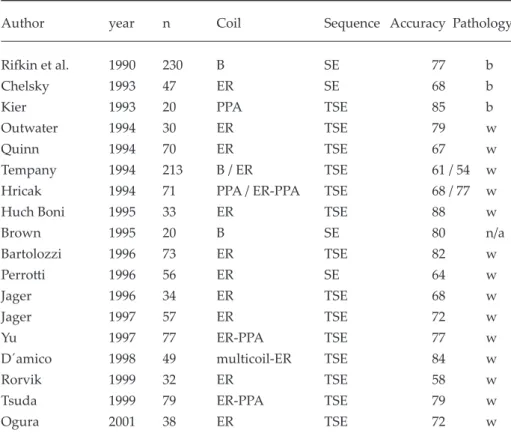
1. Determining one’s own staging performance: The role of MR imaging of the prostate is debated extensively in the literature. Initially MR imaging was performed using a conventional body coil with limited anatomical resolution [11-14]. With the introduction of new MR sequences, new coils and other technical developments numerous studies have attempted to improve local staging. The diagnostic capability of MR imaging in preoperative staging of prostate cancer is currently being established. The results, however, are diverse, as shown in table I (Table I; [15-32]).
Engelbrecht et al. performed a meta-analysis of all published studies evaluating the performance of MR imaging in the local staging of prostate cancer [33]. A wide variation in its performance was found, with a maximum combined sensitivity and specificity of 71%. This result is in agreement with a previous study by Sonnad et al. [34], who found a value of 74%. This wide variation (Table I) could only partially be explained by the use of different criteria of extraprostatic disease (strict versus lacks). If that was the case, the sensitivity-specificity pair should be located round a virtual ROC curve. Among others, the use of different techniques such as field strength, sequences, coils, contrast, patient selection, and scan direction, could not explain the wide variation in staging results. Perhaps the ideal MR of the prostate does exist, however, at this point in time, it unfortunately appears to be beyond our comprehension. In my opinion, this variation could be explained by the lack of expertise opinion, not using high specificity reading and strict criteria for extracapsular extension. However, this needs to be validated. Thus, the statement made by Jager et al. [26] in 1996 has been shown to remain appropriate: “Those who perform MR imaging in cases of prostate cancer should determine their own standard of accuracy by carefully comparing their imaging results with histopathologic findings.”
2. Consider a learning curve: An important factor in MR imaging of the prostate is its steep learning curve. It is stated that prostate MR imaging should be performed in centers where at least 25-50 patients per year are examined and where results can be compared with whole mount section histopathology [35], in order to be able for these readers to verify their own results constantly.
3. Patient selection: In general, the diagnostic capability of tests are assessed by means of sensitivity, specificity and accuracy. However, these are conditional probabilities which express the probability of a positive, respectively negative results given the fact that the patient has or does not have the disease (extra-capsular extension). However, clinicians are more interested in the probability of extra-capsular disease given a positive test result, i.e. the positive predictive value (PPV). This number is directly related with a priory chance of extra-capsular disease. The Partin tables are commonly used by urologists to determine the
likelihood of organ confined disease in patients with prostate cancer [36].
Furthermore, sensitivity and specificity may vary depending on the criteria used (strict versus lacks). The choice of lacks or strict criteria should depend on the consequences of false negative results versus false positive results. At our institution, not operating on a patient with possible curable disease is considered far worse than operating a patient who is not curable. Therefore MR is performed with high specificity being the most important goal. Langlotz has calculated that MR with a sensitivity-specificity pair of 30% - 97% is more cost-effective compared to 82 - 80% [37], although the latter has a better diagnostic capability (0.64 versus 0.81).
MR imaging may play a role in different treatment strategies. For example, it may be the final check for a patient who is considered candidate for radical prostatectomy, or it may be used to select patients with a moderate risk of extracapsular disease for either radiotherapy or surgery.
Table I: Reported staging performance of MR imaging of prostate cancer
Author year n Coil Sequence Accuracy Pathology
Rifkin et al. 1990 230 B SE 77 b
Chelsky 1993 47 ER SE 68 b
Kier 1993 20 PPA TSE 85 b
Outwater 1994 30 ER TSE 79 w
Quinn 1994 70 ER TSE 67 w
Tempany 1994 213 B / ER TSE 61 / 54 w
Hricak 1994 71 PPA / ER-PPA TSE 68 / 77 w
Huch Boni 1995 33 ER TSE 88 w
Brown 1995 20 B SE 80 n/a Bartolozzi 1996 73 ER TSE 82 w Perrotti 1996 56 ER SE 64 w Jager 1996 34 ER TSE 68 w Jager 1997 57 ER TSE 72 w Yu 1997 77 ER-PPA TSE 77 w
D´amico 1998 49 multicoil-ER TSE 84 w
Rorvik 1999 32 ER TSE 58 w
Tsuda 1999 79 ER-PPA TSE 79 w
Ogura 2001 38 ER TSE 72 w
n = number of patients; accuracy in %; B = body coil; ER = endorectal coil; PPA = pelvic phased array coil; SE = spin echo sequence; TSE = turbo spin echo sequence; b = biopsy; w = whole mount sections; n/a = not available
The effectiveness of MR imaging in other strategies is related to the prevalence of extra-prostatic disease in the group of patients undergoing MR imaging and thus to patient selection. In the literature several recommendations for patient selection are made. D’Amico et al. [29] advised the use of MR imaging of the prostate only for patients who are at intermediate risk for extracapsular extension because the probability of extraprostatic disease in this group is high enough to warrant the use of MR imaging. This was recently confirmed by Cornud et al. [38], who stated that the use of endorectal MR imaging is indicated in carefully selected patients - specifically, those with three or more positive findings in biopsy specimens, a palpable tumor, or a prostate-specific antigen level that exceeds 10 ng/mL.
With Partin’s tables (a priori chance), sensitivity and specificity on one hand, and data about life-expectancy, complication rate and cost models on the other, the cost-effectiveness of MR staging in prostate cancer can be calculated.
Pauker et al. [39, 40] developed the so-called double threshold method. They demonstrated with a decision model that if there is a high probability of having a disease the patient should be treated without testing, while if there is a low probability one should not treat and the patients should be tested. Thus, it is possible to determine two thresholds, one for testing and one for treatment without testing. These depend on the risks and costs of testing, the sensitivity and specificity of the test, the risks and benefits and costs of the treatment. Engelbrecht and colleagues [41] applied this method to MR testing. Thus, patients can be divided into three groups: a low-risk group who should undergo radical prostatectomy without MR imaging; a high-risk group of patients who should be treated by palliative radiotherapy without MR imaging and an intermediate risk group, patients in whom the selection of treatment depends on imaging results.
If one uses sensitivity and specificity of MR imaging in the assessment of stage T3 disease reported in the literature (64% and 72%, respectively), the calculated thresholds for the three categories are: radical prostatectomy without testing in the low-risk group with 45% pre-test probability of having T3 prostate cancer; MR imaging in the intermediate-risk group having a 46 - 81% pre-test probability of having stage T3 disease; and palliative radiotherapy in patients without MR in the high-risk group has a pre-test probability that exceeds 81%. Thus far, the only practical way to determine a patient’s pre-test probability of having stage T3 disease is to use the Partin tables [36, 42], the most widely used and validated nomogram [43, 44]. To be placed in the intermediate-risk group (for whom MR imaging is cost-effective), a patient should have a PSA level exceeding 10 ng/mL, a Gleason grade greater than 6, or a stage T3 tumor found on digital rectal examination.
In conclusion: data from literature suggest that high specificity MR imaging is cost-effective in the intermediate risk group. However, if results of imaging are improved such as for example reported by Fütterer et al. [45] (sensitivity of 65% and specificity 98%) the number of patients with a T3 tumour even in the low risk group (13/49) is high enough to justify MR imaging in these patients. This means with such an improved sensitiviy and specificity, all patients who are considered candidates for a radical prostatectomy should undergo MR imaging and their final treatment selection can be based on MR imaging findings.
2.3
Prostate cancer pathophysiology
Prostate cancer is a multifactorial disease. Age and a positive family history of prostate cancer are the main risk factors. Other factors are the type of diet, lifestyle-related factors, and certain genetic defects [46]. There is also a distinct geographical and racial difference in prostate cancer incidence with higher rates in Western countries and among black men, as compared to Asian countries and white men, respectively [3, 4].
On a cellular and molecular level the main task of the prostate gland is to lubricate the sperm produced in the testes during ejaculation. In healthy prostatatic epithelial cells, the enzyme aconitase is inhibited by high levels of zinc present in the cells. This, in turn, blocks the oxidation of citrate in the Krebs cycle, thus accumulating citrate in the prostatic lumina. Zinc levels decrease in prostate cancer, thereby decreasing the citrate levels. However, the causes of the loss of zinc have not yet been established and are likely to be multifactorial. [42, 48, 49]. The decrease in citrate levels is an important characteristic in spectroscopic detection of prostate cancer. (see below)
The earliest determinable pathological changes in characteristics of the healthy prostatic cells are atrophic and inflammatory changes. A cascade of these and other factors may lead to the histopathologically defined precursors of proliferative inflammatory atrophy and prostatic intraepithelial neoplasia [50].
2.3.1 Prostate anatomy
Knowledge of the zonal anatomy of the prostate is very useful considering that many prostatic diseases have a zonal distribution. More than 70% of adenocarcinomas of the prostate arise in the peripheral zone, whereas about 20% emerge in the transitional zone and 10% in the central zone. Most central gland tumors have additional tumor foci in the peripheral zone [51-53]. Since many cases of prostate cancer are multifocal, combinations of these tumor localizations frequently occur within the same patient [54]. At our institution, the prostate is divided into a central gland (the transitional and central zones) and peripheral zone (Figure 1).
There is still debate about the prostate having a capsule or not. The prostate is surrounded by a thick layer of fibromuscular tissue corresponding to the capsule. The ‘true’ prostatic capsule, however, is a thin (0.5-2mm) layer of connective tissue located external to the peripheral zone. Around this layer there is the pelvic fascia, often called the “false” prostatic capsule. Sattar et al. considered the prostate capsule as an extension of the prostate parenchyma itself [55, 56].
The periprostatic venous plexus surrounds the gland and drains into the internal iliac veins and the presacral veins. The neurovascular bundle courses along the posterolateral aspect of the gland (Figure 1) and is a preferential path for tumor spreading due to small nerve branches penetrating the prostate capsule in this area. The periprostatic fat shows high signal intensity on T2-weighted MR images, thus clearly delineating the rectum, muscles,
Figure 1. Normal prostate in a 28-year-old man. T2-weigthed MR image shows peripheral zone (PZ) with intermediate to high signal intensity. Small central gland (CG) has lower signal intensity than does the peripheral zone. The neurovascular bundle is located at the posterolateral aspect of the gland (curved arrow).
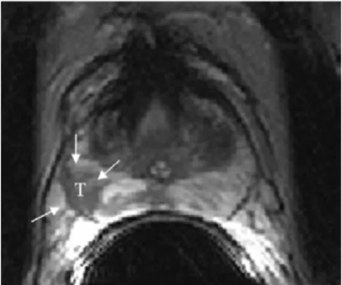
Figure 2. 56-year-old man with stage T2b prostate cancer in the right peripheral zone. T2-weighted MR image shows that the tumor (T) (arrows) has lower signal intensity than the peripheral zone.
Figure 3. 60-year-old man with stage T3a disease (T) in the left peripheral zone and central gland. T2-weighted MR image shows invasion of the neurovascular bundle (curved arrow). Obliteration of the left rectroprostatic angle (arrow), but the right neurovascular bundle and rectoprostatic angle are intact.
Figure 4A. 58-year-old man with stage T3a disease (T) in the left peripheral zone. Extracapsular extension (arrows) is clearly visible on axial T2-weighted MR image.
Figure 4B. 58-year-old man with stage T3a disease (T) in the left peripheral zone. Extracapsular extension (arrows) is clearly visible on coronal T2-weighted MR image.
Figure 5. 51-year-old man with stage T3a disease in the right peripheral zone. T2-weighted MR image shows that tumor (T) has lower signal intensity than does normal peripheral zone and shows bulging (arrows) and broad surface contact with capsule.
bones, vasculature and bladder.
On T2-weighted MR images, normal prostate tissue displays an intermediate to high signal intensity while the central gland has lower signal intensity than the peripheral zone. Conversely, the prostate has an homogeneous, intermediate signal intensity on T1-weighted images. This means differentiation between peripheral and central zone cannot be perceived.
2.3.2 Imaging of prostate carcinoma
On T2-weighted MR images, prostate carcinoma displays as a low signal intensity area in a bright normal peripheral zone (Figure 2). In addition to carcinoma, the differential diagnosis of an area of low signal intensity includes post biopsy haemorrhage, prostatitis, benign prostatic hyperplasia, effects of hormone or radiation treatment, scars, calcifications, smooth muscle hyperplasia, and fibromuscular hyperplasia. Detecting prostate carcinoma in the central gland on T2-weighted images is difficult because this area is often involved with benign prostate hyperplasia, which has signal intensity similar to that of carcinoma.
2.4 Technique
2.4.1 Imaging protocol
Interval between biopsy and MR imaging should at least be 2 to 4 weeks. At our institution regular prostate MR imaging is performed at 1.5T (Sonata, Siemens Medical Systems, Erlangen, Germany) using an integrated endorectal pelvic-phased array coil (Medrad, Pittsburgh, US). Peristalsis is suppressed by i.m. injection of 1 mg glucagon (Glucagen®; Novo Nordisk A/S, Denmark) prior to the examination. The endorectal coil is inserted and inflated with approximately 80 ml of air. The patient is then placed on the scanner table in the supine position. The scanning protocol consists of T1-weighted sagittal and axial localizer images obtained to check coil positioning. Subsequently, T2-weighted turbo spin echo images are acquired with the following parameters: 3500/132/180° [repetition time (TR) in msec/ echo time (TE) in msec/ flip angle (α)], 4-mm section thickness, 0-mm intersection gap, 15-22 slices, 280-mm field of view (FOV), 240 x 512 matrix, pixel size 0.55 x 0.55, echo train length of 15, two signals acquired). The direction of the phase encoding and read-out gradient are changed from A-P to L-R to decrease endorectal motion artifacts. In addition, axial (T1) intermediate-weighted sequence with the following parameters is required, 200 (TR), 4.4 (TE), 8° (α) and 280-320 mm FOV for detecting post biopsy hemorrhage.
2.4.2 Interpretation of MR images and pitfalls
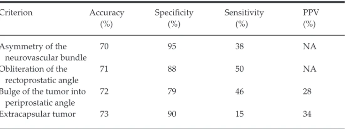
Radical retropubic prostatectomy is considered only in patients without extracapsular extension and seminal vesicle involvement [13, 28, 59]. Accurate staging is therefore especially important for the proper management of prostate cancer. The most reliable criteria for the detection of extracapsular extension of prostate carcinoma are asymmetry of the neurovascular bundle, obliteration of the rectoprostatic angle, tumor bulge into the periprostatic fat, broad tumor contact with the surface of the capsule, an extracapsular tumor, and the radiologist’s overall impression (Table II and Figure 3-5) [18, 19, 21, 28]. At our institution this is further supplemented with the charcoal criterion, a low-signal-intensity lesion with a blurring border and extension (Figure 6). The most common but misleading MR sign of extraprostatic extension, e.g. irregular bulging (especially in non-palpable tumors), is not often used. Wedge shape, diffuse extension without mass effect, and size are the morphological features of low-intensity lesions in the peripheral zone on pre-biopsy T2-weighted MR images that give the best prediction of malignancy [61]. Seminal vesicle invasion can be identified as an asymmetric area of low signal intensity in the seminal vesicles that is visible on T2-weighted MR images [18, 62]. Thickening of the tubular walls and asymmetric widening of the seminal vesicles have to be avoided as criteria, because these are non-specific signs [63]. Senile amyloidosis can mimic extraprostatic spread [64]. Prostate MR imaging should be obtained at a minimum of 2 to 4 weeks after prostate biopsy, as haemorrhage decreases staging accuracy [60]. On T1-weighted images, haemorrhages usually display high signal intensity (Figure 7). Endorectal MR imaging findings are significant predictors for detection of extracapsular disease when MR images are interpreted by genitourinary radiologists experienced with MR imaging of the prostate [65]. Engelbrecht et al. [33] suggested in their meta-analysis that the use of turbo spin echo MR sequences, an endorectal coil, and multiple imaging planes can improve staging performance of MR imaging.
Figure 6. 64-year-old man with stage T3a disease in the left peripheral zone. T2-weighted axial MR image shows a low-signal-intensity lesion with a blurring border (arrows) and extension (curved arrow), in our institution referred to as the charcoal appearance.
Figure 7. T1-weighted axial image through prostate demonstrates a high signal intensity lesion (arrow) in the right peripheral zone which represents post biopsy hemorrhage.
Table II: Criteria for Predicting Extracapsular Extension
Criterion Accuracy Specificity Sensitivity PPV
(%) (%) (%) (%)
Asymmetry of the 70 95 38 NA
neurovascular bundle
Obliteration of the 71 88 50 NA
rectoprostatic angle
Bulge of the tumor into 72 79 46 28
periprostatic angle
Extracapsular tumor 73 90 15 34
PPV = positive predictive value; NA = not applicable
2.5 Radiologic experience
2.5.1 Radiologic experience in local staging
Harris et al. [35] reported the presence of a steep learning curve in staging prostate cancer. A substantial improvement in overall staging accuracy of endorectal MR imaging can be achieved by careful pathologic correlation and by considering the anatomic features of prostate cancer.
One of the first multi-institutional studies examining detection of extracapsular extension showed no significant difference in the area under the receiver operating characteristic curve for MR imaging compared to TRUS (0.67 vs 0.62, respectively) [15]. However, this study was performed without the use of pelvic surface or endorectal coils and did not unambiguously state the imaging criteria for extracapsular extension. In 1994, the Radiologic Diagnostic Oncology Group (RDOG) study [20] utilizing the conventional body coil, and/or fat suppressed sequence with body coil and endorectal coil, reported overall accuracies for each technique of 61%, 64% and 54%, respectively. Since then only single institutional studies have been performed showing improved staging accuracies. Hricak et al. [21] found a staging accuracy of 68% using the pelvic phased array coil, which rose to 77% with integrated endorectal-pelvic phased array coil in a study population of 71 patients. In 1998, Husband et al. [66] reported superior image quality for the pelvic phased-array coil combination compared to the endorectal coil, which was mainly due to fewer artefacts. However, other groups have shown markedly improved results for endorectal coil imaging, with accuracies up to 88% [22].
In a prospective study of 82 patients with prostate cancer performed at our institution, the use of an integrated endorectal phased array coil resulted in significant improvement of anatomical details, staging accuracy, and specificity [45]. Using an endorectal-pelvic phased array coil was found to significantly reduce overstaging with equal sensitivity. Local staging
accuracy, sensitivity, and specificity using the pelvic-phased array coil alone were 59%, 54% and 62%, respectively, while using the combined endorectal-pelvic phased array coil, local staging accuracy, sensitivity, and specificity were 83%, 65%, and 98%, respectively.
2.5.2 The role of contrast enhanced MR imaging in prostate cancer
staging
Because of typical tumor contrast enhancement characteristics, cancer can be differentiated from normal tissue on fast dynamic contrast-enhanced MR imaging [67, 68]. Engelbrecht et al. [69] showed that prostate cancer demonstrated different enhancement patterns compared to both onset time, time to peak, peak enhancement and washout. Nevertheless, the number of studies evaluating the use of contrast-enhanced MR imaging in staging prostate cancer are limited, results are conflicting and difficult to compare. Fütterer et al. [70] concluded in a study of 103 patients that the use of MR imaging in prostate cancer can be incorporated in clinical practice and that multi-slice dynamic contrast-enhanced MR imaging did show significant improvement in staging performance for the inexperienced readers (88% accuracy).
2.5.3 The role of
1H-MR spectroscopy in prostate cancer staging
Image-guided proton nuclear MR spectroscopy is a technique that provides metabolic information about the prostate gland that may be used for diagnosis of prostate cancer. Prostate cancer has been shown to be characterized by a decreased level of citrate and an increased level of phosphocholine [71]. Correlations have been reported between metabolite ratios (e.g. choline and creatine over citrate ratio) and the histologic grade of prostate cancer [72]. The addition of 1H-MR spectroscopy to dynamic MR imaging can improve the tumor visualization and determination of tumor extension [73]. The addition of 1H-MR spectroscopy to MR imaging has been shown to increase staging accuracy [74] for less experienced readers and reduce interobserver variability [73].
2.6 Limitations
Hemorrhage due to biopsy can produce areas of low signal intensities that can result in discrepancies between MR imaging results and histopathology results. This has to be taken into account whenever interpreting MR images. In many papers this issue is not addressed, which is inappropriate.
An important issue in the evaluation of the data in the literature is the presence or absence of positive surgical margins. The incidence of the latter has been reported to be as high as 40% in some studies. This has major implications on the results of these studies. Are positive surgical margins T3 disease, or is this still stage T2 in case of only focal disease? Currently, no consensus has been established. In our opinion, positive surgical margins in
prostates with large tumors are considered stage T3. However, stage T2 is assigned in cases of focal disease.
There are several limitations associated with local staging of prostate cancer by MR imaging. The most important of these, as suggested by all authors, is the detection of microscopic invasion [16, 19, 45, 57]. However, none of the available imaging modalities allow the detection of microscopic invasion [75]. Positive surgical margins can contribute to the inconsistencies between imaging and pathology results. Cornud et al. [38] reported that pathologically staging tumors with positive surgical margins as stage T3, despite the fact that they appear within the limits of the prostate on MR imaging, would result in an unjustified lower accuracy of MR imaging. Epstein et al. [76] reported that patients with focal extracapsular extension showed no evidence of progression on long-term follow-up after surgery. In addition, it has been shown that endorectal MR imaging results that are negative or positive for extracapsular extension or seminal vesicle invasion help to separate patients with clinically confined disease and intermediate risk of extraprostatic spread into groups with a 78% vs. 21% 3-year rate of freedom from PSA failure [38, 77].
2.7 Future developments
One limitation of dynamic MR imaging is that the variety of MR scanners and sequences used produce different gadolinium concentration-time curves and subsequently different parametric images providing true quantitative information (Ktrans, K
ep and extracellular
volume). Therefore, development of a robust calibration method is essential. This calibration may be performed by integrating the arterial input function for correction into the imaging protocol.
As mentioned in the limitations section, the major limitation at the moment is the inability to detect microscopic invasion. The use of high magnetic field strengths offers new possibilities in prostate cancer imaging. Higher field strengths increase the signal-to-noise ratio, which may lead to a higher spatial resolution and subsequently better staging performance (see also chapter 8.8 Fütterer). Fütterer et al. [78] explored how the potential benefits of higher field strength, e.g. 3T, balances with the potential limitations for MR of the prostate. The authors stated that it is likely that imaging with an endorectal coil at a magnetic field strength of 3T expands potential clinical applications in evaluating the prostate. A voxel volume reduction from 1.21 mm3 to 0.13 mm3 could be obtained. These developments may be used to detect microscopic invasion of the prostate capsule.
2.8 Conclusions
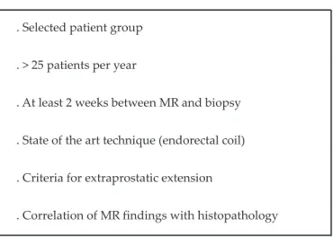
Endorectal MR imaging is the most accurate modality in staging prostate cancer and can be incorporated in the clinical work up of prostate cancer. However, several parameters have to be taken into account (Table III). The most reliable criteria for the detection of extracapsular extension of prostate cancer are asymmetry of the neurovascular bundle,
Table III: Key features for MR imaging of the prostate . Selected patient group
. > 25 patients per year
. At least 2 weeks between MR and biopsy . State of the art technique (endorectal coil) . Criteria for extraprostatic extension
. Correlation of MR findings with histopathology
obliteration of the rectoprostatic angle, tumor bulge into the periprostatic fat, broad tumor contact with the surface of the capsule, an extracapsular tumor, and the radiologist’s overall impression.
The literature reports a wide variation in the performance of MR imaging in local staging of prostate cancer. Factors contributing to improved prostate cancer MR imaging are the use of turbo spin echo MR sequence in at least two planes (axial and sagittal), an integrated endorectal-pelvic phased array coil and correlation of MR imaging result with histopathology results. MR imaging should be limited to patients with an intermediate to high probability of having extraprostatic disease. The integrated endorectal-pelvic phased array coil adds anatomical detail and improves performance of MR imaging in local staging by significantly increasing specificity.
References
1. Barry MJ. Prostate-specific-antigen testing for early diagnosis of prostate cancer. N Engl J Med 2001; 344:1373-1377
2. Jemal A, Murray T, Samuels A, et al. Cancer statistics, 2003. CA Cancer J Clin 2003; 53:5-26 3. Quinn M, Babb P. Patterns and trends in prostate cancer incidence, survival, prevalence, and
mortality. BJU Int 2002; 90:162-173
4. Quinn M and Babb P. Patterns and Trends in Prostate Cancer Incidence, Survival, Prevalence and Mortality. Part II: Individual Countries. BJU Int 2002 ; 90 :174-184
5. Frankel S, Davey Smith G, Donovan J, Neal D. Screening for prostate cancer. Lancet 2003; 361:1122-1128
6. Wang L, Mullerad M, Chen H, et al. Prostate cancer: Incremental value of endorectal MR imaging findings for prediction of extracapsular extension. Radiology 2004; 232:133-139 7. Heiken JP, Forman HP, Brown JJ. Neoplasms of the bladder, prostate and testis. Radiol Clin
North Am 1994; 32:81-98
8. May F, Truemann T, Dettmar P, et al. Limited value of endorectal magnetic resonance imaging and transrectal ultrasonography in the staging of clinically localized prostate cancer. BJU International 2001; 87:66-69
9. Sauvain JL, Palasack P, Bourscheid D, et al. Value of power Doppler and 3D vascular sonography as a method for diagnosis and staging of prostate cancer. Eur Urol 2003; 44:21-31 10. Sobin LH, Wittekind C. TNM Classification of Malignant Tumours, fifth edition, 1997, John
Wiley & Sons, Inc., New York
11. Bezzi M, Kressel HY, Allen KS, et al. Prostatic carcinoma: staging with MR imaging at 1.5 T. Radiology 1988; 169:339-346
12. Quint LA, Van Erp JS, Bland PH, et al. Carcinoma of the prostate: MR images obtained with body coils do not accurately reflect tumor volume. AJR Am J Roentgenol 1991; 156:511-516 13. Schiebler ML, Yankaskas BC, Tempany C, et al. MR imaging in adenocarcinoma of the
prostate: interobserver variation and efficacy for determining stage C disease. AJR Am J Roentgenol 1993; 158:559-562; discussion 563-564
14. Tempany CM, Rahmouni AD, Epstein JI, et al. Invasion of the neurovascular bundle by prostate cancer: evaluation with MR imaging. Radiology 1991; 181:107-112
15. Rifkin MD, Zerhouni EA, Gatsonis CA, et al. Comparison of magnetic resonance imaging and ultrasonography in staging early prostate cancer. Results of a multi-institutional cooperative trial. N Engl J Med 1990; 323:621-626
16. Chelsky MJ, Schnall MD, Seidmon EJ, Pollack HM. Use of endorectal surface coil magnetic resonance imaging for local staging of prostate cancer. J Urol 1993; 150:391-395
17. Kier R, Wain S, Troiano R. Fast spin-echo MR images of the pelvis obtained with a phased-array coil: value in localizing and staging prostatic cancer. AJR Am J Roentgenol 1993; 161:601-606
criteria for capsular extension on endorectal coil images. Radiology 1994; 193:333-339
19. Quinn SF, Franzini DA, Demlow TA, et al. MR imaging of prostate cancer with an endorectal surface coil technique: correlation with whole-mount specimens. Radiology 1994; 190:323-327 20. Tempany CM, Zhou X, Zerhouni EA, et al. Staging of prostate cancer: results of radiology
diagnostic oncology group project comparison of three MR imaging techniques. Radiology 1994; 192:47-54
21. Hricak H, White S, Vigneron D, et al. Carcinoma of the prostate gland: MR imaging with pelvic phased-array coils versus integrated endorectal-pelvic phased-array coils. Radiology 1994; 193:703-709
22. Huch Boni RA, Boner JA, Lutolf UM, et al. Contrast-enhanced endorectal coil MRI in local staging of prostate carcinoma. J Comput Assist Tomogr 1995; 19:232-237
23. Brown G, Macvicar DA, Ayton V, Husband JE. The role of intravenous contrast enhancement in magnetic resonance imaging of prostatic carcinoma. Clin Radiol 1995; 50:601-606
24. Bartolozzi C, Menchi I, Lencioni R, et al. Local staging of prostate carcinoma with endorectal coil MRI: correlation with whole-mount radical prostatectomy specimens. Eur Radiol 1996; 6:339-345
25. Perrotti M, Kaufman RP, Jennings TA, et al. Endorectal coil magnetic resonance imaging in clinically localized prostate cancer: is it accurate? J Urol 1996; 156:106-109
26. Jager GJ, Ruijter ET, van de Kaa CA, et al. Local staging of prostate cancer with endorectal MR imaging: correlation with histopathology. AJR Am J Roentgenol 1996; 166:845-852
27. Jager GJ, Ruijter ET, van de Kaa CA, et al. Dynamic TurboFLASH subtraction technique for contrast enhanced MR imaging of the prostate: correlation with histopathologic results. Radiology 1997; 203:645-652
28. Yu KK, Hricak H, Alagappan R, et al. Detection of extracapsular extension of prostate carcinoma with endorectal and phased-array coil MR imaging: multivariate feature analysis. Radiology 1997; 202:697-702
29. D’Amico AV, Schnall M, Whittington R, et al. Endorectal coil magnetic resonance imaging identifies locally advanced prostate cancer in select patients with clinically localized prostate cancer. Urology 1998; 51:449-454
30. Rorvik J, Halvorsen OJ, Albrektsen G, et al. MRI with an endorectal coil for staging of clinically localised prostate cancer prior to radical prostatectomy. Eur Radiol 1999; 9:29-34 31. Tsuda K, Yu KK, Coakley FV, et al. Detection of extracapsular extension of prostate cancer:
role of fat suppression endorectal MRI. J Comput Assist Tomogr 1999; 23:74-78
32. Ogura K, Maekawa S, Okubo K, et al. Dynamic endorectal magnetic resonance imaging for
local staging and detection of neurovascular bundle involvement of prostate cancer: correlation with histopathologuc results. Urology 2001; 57:721-726
33. Engelbrecht MR, Jager GJ, Laheij RJ, et al. Local staging of prostate cancer using magnetic resonance imaging: a meta-analysis. Eur Radiol 2002; 12:2294-2302
34. Sonnad SS, Langlotz CP, Schwartz JS. Accuracy of MR imaging for staging prostate cancer: a meta-analysis to examine the effect of technologic chance. Acad Radiol 2001; 8:149-157 35. Harris RD, Schned AR and Heaney JA. Staging of prostate cancer with endorectal MR
imaging: lessons from a learning curve. Radiographics 1995; 15:813-829
36. Partin AW, Yoo J, Carter HB, et al. The use of prostate-specific antigen, clinical stage, and Gleason score to predict pathologic stage in men with localized prostate cancer. J Urol 1993; 150:110-114
37. Langlotz CP, Schnall MD, Malkowicz SB, et al. Cost-effectiveness of endorectal magnetic resonance imaging for the staging of prostate cancer. Acad Radiol 1996; 3 Suppl 1:24-27 38. Cornud F, Flam T, Chauvenic L, et al. Extraprostatic spread of clinically localized prostate
cancer: factors predictive of pT3 tumor and of positive endorectal MR examination results. Radiology 2002; 224:203-210
39. Pauker SG, Kassirer JP. Therapeutic decision making: a cost-benefit analysis. N Engl J Med 1975; 293:220-234
40. Pauker SG, Kassirer JP. The threshold approach to clinical decision making. N Engl J Med 1980; 302:1109-1117
41. Engelbrecht MR, Laheij RJ. Patient selection for magnetic resonance imaging of prostate cancer. Eur Urol 2001; 40:300-307
42. Khan MA, Partin AW. Partin tables: past and present. BJU International 2003; 92:7-11
43. Reckwitz T, Potter SR, Partin AW. Prediction of locoregional extension and metastatic disease in prostate cancer: a review. World J Urol 2000; 18:165-172
44. Ross PL, Scardino PT, Kattan MW. A catalog of prostate cancer nomograms. J Urology 2001;
165:1562-1568
45. Fütterer JJ, Engelbrecht MR, Huisman HJ et al. MR imaging of prostate cancer: comparison of anatomical details and local staging performance using pelvis phased array and integrated endorectal pelvic phased array coils. 2004, submitted
46. Nelson WG, DeMarzo AM, Isaacs WB. Prostate cancer.N Engl J Med. 2003; 349:366-381 47. Costello LC, Franklin RB. Novel role of zinc in the regulation of prostate citrate metabolism
and its implications in prostate cancer. Prostate 1998; 35:285-296
48. Liang J-Y, Liu Y-Y, Zou J, Franklin RB, Costello LC, Feng P. Inhibitory effect of zinc on human prostatic carcinoma cell growth. Prostate 1999; 40:200-207
49. DeMarzo AM, Nelson WG, Isaacs WB, Epstein JI. Pathological and molecular aspects of prostate cancer. Lancet 2003; 361:955-964
50. DeMarzo AM, Meeker AK, Zha S, et al. Human prostate cancer precursors and pathobiology. Urology 2003; 62:55-62
51. Coakley FV, Hricak H. Radiologic anatomy of the prostate gland: a clinical approach. Radiol Clin North Am 2000; 38:15-30
52. McNeal JE. Normal and pathologic anatomy of the prostate. Urology 1981; 17:11-16
53. Billis A, Souza CAF, Piovesan H. Histologic carcinoma of the prostate in autopsies: frequency, origin, grading and terminology. Braz J Urol 2002; 28:197-206
54. Chen ME, Johnston DA, Tang K, et al. Detailed mapping of prostate carcinoma foci. Cancer 2000; 89:1800-1809
55. Sattar AA, Noël J-C, Vanderhaeghen J-J, Schulman CC, Wespes E. Prostate capsule: computerized morphometric analysis of its components. Urology 1995; 46:178-181
56. Greenhalgh R, Kirby RS. Anatomy and physiology of the prostate and benign prostatic hyperplasia. Atlas Urol Clin 2002; 10:1-9
57. Schnall MD, Imai Y, Tomaszewski J, et al. Prostate cancer ; local staging with endorectal surface coil MR imaging. Radiology 1991; 178:797-802
58. Sommer FG, Nghiem HV, Herfkens R, McNeal J, Low RN. Determining the volume of prostatic carcinoma: value of MR imaging with an external-array coil. AJR Am J Roentgenol. 1993; 161:81-86
59. Schiebler ML, Schnall MD, Pollack HM, et al. Current role of MR imaging in the staging of adenocarcinoma of the prostate. Radiology 1993; 189:339-352
60. White S, Hricak H, Forstner R, et al. Prostate cancer: effect of postbiopsy hemorrhage on interpretation of MR images. Radiology 1995; 195:385-390
61. Cruz M, Tsuda K, Narumi Y, et al. Characterization of low-intensity lesions in the peripheral zone of prostate on pre-biopsy endorectal coil MR imaging. Eur Radiol. 2002; 12:357-365 62. Chernoff DM, Hricak H. The male pelvis: prostate and seminal vesicles. Magnetic resonance
imaging of the body. New York: Lippincot-Raven, 1997; 875-900
63. Ramchandani P, Schnall MD, LiVolsi VA, et al. Senile amyloidosis of the seminal vesicles mimicking metastatic spread of prostatic carcinoma on MR images. AJR Am J Roentgenol 1993; 161:99-100
64. Jager GJ, Barentsz JO, de la Rosette JJ, Rosenbusch G. Preliminary results of endorectal surface coil magnetic resonance imaging for local staging of prostate cancer. Radiologe 1994; 34:129-133
65. Mullerad M, Hricak H, Wang L, et al. Prostate cancer: detection of extracapsular extension by genitourinary and general body radiologists at MR imaging. Radiology 2004; 232:133-139 66. Husband JE, Padhani AR, MacVicar AD, et al. Magnetic resonance imaging of prostate cancer:
comparison of image quality using endorectal and pelvic phased array coils. Clin Radiol 1998;53:673-681
67. Barentsz JO, Jager GJ, van Vierzen PBJ, et al. Staging urinary bladder cancer after transurethral biopsy: the value of fast dynamic contrast-enhanced MR imaging. Radiology 1996; 201:185-193
68. Boetes C, Barentsz JO, Mus RD, et al. MR characterization of suspicious breast lesions with gadolinium-enhanced Turbo-FLASH subtraction technique. Radiology 1994; 193:777-781 69. Engelbrecht MR, Huisman HJ, Laheij RJF et al. Discrimination of prostate cancer from
peripheral zone and central gland tissue using dynamic contrast-enhanced MR imaging. Radiology 2003; 229:248-254
70. Fütterer JJ, Engelbrecht MR, Huisman HJ et al. Dynamic Contrast-Enhanced Endorectal MR
Imaging in Staging Prostate Cancer prior to Radical Prostatectomy – Experienced versus Less Experienced Readers. Radiology, 2005; 237:541-549
71. Heerschap A, Jager GJ, van der Graaf M, et al. In vivo proton MR spectroscopy reveals altered metabolite content in malignant prostate tissue. Anticancer Res 1997; 17:1455-1460
72. Coakley FV, Qayyum A, Kurhanewicz J. Magnetic resonance imaging and spectroscopic imaging of prostate cancer. The J of Urol 2003; 170:S69-S76
73. Yu KK, Scheidler J, Hricak H, et al. Prostate cancer: prediction of extracapsular extension with endorectal MR imaging and three-dimensional proton MR spectroscopic imaging. Radiology 1999; 213:481-488
74. Fütterer JJ, Scheenen TWJ, Huisman HJ et al. Dynamic contrast-enhanced using both high spatial and temporal resolution MRI and 3D-MR spectroscopy of prostate cancer. RSNA 2002 75. Akin O, Agildere AM, Ersoy H, et al. Local staging of prostate cancer with endorectal surface
coil in a mid-magnetic field. Journal of clinical imaging 2003; 27:47-51
76. Epstein JI, Carmichael MJ, Pizov G, et al. Influence of capsular penetration on progression following radical prostatectomy: a study of 196 cases with long-term followup. J Urol 1993; 150:135-141
77. D’Amico AV, Whittington R, Malkowicz SB, et al. Critical analysis of the ability of the endorectal coil magnetic resonance imaging scan to predict pathologic stage, margin status, and postoperative prostate-specific antigen failure in patients with clinically organ-confined prostate cancer. J Clin Oncol 1996; 14:1770-1777
78. Fütterer JJ, Scheenen TWJ, Huisman HJ, et al. Initial experience of 3T endorectal coil MR imaging and 1H-spectroscopic imaging of the prostate. Invest radiol 2004; 39:671-680
3
Prostate cancer:
comparison of local staging
accuracy of pelvic phased
array coil alone versus
integrated endorectal pelvic
phased array coils
J.J. Fütterer, M.R.W. Engelbrecht, G.J. Jager,
R.P. Hartman, B.F. King,