1535-9778/09/$08.00⫹0 doi:10.1128/EC.00234-08
Copyright © 2009, American Society for Microbiology. All Rights Reserved.
Assembly of the
Trypanosoma brucei
60S Ribosomal Subunit Nuclear
Export Complex Requires Trypanosome-Specific Proteins
P34 and P37
䌤
†
Kimberly Prohaska and Noreen Williams*
Department of Microbiology and Immunology & Witebsky Center for Microbial Pathogenesis and Immunology, 253 Biomedical Research Building, University at Buffalo, Buffalo, New York 14214
Received 15 July 2008/Accepted 14 August 2008
We previously identified twoTrypanosoma bruceiRNA binding proteins, P34 and P37, and determined that
they are essential for proper ribosomal assembly in this organism. Loss of these proteins via RNA interference is lethal and causes a decrease in both 5S rRNA levels and formation of 80S ribosomes, concomitant with a decrease in total cellular protein synthesis. These data suggest that these proteins are involved at some point in the ribosomal biogenesis pathway. In the current study, we have performed subcellular fractionation in conjunction with immune capture experiments specific for 60S ribosomal proteins and accessory factors in order to determine when and where P34 and P37 are involved in the ribosomal biogenesis pathway. These studies demonstrate that P34 and P37 associate with the 60S ribosomal subunit at the stage of the nucleolar 90S particle and remain associated subsequent to nuclear export. In addition, P34 and P37 associate with conserved 60S ribosomal subunit nuclear export factors exportin 1 and Nmd3, suggesting that they are
components of the 60S ribosomal subunit nuclear export complex inT. brucei. Most significantly, the pre-60S
complex does not associate with exportin 1 or Nmd3 in the absence of P34 and P37. These results demonstrate
that, althoughT. brucei60S ribosomal subunits utilize a nuclear export complex similar to that described for
other organisms, trypanosome-specific factors are essential to the process.
Trypanosoma brucei is a parasitic protozoan and the caus-ative agent of African sleeping sickness in humans and nagana in cattle (26). It is an organism that, while extensively studied, continues to pose a serious threat to human health and eco-nomic development in sub-Saharan Africa (29). It is one of only a few diseases that is 100% fatal if left untreated and, due to the toxicity of current treatments and increasing incidence of parasite drug resistance, new chemotherapeutic agents are a necessity (1, 8). Of particular interest for drug development are trypanosome-specific factors that are required for pro-cesses that are essential to the parasite’s survival.
Ribosomal biogenesis is an essential process in all organisms ranging from bacteria to humans, and the framework of this process is highly conserved among the eukaryotic kingdoms (39). The ribosomal biogenesis pathway of the yeast Saccha-romyces cerevisiaehas been extensively studied and often serves as a paradigm for the eukaryotic pathway (Fig. 1). In this organism, the 60S ribosomal subunit consists of 46 ribosomal proteins and three rRNA species (5S, 5.8S, and 25S), whereas the 40S ribosomal subunit is comprised of 32 ribosomal pro-teins and 18S rRNA (12, 13). In addition to the ribosomal proteins that comprise the 60S and 40S ribosomal subunits, approximately 140trans-acting proteins have been identified in yeast as being required for proper ribosomal assembly to
oc-cur, most of which function in 60S biogenesis (10). Among these factors are several proteins that are required for the nuclear export of yeast pre-60S ribosomal subunits, including exportin 1 (XpoI, also known as CRM1) and Nmd3 (14, 20), Mex67-Mtr2 (44), and Arx1 (3, 21).
Following export from the nucleus, yeast pre-60S particles undergo their final maturation steps in the cytoplasm, includ-ing addition of the final large ribosomal proteins, release and recycling of nuclear export factors and, ultimately, joining with a 40S subunit/mRNA complex to form the mature 80S ribo-some (9, 17, 27, 28, 32). Thus, the work that has been per-formed inS. cerevisiaehas demonstrated that, while ribosome biogenesis is very rapid (42), it is a highly complex and multi-factorial process.
The ribosomal biogenesis pathway has not been extensively studied inT. brucei, but this organism does contain putative homologues to both 40S and 60S ribosomal proteins as anno-tated in the T. brucei gene database (http://www.genedb.org /genedb/tryp/index.jsp). It is also known that some aspects of the 60S pathway contain characteristics similar to other organ-isms, such as the existence of an L5-5S preribosomal particle (30) and the involvement of a conserved nucleolar protein, NOG1, in 60S subunit biogenesis (23, 31). However, some differences exist as well, including the processing of the 60S rRNA species (28S rRNA) into six smaller rRNA molecules (5, 43) and the involvement of the trypanosome-specific family of proteins, NOPP44/46, in 60S biogenesis (22). In addition, ribosome biogenesis and protein synthesis are developmentally regulated throughout the life cycle of the parasite (2, 4, 38), indicating that factors involved in this process may be similarly regulated themselves.
There is further evidence supporting the hypothesis that * Corresponding author. Mailing address: 253 Biomedical Research
Building, Department of Microbiology and Immunology, University at Buffalo, Buffalo, NY 14214. Phone: (716) 2279. Fax: (716) 829-2158. E-mail: [email protected].
† Supplemental material for this article may be found at http://ec .asm.org/.
䌤Published ahead of print on 22 August 2008.
77
on September 8, 2020 by guest
http://ec.asm.org/
developmentally regulated factors are involved in T. brucei ribosomal biogenesis. Our laboratory has shown that the de-velopmentally regulated, trypanosome-specific proteins, P34 and P37, associate with 5S rRNA (34), ribosomal protein L5 (K. Hellman, K. Prohaska, and N. Williams, unpublished data), and a family of nucleolar phosphoproteins, NOPP44/46 (33). More recently, our laboratory has discovered that, when P34 and P37 expression is knocked down using RNA interference (RNAi), it is lethal to the cells (18). In addition, there is a 25-fold decrease in the levels of 5S rRNA, and the polysomal profile of P34/P37 RNAi cells shows a decrease in 80S ribo-some formation, concomitant with an overall decrease in pro-tein synthesis (18). These results demonstrate that P34 and/or P37 is involved in the ribosomal biogenesis pathway and/or translation initiation.
Because ribosomal biogenesis is a highly complex process, there are several points in the pathway at which P34 and/or P37 may be involved. In Fig. 1, we present a simplified version of the yeast pathway as a model for our system, since it has been extensively studied. Beginning in the nucleolus, the 90S parti-cle is the first complex formed in the pathway (15), which includes 60S ribosomal protein L3 (27, 35). Following cleavage to form separate 66S and 43S particles, 60S ribosomal protein L11 is added to 66S in the nucleolus (16, 27), after which the particle is released into the nucleoplasm (24). Following fur-ther nucleoplasmic maturation, the pre-60S particle associates with exportin 1 through the Nmd3 adaptor protein (14, 20) and is then exported to the cytoplasm. Once in the cytoplasm, the final 60S ribosomal proteins are added as one of the last mat-uration steps (9, 27) prior to subunit joining. Finally, the ma-ture 60S subunit is joined with an mRNA-bound 40S subunit to form the mature 80S ribosome (25) and translation initiation can take place. As indicated in Fig. 1, P34 and P37 may become
associated with the maturing 60S complex at any one of these steps in the ribosomal biogenesis pathway. However, the asso-ciation of P34 and P37 with exportin 1 (19) supports a role(s) in nuclear export of the 60S ribosomal subunit for these pro-teins.
In order to determine when and where P34 and P37 are involved in ribosomal biogenesis, we have utilized subcellular fractionation together with immune capture experiments spe-cific to various ribosomal proteins and ribosomal biogenesis accessory factors based on the yeast pathway. These ex-periments demonstrate thatT. bruceirequires trypanosome-specific proteins in the otherwise highly conserved ribosomal biogenesis pathway.
MATERIALS AND METHODS
Cell culture.Procyclic-formT. brucei bruceistrain 427 was grown in Cunning-ham’s medium supplemented with 10% fetal calf serum (6). Clonal P34/P37 RNAi cells have been described previously (19). Induction of double-stranded RNA specific to P34 and P37 was initiated by addition of tetracycline (1g/ml; Sigma, St. Louis, MO) to a concentration of 1⫻106cells/ml. Cells were
har-vested 4 days postinduction and Western blot analysis was performed, probing for P34 and P37 to confirm the absence of both proteins.
Antibodies.The antibodies used for this study were as follows: P34/P37 poly-clonal antiserum (47), affinity-purified P34/P37 polypoly-clonal antibody (Bethyl Lab-oratories, Montgomery, TX), L3 polyclonal antiserum (Affinity Bioreagents, Golden, CO), L11 polyclonal antiserum (Affinity Bioreagents), L10 monoclonal antibody (Novus Biologicals, Littleton, CO), affinity-purified L5 polyclonal anti-body (Bethyl Laboratories), S6 polyclonal antiserum (Affinity Bioreagents), af-finity-purified Nmd3 polyclonal antiserum (a gift from Arlen Johnson), affinity-purifiedT. bruceiexportin 1 polyclonal antibody (Bethyl Laboratories), NOG1 and phosphoglycerate kinase polyclonal antibodies (gifts from Marilyn Parsons), and TATA binding protein polyclonal antiserum (a gift from Vivian Bellofatto).
Specificity of antibodies.The ribosomal protein antibodies used in this study were commercially obtained and were raised to recombinant human proteins. Prior to testing the specificity of these antibodies to the trypanosome proteins, we aligned each of the human protein sequences with those fromT. brucei(see Fig.
FIG. 1. Ribosomal biogenesis and the possible points of involvement of P34 and P37. This diagram depicts the biogenesis of yeast 60S ribosomal subunits, including proteins that have known associations at specific stages in this process. We have also included two of the known 60S subunit export factors, exportin 1 and Nmd3. The points at which P34 and/or P37 may become involved in the ribosomal biogenesis pathway are indicated with solid arrows. Dashed arrows indicate known biogenesis and transport processes. Data are from reference 27.
on September 8, 2020 by guest
S1 to S5 in the supplemental material). We next performed immune capture experiments (described below) with cytoplasmic and nuclear extracts specific for L3, L11, L10, S5, and S6. Subsequent Western blot analyses using the same antibodies used for the immune capture showed that each antibody demon-strated reactivity to the appropriately sized protein and that the majority of each protein was present in the pellet fraction compared to the supernatant (see Fig. S6 in the supplemental material).
We also aligned the yeast Nmd3 protein sequence with the homologousT. bruceiprotein, which revealed a high degree of identity between the two species (see Fig. S7 in the supplemental material). We utilized an antibody raised to yeast Nmd3 (a generous gift from Arlen Johnson) for Western blot analyses on nuclear and cytoplasmic extracts, the results of which demonstrated that this antibody cross-reacts with a protein of the appropriate molecular weight (data not shown). These results confirmed thatT. bruceicontains an Nmd3 homologue and that the yeast antibody was useful for further experiments.
Cellular fractionation.Cytoplasmic and whole nuclear extracts were prepared as described previously (19). Nucleolar and nucleoplasmic extracts were pre-pared as described previously (36).
Immune capture experiments.Immune capture experiments were done as described previously (19) using antibodies specific to the following proteins: P34 and P37, exportin 1, and L3, L11, L10, S5, and S6. Briefly, 500g of the indicated extract was incubated with antibody-cross-linked Dynabeads (Invitrogen, Carls-bad, CA). After incubation, the beads were washed with phosphate-buffered saline and eluted with sodium dodecyl sulfate-polyacrylamide gel electrophoresis (SDS-PAGE) sample buffer. For each immune capture experiment, the pro-tein(s) present in the pellet fractions represents the majority of the protein, compared to that in the unbound fraction (data not shown).
Western blot analyses.Following SDS-PAGE, the gels were transferred to nitrocellulose membranes (Schleicher & Schuell, Keene, NH) and blocked in 10% nonfat dry milk. Membranes were incubated with the indicated primary antibody overnight, followed by 10 washes with Tris-buffered saline with Tween and incubation with the appropriate horseradish peroxidase-conjugated second-ary antibody. Proteins were detected using SuperSignal West Pico chemilumi-nescent substrate (Pierce, Rockford, IL).
RT-PCR analyses.Immune capture experiments were performed as described above, with the modification of incubating the Dynabeads with poly(dI-dC) (Sigma, St. Louis, MO) overnight in order to decrease nonspecific RNA binding. The beads were eluted by resuspension in elution buffer (1% SDS, 50 mM dithiothreitol, and 10%-mercaptoethanol) and boiling for 5 min. Bound RNA was isolated using Trizol reagent (Invitrogen, Carlsbad, CA) as per the manu-facturer’s instructions. Subsequent reverse transcription-PCR (RT-PCR) analy-ses were performed using primers specific to internal sequences of processed 60S rRNA (28S), 40S rRNA (18S), and the entire processed 5S rRNA sequence (5). A fraction of each reaction mixture was subjected to agarose gel electrophoresis and ethidium bromide staining to visualize the RT-PCR products.
Sequential immune capture experiments.Sequential immune capture exper-iments were performed as described previously (19). Briefly, the primary immune capture against exportin 1 was performed as described for the single immune capture experiments with the modification of starting with 2 mg of nuclear extract. The beads were eluted as described for the RT-PCR analyses, followed by dilution with 1 ml of nondenaturing lysis buffer. Samples were allowed to cool to room temperature and were then used for a secondary immune capture experiment specific for L11. P34/P37 Western blot analyses were performed on the resulting pellet fractions.
RESULTS
Subcellular fractionation demonstrates that P34 and P37
are present in the nucleolar fraction of procyclic cells.
Previ-ous work in our laboratory has demonstrated that P34 and P37 are present in the 80S fractions of polysomal preparations (18) and, more recently, we have shown that they are also present in the 60S and polysomal fractions (M. Ciganda, unpublished data). We next wished to determine more specifically when and where these proteins become associated with the 60S ribo-somal subunit. Subcellular fractionation was employed in order to obtain wild-type procyclic whole nuclear, nucleoplasmic, nucleolar, and cytoplasmic extracts. It should be noted that the whole nuclear extract contains both nucleoplasmic and nucle-olar material, which is further fractionated to yield the
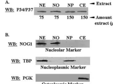
sepa-rate nucleoplasmic and nucleolar extracts. Western blot anal-yses probing for cellular compartmental markers for each fraction demonstrated that we had isolated distinct subcellular fractions (Fig. 2B), which could be used in subsequent immune capture experiments. In addition, Western blot analysis prob-ing for P34 and P37 demonstrated that both proteins were present in each of our subcellular fractions (Fig. 2A). This figure is representative of three separate experiments. The presence of these proteins in the nucleolar fraction, which had not been demonstrated previously, suggests that they may be involved in earlier nucleolar stages of ribosomal biogenesis. Thus, utilizing subcellular fractionation in conjunction with immune capture experiments, we sought to determine when and where P34 and P37 are involved in the ribosomal biogen-esis pathway. Since we had determined previously that P34 and P37 associate with both 5S rRNA (33) and ribosomal protein L5 (Hellman et al., unpublished), two components of the 60S ribosomal subunit, we focused our initial experiments on po-tential 60S ribosomal protein binding partners.
P34 and P37 associate with L3-containing 60S ribosomal
subunits.One of the earliest ribosome-associating 60S
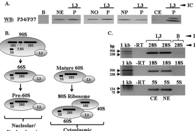
pro-teins is L3, which is added to the 90S particle in the nucleolus (27, 35). The association of a protein(s) with L3, particularly in the nucleolus, may be indicative that it functions early in ribo-somal biogenesis. Immune capture experiments specific for L3 were performed with whole nuclear, nucleolar, nucleoplasmic, and cytoplasmic extracts fromT. brucei, followed by Western blot analyses probing for P34 and P37. These experiments demonstrated that P34 and P37 associated with L3, either directly or indirectly, in all fractions tested (Fig. 3A, P lanes). Although P37 was not detected in the immune capture pellet fraction for all extracts, additional Western blot analyses dem-onstrated that it is present in each of these fractions (data not shown) but may not be seen in all blots due to its lower abundance relative to P34 (47). In addition, we confirmed that FIG. 2. P34 and P37 are present in the nucleolar fraction of pro-cyclic cells. Wild-type propro-cyclic cells were fractionated into whole nu-clear, nucleolar, nucleoplasmic, and cytoplasmic extracts. (A) A frac-tion of each extract was subjected to SDS-PAGE and Western blot analysis probing for P34 and P37. (B) A fraction of each extract was subjected to SDS-PAGE and Western blot analysis probing for specific cellular compartmental markers. NOG1, a nucleolar marker; TBP (TATA binding protein), a nucleoplasmic marker; PGK (phosphoglyc-erate kinase), a cytoplasmic marker. CE, 50g of cytoplasmic extract; NE, 50g of whole nuclear extract; NO, 100g of nucleolar extract; NP, 100g of nucleoplasmic extract.
on September 8, 2020 by guest
http://ec.asm.org/
our antibody precipitated L3 in each fraction by performing L3 Western blot analyses on each pellet fraction (data not shown). These experiments demonstrated that P34 and P37 are asso-ciated with the maturing 60S ribosomal subunit throughout the biogenesis pathway, beginning at an early, nucleolar step.
In order to ensure that L3 was being precipitated in the context of the 60S subunit and not simply free protein, we performed RT-PCR analyses on immune capture pellet frac-tions from experiments performed with cytoplasmic and nu-clear extracts. RT-PCR experiments were performed specific to 60S rRNA (28S and 5S) and 40S rRNA (18S) species.
The rRNA species found in association with L3, as demon-strated in yeast (27), are shown in Fig. 3B. L3 associates with the early 90S particle in yeast (Fig. 1), and if this is also true for T. brucei, it should be complexed with not only 28S and 5S rRNA in nuclear extracts but also 18S rRNA. In addition, L3 should associate with 28S and 5S rRNA in cytoplasmic ex-tracts, since it remains associated with mature 60S subunits, and with 18S rRNA subsequent to subunit joining. RT-PCR analyses performed on RNA extracted from nuclear and cyto-plasmic L3 immune capture experiments fromT. brucei demon-strated that L3 does associate with 28S, 18S, and 5S rRNA in both subcellular fractions (Fig. 3C). These results demonstrate that L3 in our immune capture experiments is precipitating 60S com-plexes, although we cannot rule out the possibility that some of the L3 is free protein.
P34 and P37 associate with 40S ribosomal protein S5-con-taining ribosomal particles in nucleolar and whole nuclear
extracts.An association of P34 and P37 with L3-containing 60S
particles in the nucleolar fraction may be interpreted in one of
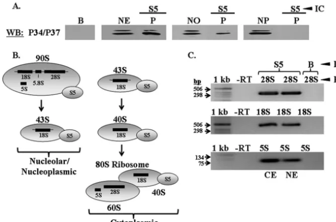
two ways: (i) P34 and P37 are associated with 90S particles or (ii) P34 and P37 associate with the subunit subsequent to the rRNA cleavage event that gives rise to the 66S and 43S sub-units. Both 90S and 66S particles are present in the nucleolus and, thus, it is important to distinguish between them to more precisely determine the 60S particle(s) with which P34 and P37 associate. In order to determine whether P34 and P37 are involved in ribosomal biogenesis at the initial 90S particle, we performed immune capture experiments specific to S5, a 40S ribosomal subunit protein that comes into the pathway at the 90S particle stage (Fig. 1) (15). Since P34 and P37 have thus far been shown to associate only with specific 60S subunit factors, an association with S5 in whole nuclear or nucleolar extracts would be indicative of P34 and P37 being present in 90S par-ticles. Immune capture experiments specific to S5 followed by Western blot analyses probing for P34 and P37 demonstrated that both proteins are complexed with S5 in whole nuclear and nucleolar extracts (Fig. 4A, P lanes), which indicates that these proteins become involved in 60S subunit biogenesis at the 90S nucleolar stage prior to subunit separation. However, neither protein associates with S5-containing particles in the nucleo-plasm (Fig. 4A, P lane), demonstrating that the association between P34 and P37 and S5 is specific to the precleaved particle and, subsequent to cleavage, they are found specifi-cally with the 60S subunit. In addition, RT-PCR experiments performed on P34/P37 immune captures using whole nuclear extracts demonstrate a complex between these proteins and 18S rRNA, most likely while it is still part of the 35S rRNA precursor (data not shown).
The rRNA species with which S5 associates in yeast are FIG. 3. P34 and P37 associate with 60S protein L3. Immune capture experiments specific to L3 were performed with whole nuclear, nucleolar, nucleoplasmic, and cytoplasmic extracts. (A) Western blot analyses were performed, probing for P34 and P37. B, immune capture beads alone; NE, 50g of whole nuclear extract; NO, 50g of nucleolar extract; NP, 50g of nucleoplasmic extract; CE, 50g of cytoplasmic extract; P, immune capture pellet fraction. (B) Diagram depicting the ribosomal complexes and rRNA species with which L3 associates. (C) RT-PCR analyses performed on RNA extracted from L3 immune capture experiments using cytoplasmic and whole nuclear extracts. 1 kb, 1-kb DNA ladder (Invitrogen); -RT, no-RT control; 28S, 28S RT-PCR; 18S, 18S RT-PCR; 5S, 5S RT-PCR; B, immune capture beads alone (control); CE, cytoplasmic extract immune capture; NE, whole nuclear extract immune capture. Results shown are representative of at least three separate experiments.
on September 8, 2020 by guest
demonstrated in Fig. 4B. Since S5 is a 40S protein that is added to the 90S particle in the nucleolus and remains associated throughout the 40S subunit maturation pathway (15, 27), it should associate with 18S and 28S rRNA species in nuclear extracts and all three rRNA species in cytoplasmic extracts (Fig. 4B). Our results demonstrate that S5 is complexed with 28S, 18S, and 5S rRNA in both nuclear and cytoplasmic ex-tracts (Fig. 4C). There is some controversy over the stage at which 5S rRNA becomes incorporated into the yeast 60S sub-unit. Some studies have indicated that it integrates into 90S particles (46), whereas others suggest that it is incorporated into the 66S particle (7). Our results demonstrate that, inT. brucei, 5S rRNA incorporates into the 90S ribosomal particle.
P34 and P37 associate with L11-containing 60S ribosomal
subunits.In order to confirm that P34 and P37 remain
com-plexed with the developing 60S ribosomal subunit, we used L11 as a marker for the late-associating proteins that are part of the 66S ribosomal particle (Fig. 1). Immune capture experiments specific for L11 demonstrated an association between P34 and P37 and L11, which is either direct or indirect, in all extracts tested (Fig. 5A, P lanes). Western blot analyses probing for L11 demonstrated that this protein was precipitated from each extract (data not shown).
Figure 5B shows the rRNA species associated with L11 throughout the yeast 60S subunit biogenesis pathway. Because L11 assembles into the maturing 60S particle at the 66S stage (Fig. 1 and 5B) (16), it should be complexed with 28S and 5S rRNA, but not 18S, inT. bruceinuclear extracts if this follows the yeast model. In cytoplasmic extracts, L11 should associate with 28S and 5S rRNA through its involvement with the
ma-ture 60S subunit and 18S rRNA in the context of the 80S ribosome (Fig. 5B). RT-PCR analyses performed on L11 im-mune capture experiments fromT. bruceidemonstrate that it associates with 28S and 5S rRNA but not 18S rRNA (Fig. 5C) in the nucleus and with all three rRNA species in cytoplasmic extracts (Fig. 5C).
The results shown in Fig. 5 demonstrate that P34 and P37 associate with the developing 60S ribosomal subunit at multi-ple steps during its biogenesis, an unusual characteristic for trans-acting factors involved in ribosomal biogenesis. The trans-acting factors typically act on one or two ribosomal par-ticle intermediates, after which they are released from the particle and replaced with subsequent factors (37).
In addition, since P34 and P37 were found to associate with 60S particles in both nuclear and cytoplasmic fractions, we next sought to determine whether they are components of the 60S nuclear export complex. Previous results demonstrated an as-sociation between P34 and P37 and exportin 1 (19), which is one of the factors required for the transport of nuclear pre-60S subunits to the cytoplasm in yeast and mammalian cells (14, 20, 40). We then hypothesized that this P34/P37-exportin 1 asso-ciation may occur in the context of the 60S ribosomal subunit. Although the 60S nuclear export complex has been elucidated in other organisms and found to contain exportin 1 and Nmd3, a nuclear export signal (NES)-containing adaptor protein (14, 20, 41), this has not been studied inT. brucei. There-fore, we first needed to determine whetherT. bruceiutilizes a nuclear export complex similar to that described for yeast and mammals.
FIG. 4. P34 and P37 associate with S5 in whole nuclear and nucleolar extracts. Wild-type procyclic whole nuclear, nucleolar, and nucleoplasmic extracts were used for immune capture experiments specific for 40S protein S5. (A) Western blot analyses were performed, probing for P34 and P37. B, immune capture beads alone; NE, 50g of whole nuclear extract; NO, 50g of nucleolar extract; NP, 50g of nucleoplasmic extract; P, immune capture pellet fraction. (B) Diagram depicting the ribosomal complexes and rRNA species with which S5 associates. (C) RT-PCR analyses performed on RNA extracted from S5 immune capture experiments using cytoplasmic and whole nuclear extracts. 1 kb, 1-kb DNA ladder (Invitrogen); -RT, no-RT control; 28S, 28S RT-PCR; 18S, 18S RT-PCR; 5S, 5S RT-PCR; B, immune capture beads alone (control); CE, cytoplasmic extract immune capture; NE, whole nuclear extract immune capture. Results shown are representative of at least three separate experiments.
on September 8, 2020 by guest
http://ec.asm.org/
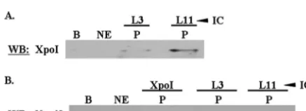
T. brucei60S subunits associate with exportin 1.In order to determine whetherT. brucei60S ribosomal subunits associate with exportin 1, we performed immune capture experiments specific for L3 and L11 using whole nuclear extracts. These experiments demonstrated that L3- and L11-containing 60S particles associate with exportin 1, although each does so to a different extent (Fig. 6A, P lanes). We do not currently know if this is due to stoichiometric differences or if it is due to our experimental procedures (e.g., differences in the efficiencies of the antibodies used in the immune capture). These results demonstrate that T. brucei 60S subunits form an exportin 1-containing nuclear export complex.
T. brucei60S subunits associate with an Nmd3 homologue.
In addition to exportin 1, it has been shown that Nmd3 is required for the nuclear export of 60S ribosomal subunits by providing the NES for the 60S-exportin 1 interaction (14, 20, 41). However, it was not known whetherT. brucei contains an Nmd3 protein homologue, so we used the yeast Nmd3 se-quence to query theT. bruceidatabase and found a
homolo-gous protein (accession number XP_845721). The two se-quences were aligned using ClustalW, which found an approximately 40% identity between the protein sequences from both organisms (see Fig. S7 in the supplemental mate-rial). We also determined that an Nmd3 antibody which had been raised to the yeast protein cross-reacted with an appro-priately sized protein inT. bruceiextracts (data not shown).
Since we had determined thatT. bruceicontains an Nmd3 homologue, we next wished to determine whether exportin 1 and 60S subunits form a nuclear export complex containing this protein. Immune capture experiments specific to exportin 1, L3, and L11 were performed using wild-type procyclic nu-clear extracts, followed by Western blot analyses probing for Nmd3. Each protein showed an interaction with Nmd3 (Fig. 6B, P lanes), demonstrating that theT. brucei60S ribosomal subunit forms a nuclear export complex containing Nmd3.
P34 and P37 associate with the 60S nuclear export complex.
Having now demonstrated that T. brucei60S ribosomal sub-units form a complex with both exportin 1 and Nmd3, we next wished to determine whether P34 and P37 are a part of this complex. We have shown previously that P34 and P37 associate with exportin 1 (19). In order to determine whether this com-plex also includes pre-60S subunits that are loaded with Nmd3, we performed immune capture experiments specific for P34 and P37 using procyclic whole nuclear extracts. Subsequent Western blot analyses probing for Nmd3 demonstrated that P34 and P37 associate with Nmd3 (Fig. 7A).
In order to confirm that P34 and P37 associate with exportin 1 and the 60S subunit simultaneously, we performed sequential immune capture experiments using nuclear extracts. We have used this technique previously to establish the complex formed between P34, P37, NOPP44/46, and exportin 1 (19). Perform-ing a series of immune captures allows for enrichment of com-plexes containing specific proteins. We performed the first FIG. 5. P34 and P37 associate with L11. Wild-type procyclic nuclear, nucleolar, nucleoplasmic, and cytoplasmic extracts were used for immune capture experiments specific for L11. (A) Western blot analyses were performed, probing for P34 and P37. B, immune capture beads alone; NE, 50g of whole nuclear extract; NO, 50g of nucleolar extract; NP, 50g of nucleoplasmic extract; CE, 50g of cytoplasmic extract; P, immune capture pellet fraction. (B) Diagram depicting the ribosomal complexes and rRNA species with which L11 associates. (C) RT-PCR analyses performed on RNA extracted from L11 immune capture experiments using cytoplasmic and whole nuclear extracts. 1 kb, 1-kb DNA ladder (Invitrogen); -RT, no-RT control; 28S, 28S RT-PCR; 18S, 18S RT-PCR; 5S, 5S RT-PCR; B, immune capture beads alone (control); CE, cytoplasmic extract immune capture; NE, whole nuclear extract immune capture. Results shown are representative of at least three separate experiments.
FIG. 6.T. brucei60S ribosomal proteins associate with exportin 1 and Nmd3. Immune capture experiments specific to 60S proteins L3 and L11 were performed using wild-type whole nuclear extracts. Sub-sequent Western blot analyses were performed probing for exportin 1 (A) and Nmd3 (B). B, immune capture beads alone; NE, 50g of whole nuclear extract; P, immune capture pellet fraction. Results shown are representative of at least three separate experiments.
on September 8, 2020 by guest
immune capture specific to exportin 1, which was eluted, re-natured, and utilized for a second capture specific to L11. Western blot analyses probing for P34 and P37 demonstrated that these proteins are present in a complex containing both exportin 1 and 60S, although P37 may have been below the level of detection in these experiments due to its lower expres-sion relative to P34 (Fig. 7B). This confirms that P34 and P37 are components of the 60S nuclear export complex and do not simply facilitate the association between 60S and exportin 1.
T. brucei60S ribosomal subunits fail to assemble with the
nuclear export machinery in the absence of P34 and P37.As
P34 and P37 are trypanosome-specific proteins associated with the 60S nuclear export complex, which is formed in organisms that lack these proteins, we hypothesized that P34 and P37 may be required for formation of this complex inT. brucei. Utilizing a previously constructed P34/P37 RNAi cell line (18), we ex-amined the 60S-Nmd3 and 60S-exportin 1 interactions in the absence of P34 and P37.
P34/P37 RNAi cells were grown to the appropriate density (1⫻106cells/ml), at which point tetracycline (1g/ml) was
used to induce double-stranded RNA expression specific for P34 and P37. The cells were harvested and fractionated into nuclear and cytoplasmic extracts at 4 days postinduction, and Western blot analysis probing for P34 and P37 confirmed the absence of both proteins in each extract (data not shown). It is important to note that the cells were harvested at a point where P34 and P37 were no longer detectable by Western blot analysis but the cells were still viable.
In order to determine whether the association between 60S subunits and Nmd3 was altered in the P34/P37 RNAi cells, we repeated our L3 and L11 immune capture experiments using the nuclear extracts prepared from these cells. These experi-ments were performed in tandem with the wild-type nuclear extracts in order to make a direct comparison of the 60S-Nmd3 association between the two cell lines. Nmd3 Western blot analyses for the L3 and L11 immune capture experiments performed with wild-type and P34/P37 RNAi nuclear extracts demonstrated that neither L3 nor L11 associates with Nmd3 in the absence of P34 and P37 (Fig. 8A, P lanes, compare wild type to RNAi).
As a control, we examined the association between L10 and
Nmd3 in P34/P37 RNAi cells. L10 is a 60S protein that asso-ciates directly with Nmd3 in the absence of 60S subunits (14). Therefore, loss of P34 and P37 should not alter the association of L10 with Nmd3. Immune capture experiments specific to L10 using wild-type and P34/P37 RNAi nuclear extracts dem-onstrated that P34 and P37 are not required for the direct protein-protein interaction between L10 and Nmd3 (Fig. 8B, P lanes, compare wild type to RNAi) but are required for the 60S-Nmd3 interaction. These data demonstrate that P34 and P37 mediate the 60S-Nmd3 interaction in the context of the whole subunit rather than individual protein-protein interac-tions.
It has been shown in both yeast and mammalian cells that Nmd3 is an adaptor protein that mediates the association be-tween the 60S ribosomal subunit and exportin 1 by providing the NES intrans(14, 20). Thus, it was of interest to examine what happens to the association between the 60S subunit and exportin 1 in the absence of P34 and P37, as we have shown in Fig. 8 that the association between 60S and Nmd3 is altered in these cells. We performed the same immune capture experi-ments as described for Fig. 8, using wild-type and P34/P37 RNAi nuclear extracts. Subsequent Western blot analyses probing for exportin 1 demonstrated that the association be-tween 60S and exportin 1 is lost in the absence of P34 and P37 (Fig. 9, P lanes, compare wild type to RNAi). Together, these results demonstrate that P34 and P37, either directly or indi-rectly, are required for proper assembly of the 60S ribosomal subunit nuclear export complex inT. brucei.
In addition to the experimental results shown, we have per-formed Western blot analyses specific to L11 using equivalent amounts of wild-type and P34/P37 RNAi nuclear extracts. Re-FIG. 7. P34 and P37 associate with the 60S nuclear export complex.
(A) Wild-type whole nuclear extract was used for immune capture experiments specific for P34 and P37. Subsequent Western blot anal-ysis was performed, probing for Nmd3. (B) Wild-type whole nuclear extract was used for sequential immune capture experiments. The first capture was performed specific to exportin 1 (XpoI), which was eluted and used for a second capture specific for L11. Subsequent Western blot analyses were performed probing for P34 and P37. B, immune capture beads alone; NE, 25g of whole nuclear extract; P, sequential immune capture pellet fraction; P2, sequential immune capture second elution. Results shown are representative of at least three separate experiments.
FIG. 8. P34 and P37 are required for the association between 60S and Nmd3. P34/P37 RNAi whole nuclear extracts were used for L3 and L11 (A) and L10 (B) immune capture experiments, and Western blot analyses were performed probing for Nmd3. B, immune capture beads alone; NE, 50g of wild-type whole nuclear extract; P, immune cap-ture pellet fraction; WT, wild type; RNAi, P34/P37 RNAi. Results shown are representative of at least three separate experiments.
FIG. 9. P34 and/or P37 is required for the association between 60S and exportin 1. Wild-type and P34/P37 RNAi whole nuclear extracts were used for L3 and L11 immune capture experiments. Subsequent Western blot analyses were performed probing for exportin 1. B, im-mune capture beads alone; NE, 100g of wild-type whole nuclear extract; P, immune capture pellet fraction; WT, wild type; RNAi, P34/P37 RNAi. Results shown are representative of at least three separate experiments.
on September 8, 2020 by guest
http://ec.asm.org/
sults from these experiments demonstrate that L11 accumu-lates in the nucleus when P34 and P37 are not present (ap-proximately 3.5-fold ⫾ 0.01-fold based on three separate experiments [data not shown]), suggesting a defect in 60S ri-bosomal subunit nuclear export. Also, preliminary results from two-dimensional difference gel electrophoresis performed with P34/P37 RNAi cytoplasmic extracts have shown that L11 pro-tein levels decrease approximately 2.5-fold in the cytoplasm (L. Wang, unpublished data), lending further support to an alter-ation in the nuclear export of 60S subunits in the absence of P34 and P37.
P34 and P37 associate with 80S ribosomes. Since we had
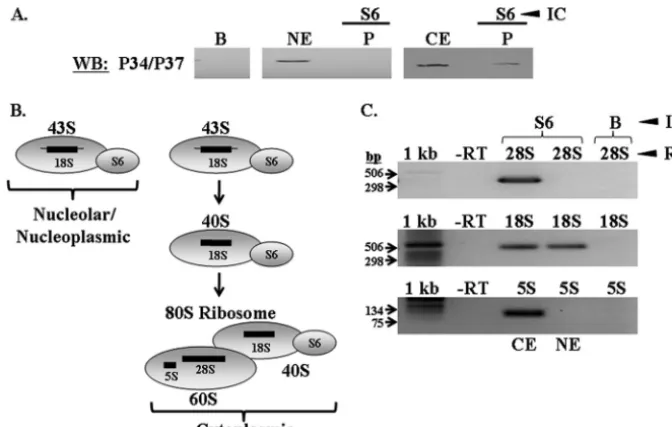
established that P34 and P37 associate with 60S subunits in the cytoplasm (Fig. 3 and 5), we next wanted to determine whether they remain so subsequent to 60S-40S subunit joining. We did not detect an association between P34 and P37 and S5 in the nucleoplasm (Fig. 6), suggesting that they act specifically on the 60S biogenesis pathway. A complex containing P34, P37, and a 40S protein in the cytoplasm would indicate that this was due to the subunits being joined to form an 80S ribosome. However, since we determined that P34 and P37 are com-plexed with S5 in whole nuclear and nucleolar extracts, we chose to use a 40S protein that does not associate with the 90S particle as our marker for 80S ribosomes (40S protein S6). Thus, a complex formed between P34 and P37 and S6 in the cytoplasm could only be due to their association with 80S ribosomes.
Thus, in order to determine whether P34 and P37 are com-plexed with 80S ribosomes, we performed immune capture experiments specific to the 40S protein S6 using cytoplasmic and nuclear extracts. Western blot analyses of the resulting
pellets demonstrated that P34 and P37 associate with S6 in cytoplasmic, but not nuclear, extracts (Fig. 10A, P lanes).
We have illustrated the rRNA species complexed with S6 in yeast in Fig. 10B. S6 incorporates into the maturing yeast 40S subunit in the nucleolus at the 43S particle (Fig. 1 and 10B) (11, 27). If this model is conserved in T. brucei, S6 should associate only with 18S rRNA in nuclear extracts, but it should associate with 28S, 18S, and 5S rRNA in cytoplasmic extracts. Our results demonstrate that these are in fact the rRNA spe-cies with whichT. brucei S6 associates in each extract (Fig. 10C), demonstrating that we efficiently precipitated 40S ribo-somal subunits.
The nuclear results from these experiments revealed that P34 and P37 play a specific role in 60S ribosomal subunit biogenesis, as there is no association with the 40S subunit. The cytoplasmic results demonstrate that P34 and P37 are com-plexed with 80S ribosomes, suggesting that they remain asso-ciated with the translational apparatus, possibly fulfilling a role in subunit joining or translation initiation.
DISCUSSION
The work presented here has more precisely defined the role(s) that two trypanosome-specific RNA binding proteins, P34 and P37, play in the process of 60S ribosomal subunit biogenesis. Ribosomal biogenesis has not been extensively studied inT. brucei, and the bulk of our current knowledge pertains to rRNA processing events (5, 43). We have shown previously that P34 and P37 associate with several 60S ribo-somal subunit components and biogenesis factors, including 5S rRNA (33), 60S ribosomal protein L5 (K. Hellman and K. FIG. 10. P34 and P37 associate with 40S protein S6 in cytoplasmic extracts. Immune capture experiments specific to S6 were performed using wild-type whole nuclear and cytoplasmic extracts. (A) Western blot analyses were performed probing for P34 and P37. B, immune capture beads alone; NE, 50g of whole nuclear extract; CE, 50g of cytoplasmic extract; P, immune capture pellet fraction. (B) Diagram depicting the ribosomal complexes and rRNA species with which S6 associates. (C) RT-PCR analyses performed on RNA extracted from S6 immune capture experiments using cytoplasmic and whole nuclear extracts. 1 kb, 1-kb DNA ladder (Invitrogen); -RT, no-RT control; 28S, 28S RT-PCR; 18S, 18S RT-PCR; 5S, 5S RT-PCR; B, immune capture beads alone (control); CE, cytoplasmic extract immune capture; NE, whole nuclear extract immune capture. Results shown are representative of at least three separate experiments.
on September 8, 2020 by guest
Prohaska, unpublished data), and a family of nucleolar phos-phoproteins, NOPP44/46 (34). In addition, cells lacking P34 and P37 show phenotypic alterations with respect to 5S rRNA levels, formation of 80S ribosomes, and overall cellular trans-lation levels (18). Thus, the goal of the current study was to more specifically determine if, when, and where P34 and P37 are involved in 60S ribosomal subunit biogenesis.
We first demonstrated that we were able to obtain distinct subcellular fractions, including cytoplasmic, whole nuclear, nu-cleoplasmic, and nucleolar extracts, as determined by Western blot analyses probing for specific compartmental markers (Fig. 2B). We had previously shown that P34 and P37 are nuclear proteins (47) and that they are also present in the cytoplasm (K. Prohaska, unpublished data). However, we had not deter-mined whether they are present in nucleolar preparations. A fraction of each of the isolated extracts was subjected to West-ern blot analysis probing for P34 and P37, which demonstrated that both proteins are present in the nucleolus (Fig. 2A). Thus, we could perform our interaction studies with each of the extracts in order to determine the extent to which P34 and P37 associate with 60S ribosomal subunits in each subcellular com-partment.
We began our studies by examining the interaction(s) be-tween P34 and P37 and L3, as it is one of the first 60S proteins to be added to the developing ribosome (35). Immune capture experiments specific for 60S subunit protein L3 demonstrated an association between P34 and P37 and L3-containing 60S subunits in whole nuclear, nucleolar, nucleoplasmic, and cyto-plasmic extracts (Fig. 3A). This was the first indication that these proteins are involved in multiple stages of 60S subunit biogenesis. As L3 is added to the 90S preribosomal particle, an association with P34 and P37 may suggest that they are also present in this early particle. In order to determine whether this is so, we performed immune capture experiments specific to S5, a 40S subunit protein that is also present in 90S particles. P34 and P37 were found in a complex with S5 in whole nuclear and nucleolar, but not nucleoplasmic, extracts (Fig. 4A), dem-onstrating that they come into the 60S biogenesis pathway at the 90S particle stage. Similar immune capture experiments specific for L11, which enters into the pathway at the late, nucleolar 66S particle, demonstrated that P34 and P37 also associate with this protein in all fractions, further confirming that they associate with 60S subunits throughout the biogenesis pathway. For both L3 and L11 immune capture experiments, we determined that 60S complexes, not just free protein, were isolated, as they contained the appropriate interacting rRNA species as demonstrated in yeast (Fig. 3B and 4B). This not only demonstrated that each antibody precipitated ribosomal complexes, but also that the overall framework of the yeast ribosomal biogenesis pathway is conserved inT. brucei.
As P34 and P37 remain complexed with L3 and L11 in both nuclear and cytoplasmic phases, we wanted to determine whether they are components of the 60S subunit nuclear ex-port complex. Although extensively studied in yeast and mam-malian cells (14, 20, 40), this complex, which contains exportin 1 and Nmd3, had not yet been demonstrated inT. brucei. We first confirmed thatT. bruceicontained an Nmd3 homologue via Western blot analysis using an Nmd3 antibody raised to the yeast protein. The antibody cross-reacted with a band of the appropriate molecular weight (data not shown), confirming
that this organism contains an Nmd3 homologue. The Camp-bell laboratory (UCLA) had already demonstrated thatT. bru-ceicontains a homologue to the nuclear export factor exportin 1 (45). Thus, we wished to determine whetherT. brucei 60S subunits associate with exportin 1 and Nmd3. Immune capture experiments specific for L3 and L11 demonstrated that both proteins associate with exportin 1 and Nmd3 (Fig. 6A and B). These results demonstrated that T. brucei utilizes a nuclear export complex similar to that described for other organisms. We have shown previously that P34 and P37 associate with exportin 1 (19). Due to their association with the 60S subunit in both nuclear and cytoplasmic preparations, we hypothesized that P34 and P37 may associate with exportin 1 in the context of the 60S subunit nuclear export complex. We performed P34/P37 immune capture experiments using whole nuclear ex-tracts, followed by Nmd3 Western blot analysis, which demon-strated that these proteins do form a complex (Fig. 7A). Ad-ditionally, we showed that P34 and P37 form a complex with exportin 1 and the 60S subunit simultaneously (Fig. 7B), dem-onstrating that these proteins are components of the 60S nu-clear export complex inT. brucei. These results led us to ask whether P34 and P37 might be required for the formation of the 60S nuclear export complex inT. brucei. Thus, we repeated the L3 and L11 immune capture experiments with wild-type and P34/P37 RNAi nuclear extracts, followed by Nmd3 and exportin 1 Western blot analyses (Fig. 8 and 9). We found that 60S subunits failed to associate with either export factor in the absence of P34 and P37, demonstrating that these proteins are not only part of, but are also required for, formation of the 60S nuclear export complex. These were significant findings, as this complex is highly conserved among other organisms. We have shown that much of the complex is conserved in T. brucei; however, P34 and P37, which are trypanosome specific, are essential to the formation of the complex as well.
In addition to their nuclear role(s) in 60S biogenesis, we have also demonstrated that P34 and P37 remain associated with mature 80S ribosomes in the cytoplasm (Fig. 10A) (18). Taken together, the results presented here demonstrate that P34 and P37 play multiple roles in ribosomal biogenesis, in-cluding assembly of the 60S export complex in the nucleus and, possibly, that they fulfill a role(s) with respect to mature 60S subunits and 80S ribosomes in the cytoplasm.
Although we have established here that P34 and P37 are required for assembly of the 60S nuclear export complex inT. brucei, further experiments need to be performed in order to confirm whether they are involved in the transport process. In yeast, multiple factors are required for nuclear export of 60S subunits in addition to exportin 1 and Nmd3, possibly due to the large size and hydrophilic nature of the 60S subunit (3, 21). These factors include the Mex67-Mtr2 heterodimer (44) and Arx1 (3, 21). We queried theT. bruceidatabase for the Mex67, Mtr2, and Arx1 protein sequences. Mex67 and Arx1 BLAST searches returned top hits that were hypothetical proteins which, when aligned with the queried sequences using ClustalW, did not show a significant extent of similarity. The Mtr2 BLAST search returned only three proteins, none of which demonstrated similarity to the Mtr2 protein. It may be that P34 and P37 represent trypanosome-specific proteins that fulfill similar roles as the proteins that have been identified in yeast, although we must consider the possibility that these proteins
on September 8, 2020 by guest
http://ec.asm.org/
ments are being performed in order to more precisely define the role(s) of P34 and P37 in 5S rRNA localization and incor-poration into 60S subunits. This will determine whether they function in the classical L5-5S pathway or whether they repre-sent a distinct pathway specific to trypanosomes.
The fact that P34 and P37 remain associated with mature 80S ribosomes suggests that they play a role(s) in the cytoplas-mic phase of ribosomal biogenesis and/or translation initiation. Polysomal profiles performed on P34/P37 RNAi cells have demonstrated an increase in free 60S subunits, a decrease in 80S ribosomes, and a decrease in total cellular translation (18). These results showed striking similarity to those obtained with yeast strains containing mutant L10 protein (9). The cause of the yeast phenotype was determined to be a defect in subunit joining, indicating that P34 and P37 may fulfill a similar cyto-plasmic role. Current work in our laboratory is focused on determining whether P34 and P37 are involved in subunit join-ing, which could account for the decrease in translation in their absence.
The requirement for trypanosome-specific proteins in a highly conserved process such as ribosomal biogenesis may be related to the life cycle ofT. bruceiand the requirement of the parasite to adapt to two separate host environments. It has been shown that protein synthesis and ribosomal biogenesis differ between procyclic and bloodstream-form trypanosomes (2, 4, 38). Thus, these developmentally regulated processes likely involve factors that are similarly regulated themselves. The levels of P34 and P37 proteins are differentially regulated throughout the life cycle ofT. brucei(47), which may, in part, relate to the differences in ribosomal biogenesis and protein synthesis between the two life cycle stages. Further experi-ments in bloodstream-form cells are required to address this possibility. Finally, the results presented in the current work have created the foundation for studying the role(s) of P34 and P37 in ribosomal biogenesis in T. brucei. The trypanosome-specific nature of these proteins and their requirement for an essential process may render them targets for future chemo-therapeutic interventions in treating African trypanosomiasis.
ACKNOWLEDGMENTS
We thank Marilyn Parsons (Seattle Biomedical Research Institute) for the antisera toT. bruceiphosphoglycerate kinase and NOG1, Viv-ian Bellofatto (University of Medicine and Dentistry of New Jersey) for the polyclonal antiserum to theT. bruceiTATA-binding protein,
some ribosomal RNA. J. Mol. Biol.196:113–124.
6.Cunningham, I.1977. New culture medium for maintenance of tsetse tissues and growth of trypanosomatids. J. Protozool.24:325–329.
7.Dechampesme, A. M., O. Koroleva, I. Leger-Silvestre, N. Gas, and S. Camier.1999. Assembly of 5S ribosomal RNA is required at a specific step of the pre-rRNA processing pathway. J. Cell Biol.145:1369–1380. 8.Delespaux, V., and H. P. de Koning.2007. Drugs and drug resistance in
African trypanosomiasis. Drug Resist. Updates10:30–50.
9.Eisinger, D. P., F. A. Dick, and B. L. Trumpower.1997. Qsr1p, a 60S ribosomal subunit protein, is required for joining of 40S and 60S subunits. Mol. Cell. Biol.17:5136–5145.
10.Fatica, A., and D. Tollervey.2002. Making ribosomes. Curr. Opin. Cell Biol.
14:313–318.
11.Ferreira-Cerca, S., G. Poll, H. Kuhn, A. Neueder, S. Jakob, H. Tschochner, and P. Milkereit.2007. Analysis of thein vivoassembly pathway of eukary-otic 40S ribosomal proteins. Mol. Cell28:446–457.
12.Ferreira-Cerca, S., G. Po¨ll, P.-E. Gleizes, H. Tschochner, and P. Milkereit.
2005. Roles of eukaryotic ribosomal proteins in maturation and transport of pre-18S rRNA and ribosome function. Mol. Cell20:263–275.
13.Fromont-Racine, M., B. Senger, C. Saveanu, and F. Fasiolo.2003. Ribosome assembly in eukaryotes. Gene313:17–42.
14.Gadal, O., D. Strauss, J. Kessl, B. Trumpower, D. Tollervey, and E. Hurt.
2001. Nuclear export of 60S ribosomal subunits depends on XpoIp and requires a nuclear export sequence-containing factor, Nmd3p, that associates with the large subunit protein Rpl10p. Mol. Cell. Biol.21:3405–3415. 15.Grandi, P., V. Rybin, J. Baszler, E. Petfalski, D. Strausz, M. Marzioch, T.
Schafer, B. Kuster, H. Tschochner, D. Tollervey, A. C. Gavin, and E. Hurt.
2002. 90S pre-ribosomes include the 35S pre-rRNA, the U3 snoRNP, and 40S subunit processing factors but predominantly lack 60S synthesis factors. Mol. Cell10:105–115.
16.Harnpicharnchai, P., J. Jakovljevic., E. Horsey, T. Miles, J. Roman, M. Rout, D. Meagher, B. Imai, Y. Guo, C. J. Brame, J. Shabanowitz, D. F. Hunt, and J. L. Woolford.2001. Composition and functional characterization of yeast 66S ribosome assembly intermediates. Mol. Cell8:505–515. 17.Hedges, J., M. West, and A. W. Johnson.2005. Release of the export adapter,
Nmd3p, from the 60S ribosomal subunit requires Rpl10p and the cytoplas-mic GTPase Lsg1p. EMBO J.24:567–579.
18.Hellman, K. M., M. Ciganda, S. V. Brown, J. L. Li, W. Ruyechan, and N. Williams.2007. Two trypanosome-specific proteins are essential factors for 5S rRNA abundance and ribosomal assembly inTrypanosoma brucei. Eu-karyot. Cell6:1766–1772.
19.Hellman, K., K. Prohaska, and N. Williams.2007.Trypanosoma bruceiRNA binding proteins P34 and P37 mediate NOPP44/46 cellular localization via the exportin 1 nuclear export pathway. Eukaryot. Cell6:2206–2213. 20.Ho, J., G. Kallstrom, and A. W. Johnson.2000. Nmd3 is a Crm1p-dependent
adapter protein for nuclear export of the large ribosomal subunit. J. Cell Biol.151:1057–1066.
21.Hung, N. J., K. Y. Lo, S. S. Patel, K. Helmke, and A. W. Johnson.2008. Arx1 is a nuclear export receptor for the 60S ribosomal subunit in yeast. Mol. Biol. Cell19:735–744.
22.Jensen, B. C., D. L. Brekken, A. C. Randall, C. T. Kifer, and M. Parsons.
2005. Species specificity in ribosome biogenesis: a non-conserved phospho-protein is required for formation of the large ribosomal subunit in Trypano-soma brucei. Eukaryot. Cell4:30–35.
23.Jensen, B. C., Q. Wang, C. Kifer, and M. Parsons.2003. The NOG1 GTP-binding protein is required for biogenesis of the 60S ribosomal subunit. J. Biol. Chem.278:32204–32211.
24.Kallstrom, G., J. Hedges, and A. W. Johnson.2003. The putative GTPases Nog1p and Lsg1p are required for 60S ribosomal subunit biogenesis and are localized to the nucleus and cytoplasm, respectively. Mol. Cell. Biol.23:
4344–4355.
on September 8, 2020 by guest
25.Kapp, D., and J. Lorsch.2004. The molecular mechanics of eukaryotic translation. Annu. Rev. Biochem.73:657–704.
26.Kennedy, P. G.2004. Human African trypanosomiasis of the CNS: current issues and challenges. J. Clin. Investig.113:496–504.
27.Kressler, D., P. Linder, and J. de la Cruz.1999. Proteintrans-acting factors involved in ribosome biogenesis inSaccharomyces cerevisiae. Mol. Cell. Biol.
19:7897–7912.
28.Lebreton, A., C. Saveanu, L. Decourty, J. C. Rain, A. Jacquier, and M. Fromont-Racine.2006. A functional network involved in the recycling of nucleo-cytoplasmic pre-60S factors. J. Cell Biol.173:349–360.
29.Matovu, E., T. Seebeck, J. C. Enyaru, and R. Kaminsky.2001. Drug resis-tance inTrypanosoma bruceispp., the causative agents of sleeping sickness in man and nagana in cattle. Microbes Infect.3:763–770.
30.Michaeli, S., and N. Agabian.1990. ATrypanosoma bruceismall RNP par-ticle containing the 5S rRNA. Mol. Biochem. Parasitol.41:7–16. 31.Park, J. H., B. C. Jensen, C. T. Kifer, and M. Parsons.2001. A conserved
nucleolar G-protein in eukaryotes. J. Cell Sci.114:173–185.
32.Pestova, T. V., V. G. Kolupaeva, I. B. Lomakin, E. V. Pilipenko, I. N. Shatsky, V. I. Agol, and C. U. Hellen.2001. Molecular mechanisms of translation initiation in eukaryotes. Proc. Natl. Acad. Sci. USA98:7029–7036. 33.Pitula, J., W. T. Ruyechan, and N. Williams.2002. Two novel RNA binding
proteins fromTrypanosoma bruceiare associated with 5S rRNA. Biochem. Biophys. Res. Commun.290:569–576.
34.Pitula, J. S., J. Park, M. Parsons, W. T. Ruyechan, and N. Williams.2002. Two families of RNA binding proteins fromTrypanosoma bruceiassociate in a direct protein-protein interaction. Mol. Biochem. Parasitol.122:81–89. 35.Rosado, I. V., C. Dez, S. Lebaron, M. Caizergues-Ferrer, Y. Henry, and J. de
la Cruz.2007. Characterization ofSaccharomyces cerevisiaeNpa2p (Urb2p) reveals a low-molecular-mass complex containing Dbp6p, Npa1p (Urb1p), Nop8p, and Rsa3p involved in early steps of 60S ribosomal subunit biogen-esis. Mol. Cell. Biol.27:1207–1221.
36.Rout, M. P., and M. C. Field.2001. Isolation and characterization of sub-nuclear compartments fromTrypanosoma brucei: identification of a major repetitive nuclear lamina component. J. Biol. Chem.276:38261–38271. 37.Saveanu, C., A. Namane, P. E. Gleizes, A. Lebreton, J. C. Rousselle, J.
Noaillac-Depeyre, N. Gas, A. Jacquier, and M. Fromont-Racine.2003. Se-quential protein association with nascent 60S ribosomal particles. Mol. Cell. Biol.23:4449–4460.
38.Shapiro, S. Z., and B. E. Kimmel.1987. Differential protein synthesis during the life cycle of the protozoan parasiteTrypanosoma brucei. J. Protozool.
34:58–62.
39.Takahashi, N., M. Yanagida, S. Fujiyama, T. Hayano, and T. Isobe.2003. Proteomic snapshot analyses of preribosomal ribonucleoprotein complexes formed at various stages of ribosome biogenesis in yeast and mammalian cells. Mass Spectrom. Rev.22:287–317.
40.Thomas, F., and U. Kutay.2003. Biogenesis and nuclear export of ribosomal subunits in higher eukaryotes depend on the CRM1 export pathway. J. Cell Sci.116:2409–2419.
41.Trotta, C. R., E. Lund, L. Kahan, A. W. Johnson, and J. E. Dahlberg.2003. Coordinated nuclear export of 60S ribosomal subunits and NMD3 in verte-brates. EMBO J.22:2841–2851.
42.Warner, J. R.1999. The economics of ribosome biosynthesis in yeast. Trends Biol. Sci.24:437–440.
43.White, T. C., G. Rudenko, and P. Borst.1986. Three small RNAs within the 10 kb trypanosome rRNA transcription unit are analogous to domain VII of other eukaryotic 28S rRNAs. Nucleic Acids Res.14:9471–9489.
44.Yao, W., D. Roser, A. Kohler, B. Bradatsch, J. Bassler, and E. Hurt.2007. Nuclear export of ribosomal 60S subunits by the general mRNA export receptor Mex67-Mtr2. Mol. Cell26:51–62.
45.Zeiner, G. M., N. R. Sturm, and D. A. Campbell.2003. Exportin 1 mediates nuclear export of the kinetoplastid spliced leader RNA. Eukaryot. Cell
2:222–230.
46.Zhang, J., P. Harnpicharnchai, J. Jakovljevic, L. Tang, Y. Guo, M. Oeffinger, M. P. Rout, S. L. Hiley, T. Hughes, and J. L. Woolford.2007. Assembly factors Rpf2 and Rrs1 recruit 5S rRNA and ribosomal proteins rpL5 and rpL11 into nascent ribosomes. Genes Dev.21:2580–2592.
47.Zhang, J., W. Ruyechan, and N. Williams.1998. Developmental regulation of two nuclear RNA binding proteins, P34 and P37, fromTrypanosoma brucei. Mol. Biochem. Parasitol.92:79–88.