AN OVERVIEW ON OPTIC NEURITIS
5
0
0
Full text
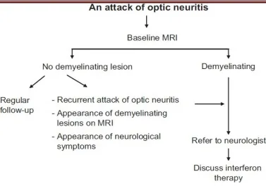
(2) Solanki Roshni et al. IRJP 2 (5) 2011 49-53 of acute ON. In this and another cross-sectional study of MS patients with ON9. RNFL was significantly reduced in the affected eye when compared with fellow eyes or disease-free controls. These and other studies have correlated RNFL thinning with impaired visual function10. OCT can be employed to monitor such progressive axonal loss in both primary and secondary progressive MS11. (B)Optic Neuritis With Ethambutol Ethambutol has been used to treat TB since the 1960s. The potential for visual impairment was recognized soon after its introduction12. The original formulation was a racemic mixture. However, when it was discovered that the L-form was predominantly responsible for its toxicity, and the D-form for its therapeutic effects, the Lform was withdrawn. Despite this, cases of irreversible visual loss have been reported in the literature and some authors have even gone on to suggest that ethambutol should not be used routinely to treat TB13,14 Ethambutol causes loss of visual acuity, colour vision and visual field. The occurrence of ocular toxicity is dose related, loss of vision most likely to occur in patients receiving 25 mg/kg/day or more. However, vision loss has been documented in approximately 1% of patients receiving the recommended therapeutic dose of 15 to 25mg/kg/day15,16,17. This rarely occurs before the patients have been on treatment for 2 months, with 7 months being the average. Two Patients with impaired renal function from renal tuberculosis may be more prone to ethambutol-associated optic neuropathy; perhaps because ethambutol depends on the kidneys for excretion18,19. It is also important for the clinician to be aware that there are reports of rapid onset, severe, bilateral visual loss despite treatment with therapeutic doses of ethambutol20,21. Patients taking ethambutol should be instructed to discontinue the drug immediately at the onset of any visual symptoms and seek medical consult. (C)Optic Neuritis With Vaccination Two types of vaccine for meningitis are distributed in Spain: A) Bivalent vaccines which include nonconjugated purified capsular bacterial polysaccharides of serogroups A and C of Neisseria meningitidis, and B) Conjugated vaccines vis-à-vis Neisseria meningitidis, C serotype, by conjugation of the oligosaccharide of the Neisseria meningitides capsule through covalent link with a carrier protein (diphtheria or tetanus toxoid) for increasing the immunogenic capacity of the vaccine. Optic neuritis (ON) can leave important visual sequels because many of these processes can be bilateral. Within etiological diagnosis there is a group of ON called post-. IRJP 2 (5) May 2011. vaccine22. Which occur in the presence of diverse viral and bacterial agents23,24. TREATMENT In most cases, visual functions return to near normal within eight to ten weeks, but they may also advance to a complete and permanent state of visual loss. Therefore, systemic intravenous treatment with corticosteroids, which may quicken the healing of the optic nerve, is often recommended, but it does not have a significant effect on the visual acuity at one year, when compared against placebo. Intravenous corticosteroids have also been found to reduce the risk of developing MS in the following two years in those patients who have MRI lesions; but this effect disappears by the third year of follow up26. Paradoxically it has been demonstrated that oral administration of corticosteroids in this situation may lead to more recurrent attacks than in non-treated patients (though oral steroids are generally prescribed after the intravenous course, to wean the patient off the medication). This effect of corticosteroids seems to be limited to optic neuritis and has not been observed in other diseases treated with corticosteroids27. Very occasionally, if there is concomitant increased intracranial pressure the sheath around the optic nerve may be cut to decrease the pressure. When optic neuritis is associated with MRI lesions suggestive of multiple sclerosis (MS) then general immunosuppressive therapy for MS is most often prescribed (IV methylprednisolone may shorten attacks; initial only oral prednisone may increase relapse rate). The above overall discussion regarding the treatment following the MRI & management protocol is followed1. (Figure 2) PROGNOSIS The pain will go away, usually in a few days. For visual recovery is usually good. The majority of patients (6580%) recover visual acuity of 20/30 or better25, 29. The vision problems will improve in the majority (92%) of patients. There are rare patients who have continued progressive loss of vision. Even in the 92% that improve, often they do not return completely to normal. Patients may be left with blurred, dark, dim, or distorted vision. Frequently colors look different or "washed out." Visual recovery usually takes place over a period of weeks to months, although both earlier and later improvement is possible. Late variations in vision are common, often associated with exercise or taking a hot shower or bath. This is known as Uhthoff's phenomena and is probably related to damage to the myelin coating. Patients who notice this problem are not more likely to get worse.. Page 49-53.
(3) Solanki Roshni et al. IRJP 2 (5) 2011 49-53 Optic neuritis can recur involving the same eye, the other eye or other parts of the central nervous system (brain and spinal cord). This may result in recurrent episodes of decreased or loss of vision or problems with weakness, numbness or other signs of brain involvement. An MRI scan can give us to give a rough guess as to the likelihood of recurrence. It will not completely exclude the possibility of future episodes or guarantee that they will happen. Other testing techniques are sometimes used to confirm the suspicion of optic neuritis. These may include visual evoked potentials (a test where you are shown a checkerboard of light and signals are recorded from electrodes on your scalp) that can show a delay in conduction due to the damage to the myelin(Neuro Ophthalmology North American Society). HERBAL TREATMENT The Ayurvedic treatment of optic neuritis is aimed at controlling the pain, treating the inflammation of the optic nerve and treating immune dysfunction of the body. Medicines like Triphala-Guggulu, Yograj-Guggulu, Punarnavadi-Guggulu, Maha-Rasnadi-Guggulu, Rasnadi-Qadha, Vat-Gajankush-Ras, Maha-VatVidhwans-Ras, Vish-Tinduk-Vati, Dashmoolarishta and Nirgundi (Vitex negundo) can be used to treat the pain in the eyes. Medicines such as Kaishor-Guggulu, PanchTikta-Ghrut-Guggulu, Triphala-Ghrut, Panch-TiktaGhrut, Tapyadi-Loh, Saptamrut-Loh and Ekang-VeerRas are used to treat the inflammation in the optic nerve28. Herbal medicines which can be used in optic neuritis are Amalaki (Emblica officinalis), Haritaki (Terminalia chebula), Behada (Terminalia bellerica), Tulsi (Ocimum sanctum), Shatavari (Asparagus racemosus), Punarnava (Boerhaavia diffusa), Rasna (Pluchea lanceolata), Guduchi (Tinospora cordifolia), Deodar (Cedrus deodara), Erandmool (Ricinus communis), Gokshur (Tribulus terrestris), Apamarga (Achyranthus aspera), Guggulu (Commiphora mukul), Shallaki (Boswellia serrata), Kuchla (Strychnos nuxvomica) and Nirgundi28. The overall prognosis following optic neuritis is generally good, with most people regaining normal vision within 2 to 6 months. Ayurvedic medicines can help to improve the therapeutic response, reduce the duration of treatment and prevent recurrent episodes of this condition. CONCLUSION In light of above review there is a comprehensive study is require for counteracting the optic neuritis association with various neurodegenerative diseases & showed some herbal options for the treatment.. IRJP 2 (5) May 2011. REFERENCES 1. Vimla Menon, Rohit Saxena, Ruby Misra, Swati Phuljhele, management of optic neuritis, Indian journal of ophthalmology, 2011;59(2):117-122. 2. Rodriguez M, Siva A, Cross SA, O'Brien PC, Kurland LT, "Optic neuritis: a population-based study in Olmsted County, Minnesota". Neurology, 1995;45(2):244–50. 3. Brewis M, Poskanzer DC, Rolland C, et al: Neurological disease in an English city. Acta Neurol Scand, 1965;42:1 4. Wray SH: Optic Neuritis. Principles and Practice of Ophthalmology, 4:2539-2568. 5. Percy AK, Nobrega FT, Kurland LT: Optic neuritis and multiple sclerosis. Arch Ophthalmol 1972;87:135. 6. Nikoskelainen E: Symptoms, signs and early course of optic neuritis. Acta Ophthalmol, 1975;53:254. 7. Lillie WI: The clinical significance of retrobulbar and optic neuritis. Am J Ophtahlmol, 1934;17:110,. 8. Costello F, Coupland S, Hodge W et al. Quantifying axonal loss after optic neuritis with optical coherence tomography. Ann Neurol, 2006;59:963–969. 9. Fisher JB, Jacobs DA, Markowitz CE et al. Relation of visual function to retinal nerve fiber layer thickness in multiple sclerosis. Ophthalmology, 2006;113:324–332. 10. Trip SA, Schlottmann PG, Jones SJ et al. Retinal nerve fiber layer axonal loss and visual dysfunction in optic neuritis. Ann Neurol, 2005;58:383–391. 11. Henderson AP, Trip SA, Schlottmann PG et al. An investigation of the retinal nerve fibre layers in progressive multiple sclerosis using optical coherence tomography. Brain, 2008;131:277–287. 12. Phillips PH. Toxic and deficiency optic neuropathies. In: Miller NR, Newman NJ, editors. Biousse V, Kerrison JB, associate editors. Walsh and Hoyt’s Clinical Neuro-ophthalmology. 6th ed. Baltimore, Maryland: Lippincott Williams and Wilkins, 005:455-6. 13. Kumar A, Sandramouli S, Verma L, Tewari HK, Khosla PK. Ocular ethambutol toxicity: is it reversible? J Clin Neuroophthalmol, 1993;13:15-17. 14. Tsai RK, Lee YH. Reversibility of ethambutol optic neuropathy. J Ocul Pharmacol Ther, 1997;13:473-7. 15. Kahana LM. Toxic ocular effects of ethambutol. Can Med Assoc J, 1987;137:212-6. 16. Barron GJ, Tepper L, Iovine G. Ocular toxicity from ethambutol. Am J Ophthalmol, 1974;77:256-60. 17. Citron KM, Thomas GO. Ocular toxicity from ethambutol. Thorax, 1986; 41:737-9. 18. Bronte SJ, Pettigrew AR, Foulds WS. Toxic optic neuropathy and its experimental production. Trans Ophthalmol Soc UK, 1976;96:355-8. 19. De Vita EG, Miao M, Sadun AA. Optic neuropathy in ethambutol treated renal tuberculosis. J Clin Neuroophthalmol, 1987;7:77-83. 20. Smith JL. Should ethambutol be barred? J Clin Neuroophthalmol, 1987;7:84-6. 21. Chatterjee VKK, Buchanan DR, Friedmann AI, Green M. Ocular toxicity following ethambutol in standard dosage. Br J Dis Chest, 1986;80: 288-90. 22. Savino PJ, Carmona O, Arbizu T, Arruga J, Sánchez Dalmau B, Vidaller A, et al. Neuritis Óptica. In: Arruga Ginebreda J, Sánchez Dalmau B. Neuropatías ópticas: Diagnóstico y tratamiento. Madrid: Sociedad Española de Oftalmología, 2002;175-206.. Page 49-53.
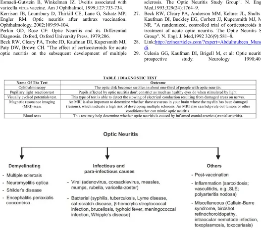
(4) Solanki Roshni et al. IRJP 2 (5) 2011 49-53 23. Esmaeli-Gutstein B, Winkelman JZ. Uveitis associated with varicella virus vaccine. Am J Ophthalmol, 1999;127:733-734. 24. Kerrison JB, Lounsbury D, Thirkill CE, Lane G, Schatz MP, Engler RM. Optic neuritis after anthrax vaccination. Ophthalmology, 2002;109:99-104. 25. Perkin GD, Rose CF: Optic Neuritis and its Differential Diagnosis. Oxford, Oxford University Press, 1979;206. 26. Beck RW, Cleary PA, Trobe JD, Kaufman DI, Kupersmith MJ, Paty DW, Brown CH. "The effect of corticosteroids for acute optic neuritis on the subsequent development of multiple. Name Of The Test Ophthalmoscopy Pupillary light reaction test Visually evoked potentials test. Magnetic resonance imaging (MRI) scan. Blood tests. sclerosis. The Optic Neuritis Study Group". N. Engl. J. Med,1993;329(24):1764–9. 27. Beck RW, Cleary PA, Anderson MM, Keltner JL, Shults WT, Kaufman DI, Buckley EG, Corbett JJ, Kupersmith MJ, Miller NR. "A randomized, controlled trial of corticosteroids in the treatment of acute optic neuritis. The Optic Neuritis Study Group". N. Engl. J. Med,1992 326(9):581–8. 28. Link:http://ezinearticles.com/?expert=Abdulmubeen_Mundewa di. 29. Celesia GG, Kaufman DI, Brigell M, et al: Optic neuritis: A prospective study. Neurology 1990;40:919.. TABLE 1 DIAGNOSTIC TEST Outcome The optic disk becomes swollen in about one-third of people with optic neuritis. Pupils affected by optic neuritis don't constrict as much as healthy eyes do when stimulated by light. This type of test is able to detect the slowing of electrical conduction resulting from damaged areas on nerves. An MRI is also important to determine whether there are areas in your brain where the myelin has been damaged (lesions), which indicate a high risk of developing multiple sclerosis. An MRI also can help rule out tumors or other conditions that can mimic optic neuritis. This test may help determine whether optic neuritis is caused by inflamed cranial arteries (cranial arteritis).. FIGURE 1 CAUSES OF OPTIC NEURITIS. IRJP 2 (5) May 2011. Page 49-53.
(5) Solanki Roshni et al. IRJP 2 (5) 2011 49-53. FIGURE 2 MANAGEMENT PROTOCOL FOR OPTIC NEURITIS PATIENT. IRJP 2 (5) May 2011. Page 49-53.
(6)
Figure
Related documents
71 Indeed, the perspective of the British governme nt was that the situation in Northern Ireland did not amount to an armed conflict of any kind in the sense of international
During the taxable periods at issue, Petitioners were engaged in the business of making retail sales of western leather goods, apparel, tack, used vehicles, trailers, and
Fuzzy integral equations play major roles in various areas, therefore a new method for finding a solution of the Fredholm fuzzy integral equation is presented. This method converts
(2012) "Because It’s a Girl Cake!:Because It’s a Girl Cake!: Fostering Dialogue About Gender Identity in Elementary Classrooms," Northwest Journal of Teacher Education :
In this paper we apply decision trees, Naive Bayesian classifiers and feature selection methods to a geriatric hospital dataset in order to predict inpatient length of stay,
We did dispose of fiscal data at the level of the fiscal unit and transferred them into a household budget survey to observe personal income taxes at different levels of the unit
An integration of the psychiatric personality disorder no- menclature with psychological models of general person- ality structure would go far in buttressing the weak con-
Staff recommends that the uniform fiduciary standard of conduct established by the Commission should provide that: [T]he standard of conduct for all brokers, dealers, and
