Antioxidant and wound healing studies on the
extracts of
Corchorus olitorius
leaf
Barku VYA
1*, Boye A
2and Quansah N
21. Department of Chemistry, School of Physical Sciences, University of Cape Coast
2. Department of Medical and Laboratory Technology, School of Physical Sciences, University of Cape Coast
*
Corresponding Author Email: [email protected] or [email protected]
ABSTRACT: The methanol extract of Corchorus olitorius leaves was subjected to antioxidant activity using 1,1-diphenyl-2-picryl hydrazyl radical (DPPH) assay and Ferric Reducing Antioxidant power (FRAP) assay. The extract exhibited a high degree of antioxidant activity. From the FRAP assay absorbance increased with increasing concentration of plant extract indicating a strong reduction potential of the plant. Phytochemical screening on both methanol and aqueous extracts revealed the following constituents’ common to both solvent extracts except alkaloids which were absent in the methanol extract: Alkaloids, terpenoids, tannins, flavonoids, cardiac glycosides. The plant powder and aqueous extracts were taken through screening for wound healing activity by excision model. Both the powder and the aqueous extract showed significant activity when compared with control and standard. Percentage of wound contraction on 18th day was found to be 100% for the powdered plant ointment, 5% powdered plant ointment and100mg/ml aqueous extract respectively. The results have significantly demonstrated the wound healing activity and the antioxidant activity of the leaves of Corchorus olitorius.
Keywords: Corchorus, shea butter, wound, povidine iodine, phytochemical screening INTRODUCTION
A wound is defined as a break in the epithelial lining of the skin and may be accompanied by disruption of the structure and function of the underlying tissue (Omale et al., 2010). Wounds are inescapable events in life. Wound may be caused by physical, chemical, thermal, microbial or immunological insult to a body tissue. Wound infection is one of the most common diseases in developing countries like Ghana because of poor hygienic conditions (Ayyanar et al., 2006). Wound infection is a major complication of injury and it accounts for 50-75 % of hospitalized deaths (Mokadas, 1998 as cited in Pirbalouti et al., 2012). Wounds are prone to infections and other complications. Infected wounds are those in which the wound area is contaminated by bacteria, which can cause suppuration or shedding of tissue. According to the World Health Organization (WHO, 2010) 5 to 7 million people in North America annually battle with chronic or complex wounds. Similarly, in Ghana 273,346 (1.64 %) of the general population suffer one or more forms of open wounds (Driscoll, 2009).
Many of the conventional wound healing drugs currently used for treating wounds are not only expensive but also pose problems such as many side effects, toxic effects, allergy and drug resistance. Most of the synthetic wound healing drugs are expensive. This situation makes it difficult for most people to afford, especially in a developing country like Ghana where the per capital income is very low. Non-treatment of wounds due to high cost of drugs can aggravate the individual’s condition and can lead to death at worst.
Additionally, most of these synthetic wound healing drugs have in one way or the other proved to be ineffective (Karukonda et al., 2000). Recent research findings indicate that, most of the bacteria which cause wound infection are showing resistance to the synthetic drugs and since these drugs cannot stop the growth or activity of these pathogens it renders the drug inappropriate for the treatment of wounds. It is also worthy to note that most of the synthetic wound healing drugs have been reported to show allergy in some patients (World Union of Wound Healing Societies, 2004). Because of this condition, the drug fails to achieve the desired effect.
In the face of the many problems associated with conventional wound healing medications, many medicinal plants have been reported for their potential in the treatment and management of wounds. These include Aloe vera,
Azadirachta indica, Carica papaya, Celosia argentea, Centella asiatica, and Cinnamomum zeylanicum which have all being reported for their wound healing potential (Dash et al., 2011; Kumar, 2007; Omale et al., 2010). According to literature, 70 % of the wound healing drugs are of plant origin, 20 % of mineral origin, and the remaining 10 % consisting of animal products, but those of plant origin are most effective (Ayyanar et al., (2006); Ayyanar & Ignacimuthu, 2009; Biswas & Mukherjee (2003)).
Medicinal plants are known to induce wound healing and regeneration of injured tissues by multiple mechanisms, one of such is Corchorus olitorius however there is not much scientific information regarding its wound healing activity. There is the need for scientific investigation and safety evaluation of plant medicines used in folklore before they could be recommended for use (Bennet, 1988). It is in the light of the above that, the present study sought to evaluate the wound healing activity of C. olitorius commonly known as “jute” a native plant used
commonly as vegetable in Ghana and extensively used by many Ghanaian tribes for managing wounds.
It is also believed that, in wound healing, antioxidants counter the excess proteases and reactive oxygen species (ROS) often formed by neutrophil accumulation in the wounded area and protect protease inhibitors from oxidative damage. Fibroblasts and other cells may be killed by excess ROS and skin lipids will be made less flexible. Antioxidants substances reduce the possibility of these adverse events occurring hence appear to be important in the successful treatment of wounds (Houghton et al., 2005). Compounds with high radical-scavenging capacity have been shown to facilitate wound-healing (Suntar et al., 2012). The present study also attempts to investigate the antioxidant potentials of the plant.
MATERIALS AND METHODS
Plant Collection, Identification, Authentication and Treatment
The leaves of C. olitorius were collected from Amamoma, a suburb of Cape Coast, Ghana. The plant was identified and authenticated by Mr. Isaac Otoo of the herbarium unit of the School of Biological Sciences, University of Cape Coast, Ghana, where a voucher specimen (SBS/UCC/H 306) was deposited. The leaves were washed and air-dried for three weeks. The dried leaves were milled into fine powder using Vivekanada Madras Mill (U.S.A) and a blender (Chefman, England, 1985).
Preparation of Methanol and Aqueous Extract
A mass of 20 g of the powdered sample was cold macerated with 40 ml of 70 % methanol. The cold macerate was filtered (Whatman filter paper, No 1). The filtrate was concentrated to remove the solvent under reduced pressure at 35-45°C using rotavapor. The resultant semi dried extract was placed in a dessicator for three days to further dry the sample. The dried extract was weighed which gave a percentage yield of 20.5 %. The same procedure was followed to obtain the aqueous extract (10.2%).
Phytochemical Screening
Both the aqueous and methanolic extracts were subjected to phytochemical screening using standard techniques of phytochemical analysis as described briefly below (Sofowora, 1993; Harbone, 1998; Trease & Evans, 1989):
Anti oxidant activity by DPPH free radical scavenging method
Antioxidant activities of C. olitorius were studied by DPPH free radical scavenging method.
The crude methanol extracts of the plants were screened for DPPH radical Scavenging activity. DPPH radical scavenging activity was measured according to the method of Braca et al., (2003) and Rajeswara et al., (2012) with little modification. Extract solutions were prepared by dissolving 0.05g of dry extract in 50ml of methanol. An aliquot of 2ml of 0.004% DPPH solution in methanol and 1ml of plant extract in methanol at various concentrations (200, 400 and 800ppm) were mixed and incubated at 25°C for 30 min. and absorbance of the test mixture was read at 517nm using a spectrophotometer (T 70 UV-VIS Spectrometer, PG Instruments Ltd) against a DPPH control containing only 1 ml of methanol in place of the extract. The DPPH solution in methanol was prepared daily before the absorbance measurements. DPPH is a purple coloured stable free radical. When reduced it becomes the yellow colored Diphenyl picryl hydrazine. All experiments were performed thrice and the results were averaged. Ascorbic acid was used as a standard (James et al., 2008 and Ramnik et al., 2008). Percent inhibition was calculated using the following expression:
% Inhibition = (Ablank – Asample /Ablank) x 100
Where Ablank and Asample stand for absorption of the blank sample and absorption of tested extract solution respectively.
Ferric Reducing Antioxidant Power Assay (FRAP)
The reducing antioxidant power of plant methanolic extracts was determined by the method of Oyaizu, (1986). Different concentrations of plant extracts (250 – 1000 ppm) in 1 ml of distilled water were mixed with phosphate buffer (3.0 ml, 0.2 M, pH 6.6) and potassium ferricyanide [K3Fe(CN)6] (2.5 ml, 1%). The mixture was incubated at 50oC for 20 min. Then, 2.5 ml of trichloroacetic acid (10%) was added to the mixture, which was then centrifuged for 10 min at 3000 rpm. The upper layer of solution (2.5 ml) was mixed with distilled water (2.5 ml) and FeCl3 (0.5 ml, 0.1%). The absorbance was measured at 700 nm against a blank using UV-Vis spectrophotometer (T 70 UV-VIS Spectrometer, PG Instruments Ltd). Increased absorbance of the reaction mixture indicates increase in reducing power. Ascorbic acid was used as standard.
Wound healing Experimental Animals
Healthy Wistar albino rats of weights (120 - 350 g) of either sex were used for the study. They were maintained under normal ambient conditions of temperature, relative humidity and day / night cycle. The rats were housed in sanitized aluminium cages (70 × 42 × 28 cm) with a base dressing of sawdust as bedding. The rats had free access to standard pellet diet (GAFCO, Tema, Ghana) and water ad libitum.
Excision wound
Excision wound model was used to evaluate the wound healing activity. Prior to the creation of excision wounds, rats in the respective groups were induced into a state of anesthesia one after the other by intramuscular injection of ketamine chloride (50 mg / kg body weight, Gracure pharmaceuticals Ltd, India, batch / lot K-022). A circular wound of 490 mm2 area was made using a surgical blade (6 mm × 1 mm), pair of scissors (8 mm × 1 mm × 2 mm) and a pair of forceps (18 mm × 1 mm), all of (Wuhan Rainbow protective products Co. Ltd, China) on the depilated ethanol sterilized dorsal region thus specifically sacral region of the rats. Excision wounds were done in the mornings. Each rat was used only once.
The animals were divided into seven groups of 5 rats per group. The various groups were treated as follows: Group 1 (5 % of povidine iodine), group 2 (shea butter), groups 3 (5 % of C. olitorius ointment, ATE), group 4 (10 % of C. olitorius ointment, ADE), groups 5 (30 mg / ml of aqueous extract, BDE), group 6 (100 mg / ml of aqueous extract, BFE), and control (untreated). The rats were treated once daily and the formulations also applied once a day till the complete epithelialization starting from the day of wounding. The rate of wound closure was determined by measuring diameter of wound using a graph paper and a meter rule on days 0, 3, 6, 9, 12, 15, 18 until complete wound healing was achieved. The percentage wound closure was calculated for each group using the measured diameter of the wounds. Wound healing property was evaluated by wound contraction percentage and wound closure time. The wound surface area was measured immediately by placing a transparent paper over the wound and tracing it out, area of this impression was calculated using the graph sheet. The same procedure was employed every third day until healing was complete (Srinivas et al., 2008 and Manjunatha et al., 2006).
employed to calculate the percentage of wound contraction, taking the initial size of the wound, 490 mm2 as 100 % by using the following equation:
% of wound contraction = initial wound size – specific day wound size x 100
Initial wound size
Determination of Microbial Load
Swabs were taken from the excision wounds on days 5, 10, and 15. The collected swabs were immediately sent to the laboratory for testing. Sterile peptone water was inoculated with the swab sample and incubated for 20 minutes at 37oC. Test tubes were arranged in a rack and filled each with 9 ml of 0.85 % physiological saline. Bacterial suspension of 1ml was pipetted after the 20 minutes of incubation into the first test tube and was mixed thoroughly. A volume of 1ml of diluted suspension was transferred from the first tube into the second tube and was mixed thoroughly. The dilution was continued in this fashion to the last tube to serially dilute the original suspension and 1ml of diluted suspension was discarded from the last tube.
A volume of 0.1ml of the bacteria suspension was transferred from each tube onto the centre of blood agar plates and spread evenly over the surface with a sterile, L-shaped glass rod. The glass rod was sterilized by dipping in 70 % isopropyl alcohol and was then flamed. The plates were incubated at 37oC for 18-24 hrs and colony counting was performed using a Quebec colony counter (Reichert Instruments, USA). The plate that gave a total colony forming units (CFU) of 30-300 was selected and used to estimate number of bacteria per ml.
To calculate the number of bacteria per ml of diluted sample the following equation was used: Bacteria per ml = Number of CFU/Volume plated (ml) x total dilution used
Total dilution = 10-3 , Volume plated = 0.1ml
Statistical analysis
The results obtained from this research was analyzed and interpreted by using Statistical package for social sciences (SPSS) version 16.0. One way analysis of variance (ANOVA) was used to determine the significance of the differences in the measured central tendencies (means) for the various groups. A confidence interval of 95% (α = 0.05) was chosen for which the probability value (P-value) obtained was compared to (α = 0.05) in other to prove or disprove the hypothesis.
RESULTS
Estimation of Antioxidant activity and phytochemical screening
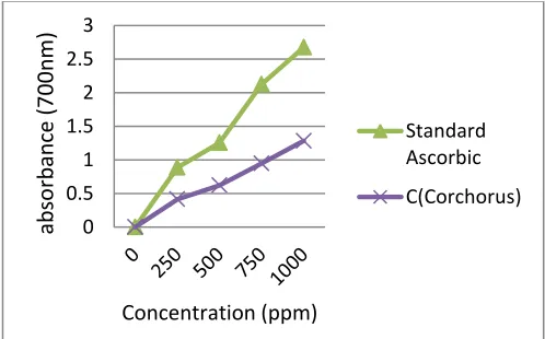
Methanol extracts were tested for the antioxidant activity in various concentrations ranging from 200 – 800 μg/ml by DPPH method. The results show the effect of these extracts to scavenge free radicals. It was observed that free radicals were scavenged by the extracts in a concentration dependent manner. The maximum percentage inhibition of DPPH for the methanol extract was 94.19 % at the highest concentration comparable to the standard ascorbic acid whose maximum was 96.78 % at the same concentration (Table 1). From the FRAP assay (fig. 1), absorbance increased with increasing concentration of plant extract. This signified the consistent reduction of Fe3+ to Fe2+ indicating the reduction potential of the plant.
Figure 1. Ferric reducing power of C. olitorius extract compared with ascorbic acid as standard
0 0.5 1 1.5 2 2.5 3
ab
so
rb
an
ce
(7
0
0
n
m
)
Concentration (ppm)
Standard Ascorbic
Both the aqueous and methanolic extracts of C. olitorius after phytochemical screening showed various phytoconstituents. Some phytoconstituents were present in only one of the extracts while some were present in both extracts (Table 2).
Wound healing activity
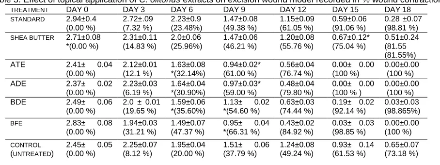
The diameter of the wound was measured in other to determine the rate of wound closure and to determine the wound healing activity of C. olitorius. The mean percentage wound closure was calculated on 0, 3, 6, 9, 12, 15 and 18 post-wounding days. The extracts healed faster than standard, shea butter and control groups. All animals in groups treated with extracts, ADE, ATE, BFE and BDE healed completely on 14, 15, 16 and 18 post-wounding days respectively. Animals treated with ointment preparation showed a faster wound healing activity as compared to the aqueous preparation and all the other treatments. The wound healing activity was dose dependent. Generally, ADE healed first followed by ATE, BFE, BDE, standard, shea butter and the control group in that order (Table 3 and Fig 2).
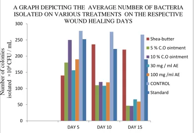
Determination of microbial load
The estimation of microbial load on the surface of the wounds had a similar trend just like the wound healing ability. Generally, the number of microbes isolated on the wounds was decreasing on days 5, 10 and 15. There was significant decrease in the number of microbes in the extract treatments and was also dose dependent. The decrease in the bacterial count on ADE, was more significant than ATE, followed by BFE, BDE, standard, shea-butter and the control group in that order (Fig. 2).
Figure 2. Comparison of the microbial load of the various rat groups that were giving different treatments. Each bar indicates the microbial load of different treated wound on the given days. AE refers to aqueous extracts while C.O refers to Corchorus olitorius
DISCUSSIONS
DPPH is relatively stable nitrogen centered free radical that easily accepts an electron or hydrogen radical to become a stable diamagnetic molecule. DPPH radicals react with suitable reducing agents as a result of which the electrons become paired off forming the corresponding hydrazine. The solution therefore loses colour stoichometrically depending on the number of electrons taken up (Mangathayaru et al., 2007). The antioxidant activity of the plant extract has been demonstrated through DPPH scavenging capacity and FRAP assay. High
0 50 100 150 200 250 300
DAY 5 DAY 10 DAY 15
Shea-butter
5 % C.O ointment
10 % C.O ointment
30 mg / ml AE
100 mg /ml AE
CONTROL
Standard
Numbe
r
of c
oloni
es
isol
ated
×
10
4
C
F
U
/ m
L
A GRAPH DEPICTING THE AVERAGE NUMBER OF BACTERIA
ISOLATED ON VARIOUS TREATMENTS ON THE RESPECTIVE
FRAP assay increases with increasing concentration of the extract. The values obtained, even though lower than the standard antioxidant, showed that C. olitorius is a moderately a good source of antioxidant compounds. The plant may therefore be rich in flavonoids and other phytoconstituents (Table 2) that are known to contribute to antioxidant properties of medicinal plants.
Table 1. Effect of methanol extracts of C. olitorius on DPPH free radical scavenging method
Drug Percentage inhibition (I %)
200ppm 400ppm 800ppm
Ascorbic acid 92.75 + 0.23 93.61+ 1.41 96.78 + 1.94
Methanol extract 88.85 + 0.12 93.56 + 0.09 94.19 + 0.06
Table 2. Results of phytoconstituents in both methanolic and aqueous extracts of C. olitorius
Substance Aqueous Extract Methanol Extract
Alkaloids + -
Terpenoids + +
Tannins + +
Flavonoids + +
Saponins - _
Cardiac glycosides + +
+ indicates presence; - indicates absence
Table 3. Effect of topical application of C. olitorius extracts on excision wound model recorded in % wound contraction
TREATMENT DAY0 DAY3 DAY6 DAY9 DAY12 DAY15 DAY18 STANDARD 2.94±0.4
(0.00 %) 2.72±.09 (7.32 %) 2.23±0.9 (23.48%) 1.47±0.08 (49.38 %) 1.15±0.09 (61.05 %) 0.59±0.06 (91.06 %)
0.28 ±0.07 (98.81 %) SHEA BUTTER 2.71±0.08
*(0.00 %) 2.31±0.11 (14.83 %) 2.0±0.06 (25.96%) 1.47±0.06 (46.21 %) 1.20±0.08 (55.76 %) 0.67±0.12* (75.04 %) 0.51±0.24 (81.55 (81.55%)
ATE 2.41± 0.04
(0.00 %) 2.12±0.01 (12.1 %) 1.63±0.08 *(32.14%) 0.94±0.02* (61.00 %) 0.56±0.04 (76.74 %)
0.00± 0.00 (100 %)
0.00±0.00 (100 %)
ADE 2.37± 0.02
(0.00 %) 2.23±0.03 (6.19 %) 1.64±0.04 *(30.90%) 0.97±0.03* (59.00 %) 0.48±0.04 (79.80 %)
0.00± 0.00 (100 % )
0.00±0.00 (100 %) BDE
2.49± 0.06
(0.00 %)
2.0 ± 0.01 (19.65 %)
1.59±0.06 *(35.60%)
1.13± 0.02
*(54.60 %)
0.63±0.03 (74.44 %)
0.19± 0.02 (92.14 %)
0.03±0.03 (98.865%)
BFE 2.83± 0.08
(0.00 %)
1.94±0.03 (31.21 %)
1.49±0.07 (47.37 %)
0.95± 0.04
*(66.31 %)
0.43±0.02 (84.92 %)
0.03± 0.03 (98.85 %)
0.00±0.00 (100 %)
CONTROL (UNTREATED)
2.45± 0.05
(0.00 %)
2.25±0.07 (8.12 %)
1.95±0.04 (20.00 %)
1.51± 0.06
(37.79 %)
1.24±0.08 (49.24 %)
0.93± 0.14 (61.53 %)
0.65±0.07 (73.18 %)
Values are mean ± SEM (n = 5). Numbers in parenthesis indicate percentage wound closure. * Significant at P < 0.05.
The four phases of normal wound healing include Haemostasis, Inflammation, Proliferation and Remodeling. Wound healing processes are well organized biochemical and cellular events leading to the growth and regeneration of wounded tissue in a special manner. Healing of wounds involves the activity of an intricate net work of blood cells, cytokines and growth factors which ultimately leads to the restoration to normal condition of the injured skin or tissue (Clark, 1991). Antioxidants counter the excess proteases and reactive oxygen species (ROS) often formed by neutrophil accumulation in the wounded area and protect protease inhibitors from oxidative damage. Fibroblasts and other cells may be killed by excess ROS and skin lipids will be made less flexible, so antioxidant substances will reduce the possibility of these adverse events occurring. Because of these several factors, overall antioxidant effects appear to be important in the successful treatment of wounds (Houghton et al., 2005). The antioxidant activity demonstrated by the extract of C. olitorius in this study is therefore of immense significance.
A significant promotion of wound-healing activity was observed in the various extracts (BDE, BFE, ATE and ADE) in the excision wound model. Result from the wound contraction indicates that both the aqueous extract and the powder of C. olitorius leaf have a significant wound healing activity.
activity. A decrease in the number of bacteria on the surface of a wound, on application of a drug indicates that the drug has antimicrobial property. Results from the microbial load suggest that the number of bacteria isolated on the surface of the wounds were decreasing. There was a greater decrease in the bacterial load of ADE, followed by ATE, BFE, BDE, standard, shea butter and the control group in a decreasing order of antimicrobial property. Phytoconstituents such as alkaloids, tannins and cardiac glycosides were found in the plant extracts. Earlier research had revealed that alkaloids, tannins, cardiac glycosides and saponins have antimicrobial property which contributes to `the medicinal property of C. olitorius (Oboh, 2009). Therefore the order of antimicrobial activity observed is in line with the phytoconstituents present in the plant.
Again, it was observed that for the same extracts higher doses caused a greater decrease in microbial loads as compared to the low doses. For instance, there was a greater decrease in the number of bacteria in ADE than ATE. The dose dependent nature of the rate of wound healing is due to the concentration of the phytoconstituents in each of the treatments.
In conclusion, we have demonstrated that C. olitorius displayed significant free-radical-scavenging activity in a concentration dependant manner in in-vitro assays. Also, the powder ointment and the water extract of C. olitorius have been shown to have a wound-healing property. The traditional uses of this plant to treat wound has therefore been confirmed.
REFERENCES
Omale J, Ayide VI. 2010. Excision and incision wound healing potential of Sabaflorida leaf extract in Rattus novergicus. International journal on
Pharmaceutical and biomedical research, 14:101-107.
Ayyanar M, Muthu C, Raja N, Ignacimuthu S. 2006. Medicinal plants used by traditional healers in Kancheepuram District of Tamil Nadu. India.
Journal of Ethnobiology and Ethnomedicine, 2:43, doi:10.1186/1746- 269-2-43.
Mokaddas E, Rotimi VO, Sanyal SC. 1998. In vitro activity of piperacillin / tazobactam versus other broad antibiotics against nosocomial gram
negative pathogens isolated from burn patients. J. Chemotherapy, 10: 208-14.
Pirbalouti Ghasemi A, Azizi S, Koohpayeh A. 2012. Healing potential of Iranian traditional medicinal plants on burn wounds in alloxan-induced
diabetic rats. Revista Brasileira de Farmacognosia, 22 (2):397-03.
World Health Organization. 2010. Wound and lymphoedema management report, 1, Geneva
Driscoll P. 2009. In clinical practice, Surgery wound management report, S247:33-5.Karukonda, S.R.K., Flynn, T.C., Boh, E.E., McBurney, E.I.,
Russo, G.G., Millikan, L.E. 2000. The effects of drugs on wound healing. International Journal of Dermatology, 39(1): 250–57.
World Union of Wound Healing Societies. 2004. Minimising pain at wound dressing-related procedures. Wounds International, 201-15.
Kumar B, Vijayakumar M, Govindarajan R, Pushpangadan, P. 2007. Ethnopharmacological approaches to wound healing exploring medicinal
plants of India. Journal Ethnopharmacol, 114: 103-13
Biswas TK, Mukherjee B. 2003. Plant medicines of Indian origin for wound healing activity: a review. Int. J. Low Extreme wounds, 2(1): 25-39. Ayyanar M, Ignacimuthu S. 2009. Herbal medicines for wound healing among tribal people in Southern India: Ethnobotanical and Scientific
evidences. International Journal of Applied Research in Natural Products, 2(3): 29-42.
Bennet RG. 1988. Fundamentals of Cutaneous Surgery, 2nd Edition, Mosby Company, USA.
Houghton PJ, Hylands PJ, Mensah AY, Hensel A, Deters AM. 2005. In vitro tests and ethnopharmacological investigations: Wound healing as
an example. J Ethnopharmacol 100: 100-107.
Süntar I, Küpeli AE, Nahar L, Satyajit D, Sarker SD. 2012. Wound healing and antioxidant properties: do they coexist in plants? Free Radicals
and Antioxidants, 2( 2): 1-7.
Sofowora A. 1993. Medicinal Plants and Traditional Medicines in Africa. 2nd edition, John Wiley & Sons, New York.
Harborne JB. 1998. Phytochemical Methods: A guide to Modern Techniques of Plant Analysis. 2nd edition, Chapman and Hall, London,
UK.
Trease GE, Evans MC. 1998. Textbook of Pharmacognosy. 13th edition, Bailliere Tindal Company, London.
Braca A, Tommasi ND, Bari LD, Pizza C, Politi M, Morelli I. 2001. Antioxidant principles from Bauhinia terapotensis. Journal of Natural
Products, 64:892-895.
Rajeswara RP, Sambasiva RE, Yasodhara B, Praneeth Dasari VS, Mallikarjuna RT. 2012. In-vitro antioxidant and antibacterial activities of
different fractions of Heliotropium indicum L.Journal of Pharmacy Research, 5(2):1051-1053
James O, Nnacheta OP. 2008. Comparative antioxidant capacity, membrane stabilization, polyphenol composition and cytotoxicity of the leaf
and stem of Cissus multi-striata African J. Biotech. 7(17): 3129-33.
Ramnik S, Narinder S, Saini BS, Harwinder SR. 2008. In-vitro antioxidant activity of pet ether extract of black pepper. Indian J. Pharmacol,
40(4):147-151.
Oyaizu M. 1986. Studies on products of browning reactions: Antioxidant activities of products of browning reaction prepared from glucosamine. Japan J. Nutr., 44: 307-315
Manjunatha BK, Vidya SM, Krishna V, Mankani KL. 2006. Wound healing activity of Leucas hirta. Indian J. Pharma. Sci.,
68(3):380-384.
Srinivas BR, Kumar RKR, Naidu VGM, Madhu SK, Agwane SB, Ramakrishna S, Diwan PV. 2008. Evaluation of antimicrobial, antioxidant and
wound healing po-tentials of Holoptelea integrifolia. J. Ethnopharmacol., 115: 249-56.
Mangathayaru K, Sravan K, Praveen AKR, Kuma RM. 2007. Invitro antioxidant studies of the aerial parts of Ori-ganum majoram Linn and
Artemesia sieversiana Ehrh. Pharmacognosy magazine, 3:10:90-4.
Clark RAF. 1991. Cutaneous wound repairs. In: Goldsmith LA(ed.) Physiology, Bio- chemistry and Molecular Bi-ology of skin. Oxford
University Press, New York.