Original Research Article
Fine Needle Aspiration Cytology of Parotid Lesions: A Study of 84 Cases
with Special Reference to Cyto-Histological Discrepancy
Subrata Pal
1, Sajeeb Mondal
2, Kingshuk Bose
3, Shubham Bhattacharya
4, Rajashree Pradhan
5,
Barnali Maiti
61MD, Demonstrator, Department of Pathology,
College of Medicine and Sagore Dutta Hospital, Kamarhati, Kolkata, West Bengal, India. 2Faculty, Department of Pathology,
College of Medicine and Sagore Dutta Hospital, Kamarhati, Kolkata, West Bengal, India. 3Assistant Professor, Department of Pathology,
Bankura Sammilani Medical College, Bankura, West Bengal, India. 4MD, Assistant Professor, Department of Pathology,
IQ City Narayana Multispeciality Hospital, Durgapur, West Bengal, India. 5MD, Faculty, Department of Pathology,
College of Medicine and Sagore Dutta Hospital, Kamarhati, Kolkata, West Bengal, India. 6MD, Assistant Professor, Department of Pathology,
IQ City Narayana Multispeciality Hospital, Durgapur, West Bengal, India.
ABSTRACT
Background: Parotid gland lesions are extremely diverse entities. Pre-operative cytodiagnosis is difficult due to heterogenesity of cellular components. However pre-operative FNAC as a diagnostic tool is till controversial.
Aims: The study was performed to evaluate the effectiveness
of FNAC in preoperative diagnosis of parotid lesions and to evaluate the cases of cyto-histological discrepancies.
Material & Methods: The study was conducted over a three years period over 84 cases of parotid lesions, where cytological diagnoses were compared with histopathological report in available cases. In all cases of cyto-histological discrepancies, possible causes of discrepancies were searched.
Results: Non neoplastic (23 cases, 29.48%) and benign (40 cases, 51.28%) cases outnumbered than malignant tumors (14 cases, 19.23%). Diagnostic sensitivity and specificity of cytology were 86.66% and 95.74% respectively. We found six cases of cyto-histological discrepancies with a diagnostic accuracy of 93.54%. Two false negative, two false positive cases were evaluated and two cases had error in typing of tumor.
Conclusion: Many of parotid tumors have one or more close
cytologic mimickers due to diverse cell types and growth pattern. However FNAC should be primary evaluating tool for diagnosis and management of parotid lesions.
Key words: Parotid, FNAC, Histopathology, Discrepancy.
*Correspondence to:
Dr. Subrata Pal,
Kalpataru Apartment, Sahid Colony, BT road, PS- Khardaha,
North 24 Pargana, West Bengal, India. Article History:
Received: 16-02-2017, Revised: 10-03-2017, Accepted: 18-03-2017
Access this article online
Website:
www.ijmrp.com
Quick Response code
DOI:
10.21276/ijmrp.2017.3.2.061
INTRODUCTION
Diverse group of non-neoplastic, benign and malignant tumors may arise in parotid glands.1,2 Parotid gland tumors comprise 3%
of all head-neck tumors.1 FNAC is a quick, simple, inexpensive,
safe and well established diagnostic procedure for different superficial lesions including parotid swellings.1,2 Open core biopsy
is no longer practiced due to risk of spillage of tumor cells and risk of damage of facial nerve.1 Many previous studies revealed
variable sensitivity, specificity and accuracy of cytology in parotid lesions.1,3,4 Though most of the salivary gland lesions have
distinctive cytomorphology, but sometimes exact cytodiagnosis very difficult due to some confounding factors.2 We focused our
MATERIAL AND METHODS
The study was undertaken in a tertiary care hospital in India over a period of three years (Jan 2011-Dec 2013). Ethical clearance was taken from institutional ethical committee. Written consents were taken from all the patients included in the study group. Data was collected retrospectively from our cytology records. All cases of parotid swellings which were undergone FNAC during the period, were included in the study group. FNAC was performed in our cytology laboratory using 22 G needle attached with 10 ml syringe. Slides were fixed in alcohol as well as air dried. Air dried
smears are stained with Leishman and Giemsa stain and alcohol fixed smears stained with PAP stain and H & E stain. Biopsy samples were received in our histopathology laboratory, were processed, sectioned and stained with H & E stain. Histopathological diagnosis was compared with cytological report and slides were reviewed to detect the cases of cyto-histological discrepancies. Sensitivity, specificity, positive predictive value and diagnostic accuracy were calculated by standard statistical methods.
Figure 1: Bar diagram showing age distribution of cases of parotid swelling.
Table 1: Distribution of cases according to cytological and histological diagnosis.
Type of lesion Diagnosis Number in Cytology (n-78) Number in histology
Non-neoplastic (n-23)
Chronic sialoadenitis 20 (25.64%) 4(5.12%)
Lymphatic cyst 1(1.28%) 2(2.56%)
Granulomatous lesion 1(1.28%)
Benign (n-40)
Pleomorphic salivary adenoma 31(39.74%) 30(38.46%)
Monomorphic adenoma 1(1.28%) 3(3.84%)
Warthin’s tumor 7(8.97%) 6(7.69%)
Oncocytoma 2(2.56%) 1(1.28%)
Malignant (n-15)
Mucoepidermoid carcinoma 6(7.69%) 5(6.41%)
Adenoid cystic carcinoma 3(3.84%) 2(2.56%)
Acinic cell carcinoma 1(1.28%) 2(2.56%)
Salivary duct carcinoma 2(2.56%) 2(2.56%)
Non Hodgkin’s lymphoma 1(1.28%) 1(1.28%)
Metastatic melanoma 1(1.28%) 1(1.28%)
Pleomorphic adenoma ex carcinoma
1(1.28%) 1(1.28%)
Polymorphous low grade adenocarcinoma
RESULTS
In the present study, we have aspirated 84 cases of parotid swelling. In 78 cases aspiration produced adequate material and in six cases, materials were inadequate even after repeated aspiration. We found 45 male patients (53.57%) and 39 female cases (46.43%) in the study group. Patients ranged in age from 8 years to 71 years with a mean age of 38.73 years. Age distribution reveals large number of cases in the age group of 21-50 years. (Figure 1) Mean age of malignant cases (53.14years) was higher than benign cases (38.19 years). In the present study, four cases (5.12%) were bilateral. We found most of the cases were benign (40 cases, 51.28%) and non-neoplastic (23 cases, 29.48%); whereas only 14 cases (19.23%) were malignant. (Table 1) Among the non-neoplastic cases, most common lesion was chronic sialoadenitis (20 cases). Among the benign parotid swellings PSA was the commonest subtype (30 cases, 38.46%). Other benign parotid tumors diagnosed are warthin’s tumor (6 cases), monomorphic adenoma (3 cases) and oncocytoma (one case). [Table 1] Most common malignant parotid tumor in our study was mucoepidermoid carcinoma (5 cases, 6.41%). Other malignancies were adenoid cystic carcinoma, acinic cell carcinoma and salivary duct carcinoma etc. We found single case of non-Hodgkin’s lymphoma, pleomorphic adenoma ex carcinoma and metastatic melanoma respectively in the present study group. [Table 1]
In comparison of cytology with final histology, we found six cases of cyto-histological discrepancies. Among these six cases two were false negative and two were false positive. In another two cases the error was in typing of the lesion for specific diagnosis. Sensitivity and specificity of FNAC in detecting malignant lesions were 86.66% and 95.74% respectively. Diagnostic accuracy of cytology was 93.54%.
DISCUSSION
FNAC is an well-established diagnostic tool for diagnosis and management of various head and neck lesions.1,4,6 In some of
previous studies, authors claimed the pre-operative FNAC has little influences on management because of high rate of false positive and false negative cases and ultimately the patients need surgery.7,8 But detection of the nature of the lesions helps in
planning of treatment of the cases. Here we focused the cases of cyto-histological discrepancies and tried to find out the causes. Mean age of parotid swelling in the present study is similar in other studies (Ali et al -44 years).1 In our study we found mostly
benign (51.28%) and non-neoplastic cases (29.48%) than the malignant tumors (17.94%). Similar observations were documented by Ali et al, Jan et al and Lurie et al.1,2,9 We found six
cases (7.14%) of inadequate aspiration in our study even after repeat aspiration. Unsatisfactory aspirations vary from 3% to 12% in different studies.1,2,4,10-14 In a meta-analysis by Schaidt et al,
non-contributory or unsatisfactory FNAC were 8.6%; similar to our finding.15 Most common benign tumor diagnosed in cytology as
well as in histology is PSA (39.74%) in the present study, similar to previous studies.1,2,9 Mucoepidermoid carcinoma is the most
common malignancy (five cases, 6.41%) in our study consistent with Ali et and Piccioni et al. But Jan et al found squamous cell carcinoma as most common malignancy in their series.1,2,16
Sensitivity and specificity of the present study are 86.66% and 95.74% respectively. In large number of studies in recent times, sensitivity of parotid lesions ranges from 54% to 92% and specificity varies from 86% to 100%.1,11,12,15,16 Diagnostic accuracy
of our study (93.54%) is also similar to Ali et al and Jan et al.1,2
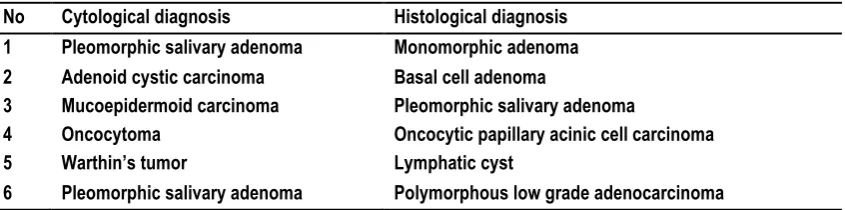
We found six cyto-histological discrepant cases (table 2), among which two were false negative for malignancy and two were false positive.
Table 2: Cases of cyto-histological discrepancy.
No Cytological diagnosis Histological diagnosis
1 Pleomorphic salivary adenoma Monomorphic adenoma
2 Adenoid cystic carcinoma Basal cell adenoma
3 Mucoepidermoid carcinoma Pleomorphic salivary adenoma
4 Oncocytoma Oncocytic papillary acinic cell carcinoma
5 Warthin’s tumor Lymphatic cyst
6 Pleomorphic salivary adenoma Polymorphous low grade adenocarcinoma
CASE 1
A 45 year male presented with 3x2 cm hard right parotid swelling for last six months. Cytology revealed hypercellular smears containing small dark basaloid cells and abundant hyaline material (Figure 2 A). The cells had very scanty cytoplasm, round to oval hyperchromatic nuclei with conspicuous nucleoli. Cytology was diagnosed as adenoid cystic carcinoma. On histopathology it was confirmed as basal cell adenoma (Figure 2B). On review of cytology smears we found that we have missed well demarcated lining of basaloid cell attached with hyaline globules, cells were in and around the hyaline globules, absence of mitosis etc. Similar error was also documented by Jurczyk et al.17,18 Absence of
stromal spindle cells, presence of many hyaline globules and hypercellularity also leaded to misdiagnosis.
CASE 2
A 62 year old male with 5x3 cm hard parotid swelling at left parotid for four months. Cytologically it was diagnosed as mucoepidermoid carcinoma but on histology it was confirmed as pleomorphic salivary adenoma with extensive squamous metaplasia.
The cytology smears showed squamoid neoplastic cells and oval to round epithelial cells and very scanty myxoid material. Paucity of chondromyxoid stroma and presence of metaplastic cells leaded to this error. Similar error has also documented in multi cystic pleomorphic salivary adenoma and where cytology lacks chondromyxoid stroma and exhibits extensive squamous metaplasia.19-21 Multiple aspirations from different sites can avoid
Figure 2: (A) Hypercellular smears containing small dark basaloid cells and abundant hyaline material in a case of basal cell adenoma, misinterpreted as adenoid cystic carcinoma [40X view, H & E stain]. (B) Histopathology of
case 1 (respective of figure 2) diagnosed as basal cell adenoma [40X view, H& E stain].
Figure 3: (A) Neoplastic oncocytic cells and occasional cyst-macrophages in the background of blood, misinterpreted as oncocytoma (case 3), diagnosed as oncocytic papillary acinic cell carcinoma in histology
[L & G stain, 40X view]. (B) Histopathology of case 3 (respective of figure 3A) diagnosed as oncocytic papillary acinic cell carcinoma (H& E stain, 10X view).
CASE 3
51 year male presented with a firm mass at left parotid region for eight months. Aspiration material was altered blood mixed. The smears exhibit clusters of oncocytic cells and occasional cyst macrophages in the background of blood. The oncocytic cells had abundant eosinophilic granular cytoplasm, round nuclei with mild pleomorphism and conspicuous nucleoli (Figure 3A). On histopathology it revealed a tumor composed of complex branching papillary configuration with invasion into surrounding stroma (Figure 3B). The neoplastic cells are oncocytic cells with abundant eosinophilic cytoplasm, round vesicular nuclei with prominent nucleoli. Histologically it was diagnosed as oncocytic papillary acinic cell carcinoma of parotid. In previous studies, the authors shown that oncocytic acinic cell carcinoma closely mimics
oncocytoma specially the papillary pattern.22 Absence of
significant cytological atypia, pleomorphism, stippled nuclei and overlooking of cytoplasmic vacuoles were the causes of misdiagnosis.
CASE 4
adenocarcinoma. Overlooking of palisading neoplastic cells around myxoid stroma leaded to such false negative diagnosis. Similar findings and error have been experienced by Sing et al, Sahik et al and Wantanabe et al.23-25
CASE 5
A 45 year female presented with firm left parotid mass for last 6 months. Aspirated smears revealed cohesive benign epithelial cells with fragments of myxoid matrix and occasional spindle cells, diagnosed as PSA in cytology. In histology it was diagnosed as basal cell adenoma. Basal cell adenomas have high rate of erroneous diagnosis in cytology and main differential diagnosis is epithelial rich PSA.26,27 We have missed the streaming palisaded
basaloid cells at edge of cohesive cell clusters and acellular matrix ribbons which help to distinguish as basal cell adenoma.
CASE 6
A young male of 22 years presented with 3x2 cm cystic parotid swelling for last 5 months. Aspiration was dark brown fluid and cytology revealed plenty of lymphoid cells with occasional oncocytic cells in a dirty fluidy background. Cytologically it was diagnosed as warthin’s tumor but in histology it was diagnosed as lymphatic cyst. The histiocytes and metaplastic lining epithelial cells were misinterpreted as oncocytic cells. Oncocytic metaplasia of lining epithelium of lymphatic cyst is a close differential
diagnosis of warthin’s tumor in cytology.28
CONCLUSION
FNAC is a safe rapid cost effective and easy diagnostic tool with very good accuracy (93.54%). Despite some limitations and close mimickers in parotid gland tumors, we appreciate cytodiagnosis to avoid unnecessary surgery in non-neoplastic parotid lesions and to determine the nature of neoplastic lesions in management of benign and malignant parotid tumors. Our discussion regarding the cyto-histological discrepancies will help the cytologists for better differentiation and categorization of parotid gland cytology.
REFERENCES
1. Ali NS, Akhtar S, Junaid M, Awan S and Aftab K. Diagnostic accuracy of fine needle aspiration cytology in parotid lesions. Int Sch Res Net Surg 2011. 2011:721525.
2. Jan IS, Chung PF, Weng MH, Huang MS, Lee YT, Cheng TY, et al. Analysis of fine-needle aspiration cytology of the salivary gland. J Formos Med Assoc 2008; 107(5):364–70.
3. Costas A, Castro P, Martin-Granizo R, Monje F, Marron C, Amigo A. Fine needle aspiration biopsy (FNAB) for lesions of the salivary glands. Br J Oral Maxillofac Surg. 2000; 38(5):539–42. 4. Stewart CJ, MacKenzie K, McGarry GW, Mowat A. Fine-needle aspiration cytology of salivary gland: a review of 341 cases. Diagn Cytopathol 2000; 22(3):139–46.
5. Stanley, MW. Selected problems in fine needle aspiration of head and neck masses. Mod Pathol.2002;15(3):342–350. 6. Atula T, Greenman R, Laippala P, Klemi PJ. Fine needle aspiration biopsy in the diagnosis of parotid gland lesions: evaluation of 438 biopsies. Diagn Cytopathol 1996;15(3):185–90. 7. Salgarelli AC, Capparè P, Bellini P, Collini M. Usefulness of fine-needle aspiration in parotid diagnostics. Oral Maxillofac Surg 2009;13(4):185–90.
8. Batsakis JG, Sueige N, el-Naggar AK. Fine-needle aspiration of salivary glands: its utility and tissue effects. Ann Otol Rhinol Laryngol1992;101(2):185–8.
9. Lurie M, Misselevitch I and Fradis M et al. Diagnostic value of fine needle aspiration from parotid gland lesions. Isr Med Assoc J 2002;4(9):681-3.
10. Boccato P, Altavilla G, Blandamura S. Fine needle aspiration biopsy of salivary gland lesions: a reappraisal of pitfalls and problems. Acta Cytol 1998;42(4):888–98.
11. Fakhry N, Antonini F, Michel J, Penicaud M, Mancini J and Lagier A et al. Fine needle aspiration cytology in the management of parotid masses: evaluation of 249 patient. Eur Ann Otorhinolaryngol Head Neck Dis 2012;129(3):131–5.
12. Zbaren P, Schar. C, Hotz MA, Loosli H. Value of fine-needle aspiration cytology of parotid gland masses. Laryngoscope 2001;111(11):1989–1992.
13. Wong DS, Li GK. The role of fine-needle aspiration cytology in the management of parotid tumors: a critical clinical appraisal. Head Neck 2000; 22(5):469–73.
14. Bartels S, Talbot JM, DiTomasso J, Everts EC, Andersen PE, Wax MK, et al. The relative value of fine-needle aspiration and imaging in the preoperative evaluation of parotid masses. Head Neck 2000,22(8):781–6.
15. Schmidt RL, Hall BJ, Wilson AR, Layfield LJ. A systematic review and meta-analysis of the diagnostic accuracy of fine-needle aspiration cytology for parotid gland lesions. Am J Clin Pathol2011;136(1):45-59.
16. Piccioni LO, Fabiano B, Gemma M, Sarandria D, Bussi M. Fine needle aspiration cytology in the diagnosis of parotid lesions. Acta Otorhinolaryngol Ital 2011;31(1):1–4.
17. Jurczyk M, Peevey JF, Vande Haar MA, Lin X. Pitfalls of fine-needle aspiration cytology of parotid membranous basal cell adenoma—A review of pitfalls in FNA cytology of salivary gland neoplasms with basaloid cell features. Diagn Cytopathol 2015;43(5):432-7.
18. Gupta N, Bal A, Gupta AK, Rajwanshi A. Basal cell adenoma: A diagnostic dilemma on fine needle aspiration cytology. Diagn Cytopathol 2011;39(12):913–6.
19. Batrani M, Kaushal M, Sen AK, Yadav R, Chaturvedi NK. Pleomorphic adenoma with squamous and appendageal metaplasia mimicking mucoepidermoid carcinoma on cytology. Cytojournal 2008;6:5.
20. Brachtel EF, Pilch BZ, Khettry U, Zembowicz A, Faquin WC. Fine-needle aspiration biopsy of a cystic pleomorphic adenoma with extensive adnexa-like differentiation: differential diagnostic pitfall with mucoepidermoid carcinoma. Diagn Cytopathol 2003;28(2):100-3.
21. Daneshbod Y, Daneshbod K, Khademi B. Diagnostic difficulties in the interpretation of fine needle aspirate samples in salivary lesions: Diagnostic pitfalls revisited. Acta Cytol 2009;53(1):53–70.
22. Jayaram G, Dashini M. Evaluation of the fine needle aspiration cytology of the salivary glands: an analysis of 141 cases. Malaysian J Pathol 2001;23(2):93- 100.
23. Singh S, Garg N, Marwah N, Kalra R, Singh V, Sen R. Fine needle aspiration cytology in lesions of oral and maxillofacial region: Diagnostic pitfalls. J Cytol 2011;28(3):93–7.
cytology of minor salivary gland tumours of the palate. Cytopathol 2002;13(5):309–16.
25. Wantanabe K, Ono N, Saito K, Saito A, Suzuki T. Fine-needle aspiration cytology of polymorphous low-grade adenocarcinoma of the tongue. Diagn Cytopathol 1999;20(3):167–9.
26. Vicandi B, Jiménez-Heffernan JA, López-Ferrer P, González-Peramato P, Patrón M and Viguer JM. Fine needle aspiration cytology of basal cell adenoma of the salivary gland: a cytohistological correlation study of 35 cases. Cytopathol 2012;23(5):315-9.
27. Midi A, Aydin O, Comunoglu C, Boyaci Z, Peker O. Basal cell adenoma of salivary gland; cytological features and differential diagnosis. 2009. [Last accessed on 2014 Sep 12]. p. 8. [Available from:www.KBB.Forum.net]
28. Mukunyadzi P. Review of fine-needle aspiration cytology of salivary gland neoplasms, with emphasis on differential diagnosis. Am J Clin Pathol 2002;118(suppl 1):S100–15.
Source of Support: Nil. Conflict of Interest: None Declared.
Copyright: © the author(s) and publisher. IJMRP is an official publication of Ibn Sina Academy of Medieval Medicine & Sciences, registered in 2001 under Indian Trusts Act, 1882. This is an open access article distributed under the terms of the Creative Commons Attribution Non-commercial License, which permits unrestricted non-commercial use, distribution, and reproduction in any medium, provided the original work is properly cited.
Cite this article as: Subrata Pal, Sajeeb Mondal, Kingshuk Bose, Shubham Bhattacharya, Rajashree Pradhan, Barnali Maiti. Fine Needle Aspiration Cytology of Parotid Lesions: A Study of 84 Cases with Special Reference to Cyto-Histological Discrepancy. Int J Med Res Prof. 2017; 3(2):285-90.

![Figure 2: (A) Hypercellular smears containing small dark basaloid cells and abundant hyaline material in a case of basal cell adenoma, misinterpreted as adenoid cystic carcinoma [40X view, H & E stain]](https://thumb-us.123doks.com/thumbv2/123dok_us/1431931.1657104/4.595.58.541.324.555/figure-hypercellular-containing-basaloid-abundant-material-misinterpreted-carcinoma.webp)
