NEW RESEARCH METHODOLOGIES USED FOR THE INVESTIGATION OF IMMUNE SYSTEM IN VETERINARY SPECIES
Marek Sinkora, Jana Sinkorova
Institute of Microbiology of the Academy of Sciences of the Czech Republic, v.v.i., Department of Gnotobiology, Doly 183, 549 22 Novy Hradek, Czech Republic
Abstract
Financial supports for basic research in veterinary immunology are often limited due to interdisciplinary nature. Grant agencies supporting basic research argue that subjects are applied while agriculture agencies argue that it belongs to basic research. Consequences of this approach are a loss of financial resources for veterinary projects. We have shown that this limitation can be precluded by using of state-of-the-art technologies. These include high-speed flow cytometry sorting, high-throughput molecular analysis of single cells, microdissection and single cell recovery by micromanipulation. We have recently used these high-tech methodologies to (1) disprove the concept that porcine hind-gut follicles are a site of B cell lymphogenesis, (2) characterize B cell development in the bone marrow, (3) characterize lineages, developmental and activation markers of γδ T cells and (4) show negative modulation of immune system by some viruses. This work was supported by Czech Science Foundation grant P502/12/0110 and P502/10/0038.
Key words: State-of-the-art Technologies, Veterinary Immunology, High-tech Methodologies, Applied and Basic Research
1. INTRODUCTION
Studies in veterinary immunology involve large animals that offer the advantage of large sample volumes, the ability to perform surgical procedures that are difficult in rodents and allows greater experimental control over maternal and environmental factors by taking advantage of differences in placentation and the precocial nature of many large animal offspring. These advantages are offset by animal acquisition and maintenance costs, facility limitations on numbers, the outbred genetics of nearly all large animals and the limited number of immunochemical reagents available. Thus a balance must be established to justify their use in advancing frontiers in immunology and to defend their usage for basic research. In opposite case, researchers take a chance that their research will be refused or replaced by superior technologies. This article reviews our recent technologies, procedures and findings that result in sufficient financial support and highly relevant publications that assure sustainability of veterinary science.
2. USED STATE-OF-THE-ART TECHNOLOGIES
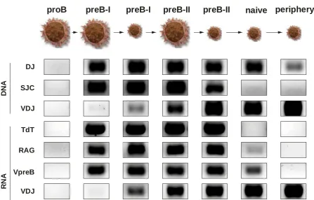
The most common procedure used in our laboratories is flow cytometry analysis and high-speed flow cytometry sorting. Flow cytometry is a laser-based technology employed in analysis of suspensions of cells in a stream of fluid and passing them by an electronic detection apparatus. It allows simultaneous multiparametric analysis of the physical and chemical characteristics of up to thousands of cells per second. Although flow cytometry is routinely used in the diagnosis it has many other applications namely in basic research. A common variation is to physically sort cells based on their properties, so as to purify populations of interest. We have used this technology in many of our studies (Sinkora et al. 1998, Sinkora et al. 2000, Sinkora et al. 2002, Sinkora et al. 2005, Sinkora et al. 2007, Sinkora et al. 2011, Sinkora et al. 2013, Sinkora & Sinkorova 2014, Sinkora et al. 2014, Stepanova & Sinkora 2012, Stepanova & Sinkora2013). Example of our recent findings is analysis that defines early B cell developmental stages in swine and connects them with the expression of B lineage associated genes and status of immunoglobulin heavy chain rearrangement (Fig. 1).
proB
preB-I
preB-I
preB-II
preB-II
naive periphery
VDJ DJ
SJC
VDJ TdT
RAG
D
N
A
R
N
A VpreB
Figure 1: Example of results from high-speed flow cytometry sorting. Figure show a detection of rearrangement-specific products and transcripts from sorted bone marrow cells. According to phenotype studies, seven individual subsets (large proB, large preB-I, small preB-I, large preB-II, small preB-II, naive and peripheral B cell-lineage cells) were sorted by flow cytometry. Each sorted subset was thereafter examined for the presence of rearrangement-specific products (DNA) and transcripts (RNA) by two-round polymerase chain reaction using specific primer sets. Rearrangement-specific products and transcripts include: DJ = partial VDJ rearrangement for immunoglobulin heavy chain, SJC = signal joint circles, VDJ = complete VDJ rearrangement for immunoglobulin heavy chain, TdT = terminal deoxynucleotidyl transferase, RAG = recombination activation genes and VpreB = invariable immunoglobulin iota chain of surrogate immunoglobulin light chain.
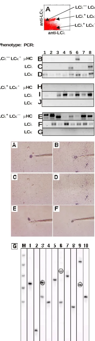
The second procedure used in our lab is high-throughput molecular analysis of single cells. This methodology is based on flow cytometry sorting of single cells into individual tubes so that each tube contain one cells. This allows analysis of single cells by molecular approaches to define genetic propensities of individual cells. Single cells are sorted into 96-wells PCR strips in about minutes so that analysis of thousands of cells can be done in a hour. Example of this analysis is shown in Figure 2. In this specific example, B cell lineage cells from the bone marrow were sorted according to expression of light chain as single cells and analyzed for expression of immunoglobulin light (LC) and heavy (HC) chain genes.
The last procedure that is routinely used in our lab include similar experimental approach for analysis of single cells as in the case of flow cytometry sorting. However, tissue sections are used instead of cell suspensions and mechanical microdissection of cells from tissue sections is performed by micromanipulator attached to inverted microscope and equipped by a glass tip. Advantage of this procedure is precise and known location of harvested cells in a tissue. Example of this analysis is shown in Figure 3. This particular analysis shows detection of immunoglobulin heavy chains in B cells captured from section of the thymus.
LC LCλ+ κ— LC LCλ+ κ+
LC LCλ+ κ— µHC LCλ LCκ
E
F
G
LC LCλ+ κ+ µHCLCλ LCκ
H
I
J
Phenotype: PCR: LCλ— LCκ+ µHCLCλ LCκ
1 2 3 4 5 6 7 8
B
C
D
a n ti -L C κ anti-LCλLCλ LCκ
A
Figure 2: Example of results from high-throughput molecular analysis of single cells. Bone marrow cells were stained for immunoglobulin light chain lambda (LCλ) and kappa (LCκ) and sorted as individual cells into separate tubes (A). After sorting, cDNA was prepared using random hexamer primer and two-round polymerase chain reaction (PCR) was performed using specific primer sets for LCλ, LCκ and immunoglobulin heavy chain (HC). Result of amplification was analyzed in agarose gels and representaite results from eight different cells (1-8 above gel strips) for each amplification is shown. The results include all three population of LCλ—LCκ+ (B-D), LCλ+LCκ+(H-J) and LCλ+LCκ—
cells (E-G). These three population were always analyzed for presence of HC (B, H, E), LCλ (C, I, F) and LCκ (D, J, G) genes.
Figure 3: Example of results from microdissection and single cell recovery by micromanipulation. Figure show a sections of tissue with labeled cell of interest before (A-B) and after (C-D) capture of cells by micromanipulator. Showed is also a top of the glass tip with attached cell after microdissection (E-F). Top of the tip was broken to a tube after cell capture and two-round polymerase chain reaction was performed in each tube using specific primer sets for immunoglobulin heavy chains. The result of PCR amplification is shown on sequencing gel for ten different cells (1-10 above gel strips; G). Extreme left line marked by "M" is a molecular marker composed of five defined lengths of genes for immunoglobulin heavy chain rearrangement. Note that all bands from single cell amplification were subsequently sequenced and only bands that are circled were found non-productive. These rusults confirm that single B cell can only contain one productive rearrangement (only one band) or two rearrangements, one of which is productive while the second is non-productive (two bands from one of which is circled).
3. PARADIGMS THAT WERE CHANGED OR CREATED
3.1. Disproving the concept that porcine hind-gut follicles are a site of B cell lymphogenesis
Artiodactyls possess gut-associated lymphoid tissue that appears in fetal life and which is located at the extreme end of the ileum. These ileal Peyer’s patches (IPP) contain mostly B cells and involute early in postnatal life. Rabbits have a similarly-located lymphoid organ called the sacculus rotundus. Studies in sheep and rabbits have lead to the concept that the lower hindgut represent primary lymphoid tissue for B cells and is necessary for normal B cell development, analogous to the bursa of Fabricius in chicken (Yasuda et al. 2006). Since bursectomy results in B cell deficiency, we wondered if resection of the IPP of piglets would have a similar effect. Comparison of IPP resected, surgical shams and untreated germ-free piglets, all of which were later colonized with a defined commensal flora demonstrates that resection of the IPP did not alter the level and phenotype of B and T cells in lymphoid tissues and the blood during 10 weeks after surgery (Sinkora et al. 2011). The resection of IPP also did not alter antibody levels in serum and secretions or retard diversification of the antibody repertoire (Butler et al. 2011). On the other hand, colonization of IPP caused shift from the fetal type of lymphocyte distribution to the adult type that is characteristic by prevalence of effector and IgA switched B cells (Sinkora et al. 2011). Moreover, colonization leads to appearance of effector αβ and γδ T cells. Colonization also caused >10-fold greater diversification of antibody repertoire and 3- to 5-fold increase in the frequency of somatic hypermutation (Butler et al. 2011). Comparison of germ-free with colonized pigs and experiments utilizing surgical transposition of JPP into terminal ileum or construction of isolated ileal loops indicate that lymphocyte development in IPP is dependent on colonization (Butler et al. 2011; Sinkora et al. 2011). Although our studies confirmed higher mitotic and apoptotic rate in IPP, they failed to identify any cell populations that resemble developing B lineage cells in the bone marrow (Sinkora et al. 2011). These results indicate that porcine IPP are not required for systemic B cell generation or maintenance, but are secondary lymphoid tissue that appear important in immune responses to colonizing bacteria.
3.2. Characterization of B cell development in the bone marrow
Since our previous findings showed that at least in swine (but probably also in other ungulates), ileal Peyer’s patches are not a significant source of B cells, are not required for maintenance of the systemic B cell pool and are not a site of primary B cell lymphogenesis (Butler et al. 2011; Sinkora et al. 2011; Butler & Sinkora 2013), we have analyzed porcine bone marrow, which is the primary lymphoid organ also for mouse and human (Ghia et al. 1996; Hardy & Hayakawa, 2001). According to expression of MHC class II, CD2, CD21, CD25, CD45RC, CD172a , SWC7 and μHC, porcine bone marrow cells were resolved into seven subsets representing sequential stages of B cell development (Sinkora & Sinkorova, 2014). Profile of rearrangement-specific products and transcripts from sorted bone marrow cells confirmed the proposed developmental pathway (Fig. 1). The same developmental pathway was further proven by analysis of selection for productive rearrangements in immunoglobulin heavy chains and also by cultivation studies (Sinkora & Sinkorova, 2014). Cultivation also showed that earliest precursors with incomplete DJ rearrangements can still revert their B cell differentiation and develop along myeloid lineage, while this is impossible for later developmental stages. Proliferation and the apoptotic potential of individual developmental stages as well as critical check-points were also identified. Colocalization experiments showed that early molecular complex MHC-II / CD2 / CD172a / SWC7 is replaced by complex MHC-II / CD2 / CD21 / SWC7 / IgM in immature cells, while CD25 and CD45RC did not colocalize with any other studied molecules. This approach finally proves that the bone marrow in pigs is fully functional in adult animals and that B lymphogenesis occurs there throughout life. Therefore, a course and a direct site of B cell lymphogenesis in swine was described (Sinkora & Sinkorova, 2014).
3.3 Characterization of lineages, developmental and activation markers of γδ T cells
T lymphocytes of γδ lineage are evolutionary conserved cells which develop in the thymus similarly to αβ T cells (Xiong & Raulet 2007). However, γδ T cells do not need any selection for pre-antigen receptors (like pre-BCR or pre-TCRαβ) and therefore mature faster than αβ T cells, develop without any TCRlow transitional stage and are released much earlier to the periphery (Sinkora et al. 2000, Sinkora et al. 2005, Sinkora et al. 2007, Xiong & Raulet 2007). Swine together with ruminants and birds belongs to the group of γδ high species in which γδ T cells are not preferentially limited to epithelia and may account for >70% of all T cells (Hein & Dudler, 1993). Traditionally, γδ T cells in swine are subdivided into three subsets based upon their expression of CD2 and CD8 and include CD2—CD8—, CD2+CD8— and CD2+CD8+ cells (Sinkora et al. 1998, Sinkora et al. 2005, Sinkora et al. 2007, Yang & Parkhouse, 1996). Our previous studies revealed basic distribution of porcine γδ T cells (Sinkora et al. 1998), their ontogeny (Sinkora et al. 1998, Sinkora et al. 2005), development in the thymus (Sinkora et al. 2000, Sinkora et al. 2005, Sinkora et al. 2007) and the repertoire diversification of their TCR (Holtmeier et al. 2004). However, none of these studies focused on a detailed analysis of peripheral γδ T cells, and no other studies have been performed to explain differences in the phenotypic profile of γδ T cells subsets. In our recent studies we have shown that many γδ T cells can constitutively express CD25 and MHC-II and that the frequency of γδ T cells positive for CD25, CD11b, SWC1 and SWC7 can be increased by stimulation (Stepanova & Sinkora 2012). A diversified TCRδ repertoire was found inside CD25+
, CD11b+, SWC1— and CD45RA— γδ T cells. Ontogenetic studies revealed various age and/or colonization dependency for expression of all studied molecules except of SWC7. Findings generally indicate that CD25 represent an activation molecule that probably marks a functionally distinct subsets, expression of CD11b is perhaps connected to early functions of naive γδ T cells in the periphery, SWC1 is lineage specific marker, SWC7 may represent an activation molecule with intrinsic or transient expression, and the expression of CD45RA/RC most likely defines naive and terminally differentiated cells (Stepanova & Sinkora 2012). We have also showed that porcine γδ T cells have two levels of TCRγδ expression (Stepanova & Sinkora 2013). While TCRγδmed
cells are mostly CD2+CD8— and CD2+CD8+, TCRγδhi cells are highly enriched for CD2—CD8—. This distribution is independent of bacterial colonization and it is already established in the thymus prior to export of γδ T cells to the periphery. Sorting and cultivation experiments revealed that CD2—
CD8—γδ T cells are unable to acquire CD2 and CD8, while CD2+ subsets can gain or loose CD8 (Stepanova & Sinkora 2013). There is also differential susceptibility for proliferation between CD2+ and CD2—γδ T cells. Population of CD2— γδ T cells is also absent in immature thymocytes. In addition, subpopulations of CD2+ and CD2— γδ T cells in the thymus differ in expression of auxiliary surface molecules such as CD25, CD45RA/RC and MHC-II. Moreover, TCRγδhi cells can generate TCRγδmed cells but never the opposite. The only exception is the thymus where a few TCRγδmed cells can be induced to TCRγδhi but only under IL-2 influence. Repertoire of TCRδ is polyclonal in all subsets indicating there is the same extent of diversification and equal capability of immune responses (Stepanova & Sinkora 2013). Results collectively indicate that CD2 expression determines two lineages of γδ T cells that differ in many aspects. Because CD2—γδ T cells are missing in the blood of humans and mice but are obvious in other members of γδ high species such as ruminants and birds, our findings support the idea that circulating CD2—γδ T cells are a specific lineage.
3.4. Showing negative modulation of immune system by some viruses.
Swine influenza virus (SIV), porcine reproductive and respiratory syndrome virus (PRRSV) and porcine circovirus type 2 (PCV2) are leading causes of disease in young pigs worldwide and are responsible for significant economic losses with an estimated annual loss to PRRSV alone approaching 1 billion dollars just in the USA (Straw et al. 2006). Vaccines are available for each of these viruses but they have variable efficacy. Currently, all subunit vaccines for PRRSV have proven ineffective. Vaccines for PCV2 protect animals from clinical signs but the virus is not eliminated (Darwich & Mateu, 2012). Limitation of vaccines against SIV that uses genetic reassortment is known (Kreijtz et alo., 2011). Nevertheless, even germ-free piglets lacking passive antibodies can resolve SIV infection within 6-7 days post challenge (Butler et al. 2012) whereas resolution of PRRSV (Butler et
al. 2008) and PCV2 (Gauger et al. 2011) infections is delayed. This delay may result from the ability to block, postpone or dysregulate an effective host immune response allowing the diseases to become pandemic. Since the mechanism of the successful resolution of SIV infection are well described but no such information exist for delayed resolution of PRRSV and PCV2 infections, we wished to compare the lymphocyte profile of germ-free and SIV infected piglets with those infected with PRRSV and PCV2 in a setting in which only the virus can be responsible for the changes. Our results showed that PRRSV caused a large increase in the proportion of lymphocytes at the site of infection and rapid differentiation of B cells leading to a high level of Ig-producing cells but a severe reduction in primed B cells (Sinkora et al. 2014). Unlike SIV and PCV2, PRRSV also caused an increase in terminally differentiated subset of γδ T cells and polyclonal expansion of major Vβ families suggesting that non-specific helper T cells drive swift B cell activation. Distinct from infections with SIV and PRRSV, PCV2 infection led to the: (a) prevalence of MHC-II+ T cytotoxic cells, (b) restriction of the T helper compartment in the respiratory tract, (c) generation of a high proportion of FoxP3+ T cells in the blood and (d) selective expansion of IgA and IgE suggesting this virus elicits a mucosal immune response. These findings suggest that PRRSV and PCV2 may negatively modulate the host immune system by different mechanisms which may explain their persistence (Sinkora et al. 2014).
4. CONCLUSION
Using state-of-the-art technologies in multidisciplinary research areas of basic research now make it possible to overcome limitations that prevent a loss of financial resources for veterinary projects. Our recent scientific activities showed that usage state-of-the-art technologies lead to acquiring of sufficient financial supports for basic research in veterinary immunology and result in publications of findings in respected international journals.
5. REFERENCES
Butler, JE, Weber, P, Wertz, N & Lager, KM 2008, 'Porcine reproductive and respiratory syndrome virus (PRRSV) subverts development of adaptive immunity by proliferation of germline-encoded B cells with hydrophobic HCDR3s', Journal of Immunology, vol. 180, no. 4, pp. 2347-2356.
Butler, JE, Santiago-Mateo, K, Sun, XZ, Wertz, N, Sinkora, M & Francis DH 2011', ,'Antibody repertoire development in fetal and neonatal piglets. XX. B cell lymphogenesis is absent in the ileal Peyer's patches, their repertoire development is antigen dependent, and they are not required for B cell maintenance', Journal of Immunology, vol. 187, no. 10, pp. 5141-5149.
Butler, JE, Sun, XZ, Wertz, N, Vincent, A, Zanella, E & Lager, KM 2012, 'Antibody repertoire development in fetal and neonatal piglets. XVI. Influenza stimulates adaptive immunity, class switch and diversification of the IgG repertoire encoded by downstream Cγ genes', Immunology, vol. 138, no. 2, pp. 134-144.
Butler, JE & Sinkora, M 2013, 'The enigma of the lower gut-associated lymphoid tissue (GALT)', Journal of Leukocyte Biology, vol. 94, no. 2, pp. 259-270.
Darwich, L & Mateu, E 2012, 'Immunology of porcine circovirus type 2 (PCV2)', Virus Research vol. 164, no. 1-2, pp. 61-67.
Gauger, PC, Lager, KM, Vincent, AL, Opriessnig, T, Kehrli Jr., ME & Cheung, AK 2011, 'Postweaning multisystemic wasting syndrome produced in gnotobiotic pigs following exposure to various amounts of porcine circovirus type 2a or type 2b', Veterinary Microbiology, vol., 153, no. 3-4, pp. 229-239.
Ghia, P, ten Boekel, E, Sanz, E, de la Her, A, Rolink, A & Melchers, F 1996, 'Ordering of human bone marrow B lymphocyte precursors by single-cell polymerase chain reaction analyses of the rearrangement status of the immunoglobulin H and L chain gene loci', Journal of Experimental Medicine, vol. 184, no. 6, pp. 2217-2229.
Hardy, RR & Hayakawa, K 2001, 'B cell development pathways', Annual Review of Immunology, vol. 19, pp. 595-621.
Hein, WR & Dudler, L 1993, 'Divergent evolution of T cell repertoires: extensive diversity and developmentally regulated expression of the sheep γδ T cell receptor', EMBO Journal, vol. 12, no. 2, pp. 715–724.
Holtmeier, W, Geisel, W, Bernert, K, Butler, JE, Sinkora, M, Rehakova, Z, Sinkora, J & Caspary, WF 2004, 'Prenatal development of the porcine TCRδ repertoire: dominant expression of an invariant T cell receptor Vδ3-Jδ3 chain', European Journal of Immunology, vol. 34, no. 7, pp. 1941-1949.
Kreijtz, JH, Fouchier, RA & Rimmelzwaan, GF 2011, 'Immune responses to influenza virus infection', Virus Research, vol. 162. no. 1-2, pp. 19-30.
Sinkora, M, Sinkora, J, Rehakova, Z, Splichal, I, Yang, H, Parkhouse, RM & Trebichavsky, I 1998, ' Prenatal ontogeny of lymphocyte subpopulations in pigs', Immunology, vol. 95, no. 4, pp. 595-603.
Sinkora, M, Sinkora, J, Rehakova, Z & Butler JE 2000, ' Early ontogeny of thymocytes in pigs: sequential colonization of the thymus by T cell progenitors', Journal of Immunology, vol. 165, no. 4, pp. 1832-1839.
Sinkora, M, Sinkorova, J & Butler, JE 2002, 'B cell development and VDJ rearrangement in the fetal pig', Veterinary Immunology and Immunopathology, vol. 87, no. 3-4, pp. 341-346.
Sinkora, M, Butler, JE, Holtmeier, W & Sinkorova, J 2005, 'Lymphocyte development in fetal piglets: facts and surprises', Veterinary Immunology and Immunopathology, vol. 108, no. 1-2, pp. 177-184. Sinkora, M, Sinkorova, J, Cimburek, Z & Holtmeier, W 2007, 'Two groups of porcine TCRγδ+
thymocytes behave and diverge differently', Journal of Immunology, vol. 178, no. 2, pp. 711-719.
Sinkora, M, Stepanova, K, Butler, JE, Francis, D, Santiago-Mateo, K, Potockova, H, Karova, K & Sinkorova, J 2011, 'Ileal Peyer's patches are not necessary for systemic B cell development and maintenance and do not contribute significantly to the overall B cell pool in swine', Journal of Immunology, vol. 187, no. 10, pp. 5150-5161.
Sinkora, M, Stepanova, K & Sinkorova, J 2013, 'Different anti-CD21 antibodies can be used to discriminate developmentally and functionally different subsets of B lymphocytes in circulation of pigs', Developmental and Comparative Immunology, vol. 39, no. 4, pp. 409-418.
Sinkora, M & Sinkorova, J 2014, 'B cell lymphogenesis in swine is located in the bone marrow', Journal of Immunology, in press.
Sinkora, M, Butler, JE, Lager, KM, Potockova, H & Sinkorova, J 2014, 'The comparative profile of lymphoid cells and the T and B cell spectratype of germ-free piglets infected with viruses SIV, PRRSV or PCV2', Veterinary Research, in press.
Stepanova, K & Sinkora, M 2012, 'The expression of CD25, CD11b, SWC1, SWC7, MHC-II, and family of CD45 molecules can be used to characterize different stages of γδ T lymphocytes in pigs', Developmental and Comparative Immunology, vol. 36, no. 4, pp. 728-740.
Stepanova, K & Sinkora, M 2013, 'Porcine γδ T lymphocytes can be categorized into two functionally and developmentally distinct subsets according to expression of CD2 and level of TCR', Journal of Immunology, vol. 190, no. 5, pp. 2111-2120.
Straw, EB, Zimmerman, JJ, D’Allaire, S & Taylor, DJ 2006, Diseases of Swine, 9th edn, Blackwell Publishing Professional, Oxford.
Sun, X, Wertz, N, Lager, K, Sinkora, M, Stepanova, K, Tobin, G & Butler, JE 2012, 'Antibody repertoire development in fetal and neonatal piglets. XXII. λ Rearrangement precedes κ rearrangement during B-cell lymphogenesis in swine', Immunology, vol. 137, no. 2, pp. 149-159.
Xiong, N & Raulet, DH 2007, 'Development and selection of γδ T cells', Immunological Reviews, vol. 215, pp. 15-31.
Yang, H & Parkhouse, RME 1996, 'Phenotypic classification of porcine lymphocyte subpopulations in blood and lymphoid tissues', Immunology, vol. 89, no. 1, pp. 76-83.
Yasuda, M, Jenne, CN, Kennedy, LJ & Reynolds, JD 2006, "The sheep and cattle Peyer's patch as a site of B-cell development', Veterinary Research, vol. 37, no. 3, pp. 401-415.