University of South Carolina University of South Carolina
Scholar Commons
Scholar Commons
Theses and Dissertations Spring 2020
The Development of Polymer Constructs for Adipose Tissue
The Development of Polymer Constructs for Adipose Tissue
Engineering Applications
Engineering Applications
Kendall Murphy
Follow this and additional works at: https://scholarcommons.sc.edu/etd
Part of the Chemical Engineering Commons Recommended Citation
Recommended Citation
Murphy, K.(2020). The Development of Polymer Constructs for Adipose Tissue Engineering Applications. (Doctoral dissertation). Retrieved from https://scholarcommons.sc.edu/etd/5828
This Open Access Dissertation is brought to you by Scholar Commons. It has been accepted for inclusion in Theses and Dissertations by an authorized administrator of Scholar Commons. For more information, please
THE DEVELOPMENT OF
P
OLYMER CONSTRUCTS FOR ADIPOSE TISSUE ENGINEERING APPLICATIONS by Kendall Murphy Bachelor of Science University of Kentucky, 2015Submitted in Partial Fulfillment of the Requirements For the Degree of Doctorate of Philosophy in
Chemical Engineering
College of Engineering and Computing University of South Carolina
2020 Accepted by:
R. Michael Gower, Major Professor Mark Uline, Committee Member Melissa Moss, Committee Member
Marj Pena, Committee Member
Esmaiel Jabbari, Committee Member
© Copyright by Kendall Murphy, 2020 All Rights Reserved.
ABSTRACT
The adipose tissue functions as the body’s main energy reservoir and plays a central role in maintaining whole body energy homeostasis. The ability to modulate this tissue’s inherent endocrine and metabolic functions has promising implications in treating disease associated with adipose tissue dysfunction. This work revolves around two diseases where adipose tissue inflammation and metabolic dysfunction drive the disease, obesity and cachexia. Both diseases impact a significant population of U.S. adults and substantially reduce patient quality of life.
In this study, we first demonstrate the use of novel therapeutic platforms engineered to specifically target adipose tissue inflammation and lipid catabolism through localized drug delivery for the treatment of obesity. Specifically, we developed poly(lactide-co-glycolide) scaffolds loaded with resveratrol, a small molecule with promising anti-obesity and anti-inflammatory properties, but one that suffers from poor bioavailability. Implant into the epididymal fat of lean mice indicates that resveratrol augments an anti-inflammatory environment established by PLG scaffolds without drug. Furthermore, this strategy protected against inflammatory stimuli, such as mice fed a high fat diet and adipocytes treated with pro-inflammatory cytokines. Additionally, mice pre-treated with resveratrol loaded scaffolds and then fed a high fat diet gained significantly less body weight and adipose tissue mass compared to mice that received scaffolds without the drug.
Collectively, this shows that PLG scaffolds are a promising platform for the treatment of metabolic diseases.
Secondly, we characterize the impact of chemotherapy treatment on adipose tissue remodeling as a model for cancer associated cachexia. Here we report for the first time that a clinically relevant bolus of doxorubicin significantly reduces animal body weight and induces fibrosis in subcutaneous adipose tissue in female rats. Similar to cachexic patients, this response was associated with an increase in collagen 1 and a marker of activated fibroblasts. Finally, we indicate that the subcutaneous adipose tissue exhibited greater fibrosis compared to visceral adipose tissue. This work is expected to provide greater understanding of doxorubicin’s potential role in promoting cancer-associated cachexia and provide insight for the development of future strategies to sustain adipose tissue health during chemotherapy treatment.
TABLE OF CONTENTS
Abstract ... iii
List of Tables ... vii
List of Figures ... viii
Chapter 1: Background and Significance ...1
1.1 Obesity Epidemic ...1
1.2 Adipose Tissue Function...2
1.3 Adipose Tissue Dysfunction During Metabolic Disease ...4
1.4 Current Obesity Treatment Strategies ...5
1.5 Biomaterials for Tissue Engineering Applications ...6
1.6 Host Response to Biomaterial Implants ...7
1.7 Fundamentals of PLG Drug Delivery Devices ...9
1.8 Resveratrol ...11
1.9 Resveratrol Loaded Polymer Scaffolds for Adipose Tissue Engineering ...12
1.10 Understanding Adipose Tissue Remodeling in Response to Chemotherapy ..14
Chapter 2: Resveratrol Delivery from Porous Poly(lactid-co-glycolide) Scaffolds Promotes an Anti-Inflammatory Environment within Visceral Adipose Tissue ...17
2.1 Introduction ...17
2.2 Materials and Methods ...19
2.5 Tables ...39
2.6 Figures ...40
Chapter 3: Modulation of Adipocyte Size and Fat Pad Weight via Resveratrol Releasing Scaffolds Implanted into Epididymal AdiposeTissue ...53
3.1 Introduction ...53
3.2 Materials and Methods ...56
3.3 Results ...62
3.4 Discussion ...65
3.5 Figures ...71
Chapter 4: Single Bolus Doxorubicin Administration Induces Fibrosis in Subcutaneous Adipose Tissue: A Therapeutic Opportunity ...81
4.1 Introduction ...81
4.2 Materials and Methods ...84
4.3 Results ...87
4.4 Discussion ...90
4.5 Figures ...94
Chapter 5: Concluding Remarks and Future Directions ...98
References ...104
Appendix A Best Practices for Western Blotting ...119
Appendix B Best Practices for Flow Cytometry...131
LIST OF TABLES
LIST OF FIGURES
Figure 2.1 Resveratrol wavelength scan ...40
Figure 2.2 Resveratrol PLG standard curve reproducibility ...41
Figure 2.3 Gating scheme for flow cytometry ...42
Figure 2.4 SEM images of resveratrol loaded PLG particles and scaffolds ...43
Figure 2.5 Resveratrol loading capacity of PLG particles and scaffolds ...44
Figure 2.6 Resveratrol loading capacity and activity after year long storage ...45
Figure 2.7 Resveratrol release from PLG scaffolds ...46
Figure 2.8 Effect of resveratrol on immune cell infiltration following scaffold implant ..47
Figure 2.9 Effect of resveratrol on expression of proteins associated with M2 macrophage polarization ...48
Figure 2.10 Effect of resveratrol on cytokine levels following scaffold implant ...49
Figure 2.11 Detection of IL-6 in scaffold environment using western blot ...50
Figure 2.12 Effect of resveratrol on Bax and Mcl-1 following scaffold implant ...51
Figure 2.13 Ability of resveratrol to induce anti-inflammatory response in proinflammatory environments ...52
Figure 3.1 Scaffold characterization ...71
Figure 3.2 Resveratrol release profile ...72
Figure 3.3 Epididymal adipose tissue remodeling after resveratrol scaffold implant ...73
Figure 3.4 Effects of resveratrol scaffolds on key lipolysis and fatty acid oxidation protein expression 28 days after implant in lean mice ...74
Figure 3.5 Effects of resveratrol scaffolds on key lipolysis and fatty acid oxidation
protein expression 14 days after implant in lean mice ...75 Figure 3.6 Spatial analysis of CPT1 in the epididymal fat pad after scaffold implant ...76 Figure 3.7 Effect of resveratrol on CPT1 expression in RAW 264.7 macrophages ...77 Figure 3.8 Impact of scaffolds on weight gain and epididymal fat pad mass in mice
challenged with a high fat diet ...78 Figure 3.9 Effects of resveratrol scaffolds on epididymal fat adipocyte size after high fat diet challenge ...79 Figure 3.10 Effects of resveratrol scaffolds on key lipolysis and fatty acid oxidation protein expression after high fat diet challenge ...80 Figure 4.1 Study timeline, body weight change, and fat pad mass 9 days after Dox
administration ...94 Figure 4.2 OWAT and IWAT histological analysis ...95 Figure 4.3 Effect of Dox administration on total protein content 9 days after injection ..95 Figure 4.4 Effect of Dox administration on profibrotic proteins ...96 Figure 4.5 OWAT and IWAT adipocyte area and number 9 days after Dox administration ...96 Figure 4.6 Effect of Dox administration on triglyceride content 9 days after
administration ...97 Figure C.1 Screenshot of permission from ACS Journals to reuse published work in this dissertation ...139
CHAPTER
1
BACKGROUND AND SIGNIFICANCE
1.1Obesity Epidemic
Obesity is a metabolic disease defined as excessive fat accumulation1. A patient is
clinically considered obese when his or her body mass index (BMI), calculated as weight (kg) / height (m2), is greater than or equal to 30 and severely obese when his or her BMI is
greater than or equal to 402. The Center for Disease Control and Prevention’s recent report
states that 42.4% of U.S. adults (age 18 and older) are considered obese and 9.2% are considered severely obese based on the previously stated definitions2. In 2000, the
prevalence of obesity and severe obesity in U.S. adults were 30.5% and 4.7%, respectively, indicating a striking increase in the percent of both populations over the last two decades2.
Therefore, a significant portion of the U.S population deals with this disease and its associated diseases, as obesity is a key risk factor for several chronic and life-threatening diseases, including heart disease, stroke, type 2 diabetes, and cancer3. As the prevalence of
obese patients increase, it is likely that the prevalence of these comorbidities will increase as well. Relevant to the current global public health crisis, obesity was determined to be a risk factor for more severe outcomes during the influenza A H1N1 pandemic of 2009 and therefore, is likely to also be a risk factor for COVID-194,5. In addition, not only does
healthcare costs of obese patients are approximately 30% higher than patients with a normal weight6. Furthermore, annual medical cost from treating obesity-related disorders
in the U.S. is projected to increase by $28 billion per year by 2020 and $66 billion per year by 2030 due to the increase in prevalence of obesity and obesity related disorders6.
Collectively, continued increase in obesity prevalence and the disease’s overwhelming health and economic burdens signify the need for continued research activities to further our understanding of the biological events driving the disease and to develop novel therapeutic platforms.
1.2Adipose Tissue Function
Obesity is associated with excess accumulation of fat, or adipose tissue, which is the main organ involved in the progression of this disease. This tissue functions as the body’s main energy reservoir and plays a central role in maintaining whole body energy homeostasis7. Adipocytes, the tissue’s parenchymal cell, accomplish this by storing excess
caloric energy in the form of triglycerides in specialized lipid droplets8. As other organs in
the body require energy, triglycerides are broken down into fatty acids, via a process called lipolysis, released into the blood stream and are ultimately taken up by the energy demanding organs9.
Decades of research indicate a key function of adipose tissue as a storage site for lipids; however, in the last two decades, it has become well accepted that this tissue also functions as an endocrine organ10. Indeed, the adipose tissue regulates metabolism,
immunity, reproduction, and many other processes required for homeostasis through the hormones, cytokines, and other factors it secretes8,11,12. For example, the adipose tissue
adipocytes which travel to distant organs via the blood stream to promote certain responses critical to metabolic health. Commonly studied adipokine examples include adiponectin, an adipokine involved in increasing glucose uptake and lipid metabolism13, and leptin,
which controls satiety10.
The adipocyte is not the only endocrine acting cell in the adipose tissue. In fact, the adipose tissue is comprised of several other cell types, such as immune cells and stromovascular cells, that secrete various factors that can signal in both an endocrine and paracrine manner, impacting distant tissues as well as local adipocytes. Macrophages, an innate immune cell, make up 5% of the total cells within the adipose tissue and is the largest immune cell population residing within the tissue14. Under lean conditions, adipose tissue
resident macrophages possess an inflammatory phenotype in which they produce anti-inflammatory cytokines, lipid buffering, and promote insulin sensitivity15. In addition,
adipose tissue macrophages also play a key role in immune surveillance and removal of dying cells via efferocytosis within the tissue14,16. Furthermore, macrophages and their
secreted factors control adipocyte metabolic function, including lipolysis and differentiation16. For example, anti-inflammatory macrophages enhance mitochondrial
activity in adipocytes in vitro via both direct-contact and non-contact mechanisms, suggesting the role of soluble factors17. Undoubtedly, adipose tissue function is
inextricably linked to the immune cells present within it and their phenotype. The following section will further elaborate on the role macrophages play during obesity induced adipose tissue dysfunction.
1.3Adipose Tissue Dysfunction During Metabolic Disease
Looking at lipid metabolism through the eyes of a chemical engineer, one should imagine an energy balance, where the amount of energy taken into the system (i.e. eaten) should balance with the amount of energy needed by the system to function (energy expenditure). In the case of chronic overnutrition, there is a higher amount of energy going into the body than what the body requires; therefore, this energy must be stored. Adipocytes store this energy in the form of lipids in specialized droplets18. Therefore, during obesity,
lipid droplets become larger and in turn increase the size of the adipocytes, termed hypertrophy. However, the adipose tissue has limited storage capacity; therefore, chronic overnutrition can lead to an accumulation of lipid metabolites in other tissues, such as the liver and skeletal muscle.
Lipid accumulation in the adipose tissue leads to local inflammation as the number of macrophages are significantly increased in the adipose tissue during obesity, making up to 40% of all adipose tissue cells in obese mice19. Environmental changes that occur in the
adipose tissue during obesity development, such as an increase in lipid exposure and hypoxia, activates resident macrophages and adipocytes to secrete pro-inflammatory chemokines and cytokines, such as monocyte chemoattractant protein-1 (MCP-1), TNF-α, and IL-620. Pro-inflammatory cytokine signaling can significantly modulate adipocyte
metabolism. For example, TNF-α signaling leads to reduced glucose uptake, increased lipolysis, and potentially reduced adipocyte differentiation and lipogenesis21. Through the
release of these soluble factors, the increase in the number of proinflammatory macrophages in the tissue has been suggested to contribute to local and systemic insulin resistance, potentially leading to a type 2 diabetic state21.
1.4Current Obesity Treatment Strategies
Current methods to mitigate this disease include lifestyle changes, pharmaceuticals, and bariatric surgery. Each of these strategies pose challenges that hinder their effectiveness. First, diet and exercise alone do not typically result in long term outcomes for those with severe obesity. For example, severely obese patients who participated in a dietary and physical activity intervention plan and group support sessions lost approximately 9% of their body weight within the first year; however, these participants gained almost half of their loss back by year four22.
There are several FDA approved medications that are prescribed to patients to be taken in combination with lifestyle interventions as a weight loss regimen. Some of these medications work to make patients feel satiated faster, such as Lorcaserin, and others work to reduce the amount of fat absorbed after eating, such as Orlistat23. In fact, patients who
supplement their lifestyle intervention regimen with FDA approved weight loss drugs lose 3-9% more of their starting weight compared to those patients who do not24. However, the
severe side effects associated with these drugs, including increased heart rate and diarrhea among several others, deters their use. Furthermore, in February 2020, the FDA requested to withdrawal Lorcaserin from the market due to an increase in potential cancer risk21.
Therefore, these drugs are considered effective in achieving weight loss; however, their negative effects likely out ways their benefits.
Finally, bariatric surgery, an option for the morbidly obese, is highly invasive and comes with high risks of post-operative or chronic complications25. For example, sleeve
gastrectomy is a common bariatric surgery where the patient’s overall stomach size is reduced by 25%. One in every 5 sleeve gastrectomy patients develop gastroesophageal
reflux disease (GERD), which is associated with frequent and severe heartburn, vomiting, and difficulty swallowing, leading to maladaptive eating and possible weight gain25. If
GERD does not improve naturally, medications are used to manage the severe symptoms26.
Due to the ineffectiveness and inconveniences of the current obesity management strategies, it is quite evident that novel therapeutic strategies must be developed. These strategies should be minimally invasive, prevent unwanted side effects associated with systemically administered drugs and bariatric surgeries, and designed to also be combined with diet and exercise.
1.5Biomaterials for Tissue Engineering Applications
Tissue engineering is a sophisticated collaboration between engineering, life sciences, pharmaceutical science, and material science. The primary goal of tissue engineers is to use a combination of biocompatible materials, drugs, and biological products, such as cells and proteins, to repair injured tissue or direct tissue function27.
Generally, biomaterials developed for these applications are designed to allow for cell mobility and extracellular matrix deposition in order to enhance cell survival and integrate the material with the host tissue28. Therefore, a common construct for tissue engineering
applications is in the form of porous scaffolds fabricated from either biological or synthetic materials which can be loaded with drugs, proteins, cells, or a combination thereof to promote desired tissue events29. Undoubtedly, one of the most commonly used synthetic
polymers for this application is poly(lactide-co-glycolide) (PLG), likely due to its biocompatibility, biodegradability, and previous FDA approval30. One example of an FDA
used as a prostate cancer treatment31. A central focus of this dissertation is the use of porous
PLG scaffolds for adipose tissue engineering.
Polymer scaffolds have been successfully utilized as platforms for adipose tissue engineering. For example, they have been supplemented with adipose derived stem cells, immune cells, and/or a host of different stimuli to generate healthy adipose tissue in vivo as a repair mechanism for soft tissue defects as well as to develop 3-dimensional in vitro models to better study adipose tissue biology and potential obesity therapeutics in the dish32–34. Furthermore, polymer scaffolds have extensively been used to enhance the
survival of endocrine cell (islets) grafts upon implant into the adipose tissue28,35–37.
However, to our knowledge, little efforts have been taken to use these devices to directly repair injured adipose tissue or impact its inherent metabolic or endocrine functions. Furthermore, little research has tested the benefit of these scaffolds as drug delivery vehicles to directly target the adipose tissue. We propose that polymer scaffolds could be a very convenient strategy to deliver promising anti-obesity drugs whose success may otherwise be hindered by poor bioavailability or harsh side effects. Furthermore, while scaffolds have been implanted into the adipose tissue previously, their effects on the adipose tissue immune compartment and lipid metabolism remains uncharacterized. 1.6Host Response to Biomaterial Implants
A central aim for tissue engineers is to understand the immune response to biomaterial implants and develop strategies to mitigating or direct this response that ensues upon implant of foreign materials. The immune response to biomaterial implants within the adipose tissue remains largely uncharacterized; however, studies of subcutaneous biomaterial implant indicates a cascade of events that make up an acute inflammatory
phase, chronic inflammatory phase, and wound healing phase. Upon implant, injury disrupts homeostasis within the tissue and the adsorption of proteins on the surface of the material induces the acute inflammation phase38–40. During this phase, proinflammatory
cytokines such as tumor necrosis factor-alpha (TNF-α) and interleukin-6 (IL-6) as well as chemokines, such as CXCL1 and monocyte chemotactic protein 1 (MCP-1), are secreted to recruit neutrophils and monocytes from the blood stream and into the implant site38–40.
If the material is not fully degraded during the acute phase, the chronic inflammation phase follows and is characterized by the presence of macrophages, monocytes, lymphocytes, such as natural killer (NK) cells, T cells, and B cells at the implant site38–40. The wound
healing process begins with the resolution of inflammation through the increase of anti-inflammatory cytokines, such 10 (IL-10), 4 (IL-4) and interleukin-13 (IL-interleukin-13), downregulation of inflammatory mediators, and apoptosis of immune cells38–
40. IL-4 and IL-13 stimulate macrophages to fuse together to form multinucleated giant
cells at the biomaterial interface38–40. Furthermore, anti-inflammatory macrophages
stimulate extracellular matrix remodeling and fibroblast activation resulting in the encapsulation of the material38–40. In terms of developing implantable devices to modulate
adipose tissue function for the treatment of metabolic disease, it would be beneficial to limit inflammatory cytokine production upon implant while simultaneously enhancing anti-inflammatory cytokines that may improve adipose tissue health. This is particularly important since excess adipose tissue inflammation characterized by inflammatory immune cell infiltration and inflammatory cytokine production is associated with metabolic disease progression.
1.7Fundamentals of PLG Drug Delivery Devices
1.7.1 Fabrication of PLG Microparticles and Scaffolds
PLG is an attractive polymer to use in the development of drug delivery devices due to its biodegradability, biocompatibility, and its easily tailored characteristics, such as molecular weight and lactide:glycolide ratio (composition) 41. In addition, it is easily
fabricated into drug delivery devices42. Extensively used PLG drug delivery constructs are
in the forms of microparticles and scaffolds41. Our work revolves around loading drugs
into PLG microparticles via a single emulsion (oil in water) solvent evaporation process43.
Briefly, PLG and drug of choice are dissolved in an organic solvent and then this mixture is emulsified using a homogenizer in the presence of an emulsifier (such as polyvinyl alcohol). This mixture is added to water in which the organic solvent is extracted thus hardening the oil droplets into particles. The particles are washed and removed via centrifugation and subsequent lyophilization. Certainly, one can directly inject these particles for various applications, but in the case of our work, we then mix these particles with a sacrificial porogen, salt, to form our porous drug loaded PLG scaffolds. Briefly, the particle salt mixture is pelleted under a ton of pressure in a die of specific dimensions and this pellet is then placed in a custom-made pressure vessel exposed to high-pressure CO2
gas. In this environment, the glass transition temperature of PLG is reduced, therefore liquifying the polymer surrounding the salt crystals44. After a pressurization and
depressurization sequence, the particles return to a solid state fused around the salt crystals44, which is leached from the scaffold after washing in deionized water leaving a
porous drug loaded matrix. Once fabricated, scaffolds are dissolved in an organic solvent and measured against a standard curve using a spectrophotometer to determine initial drug
loading. Furthermore, drug release kinetics are determined by incubating drug loaded scaffolds in deionized water at 37 °C and 5% CO2 and then measuring the amount of drug
in the scaffold after certain time frames. Several polymer properties can be modified prior to fabrication that will impact drug release from the scaffold. PLG degradation fundamentals and how polymer properties alter degradation are further discussed in the next section.
1.7.2 Factors that Effect Release Kinetics
PLG is deemed biodegradable as it degrades in water into lactic acid and glycolic acid via hydrolysis of its ester linkages45. Degradation highly depends on bulk erosion such
that water penetrates the matrix and hydrolyzes the polymer into oligomers and then monomers41. A biphasic drug release profile is commonly observed using PLG devices
where an initial burst of drug is followed by a stable release phase. The initial burst is due to drug on the surface as well as initial penetration of water that begins to decrease the molecular weight of the polymer, but has not hydrolyzed the polymer to the point of monomer production41. In the second phase, drug diffuses through the polymer layer as
water hydrolizes the polymer into oligomers and monomers41. Polymer degradation can be
modified by altering polymer characteristics, such as composition, molecular weight, and matrix size and geometry42. For example, degradation rate increases with the glycolic
portion of the polymer composition; therefore, a 65:35 lactic acid to glycolic acid ratio degrades faster than a 75:25 ratio41. Furthermore, higher molecular weight polymers
generally exhibit slower degradation rates. Lastly, the size and shape of the matrix will impact the surface area to volume ratio, which increases the degradation rate42. Therefore,
PLG is a suitable material to achieve desired delivery kinetics of various drugs as the degradation kinetics can be easily altered by changing polymer properties.
1.8Resveratrol
Resveratrol (trans-3,5,4’-trihydroxystilbene) is a naturally occurring polyphenol found in various plants, such as grape skins and peanuts, that, in plants, protects the plant against ultraviolet rays, pathogens, injury, and abiotic stresses46. Since the “French
paradox” was termed, a proposal that wine consumption protected French people from heart disease despite their consumption of a high fat diet, resveratrol has established a reputation of providing health benefits47. Relevant to our overall work, this molecule has
been extensively studied for its anti-inflammatory and anti-obesity properties48. For
example, cultured adipocytes treated with conditioned media from resveratrol and LPS treated RAW 264.7 macrophages protected cells from elevated levels of TNF-α and IL-6 and inflammation-related changes in adipokine gene expression49. In addition, oral
resveratrol treatment significantly reduced body weight and adipose tissue mass in high fat diet fed and genetically obese rodents50–53. Much research indicates that resveratrol
enhances activity of proteins that lead to lipid mobility via lipolysis and lipid catabolism via fatty acid oxidation54–59. For example, resveratrol has shown to increase the expression
of adipose triglyceride lipase (ATGL)52,54,55,60 and carnitine palmitoyl transferase 1
(CPT1)56–58,61, the rate limiting enzymes involved in lipolysis and fatty acid oxidation,
respectively18. Furthermore, these effects are likely a result of resveratrol activation of
AMP kinase (AMPK)54,59. Therefore, through its cellular actions in adipocytes and
However, resveratrol clinical trials have resulted in modest or no effects on metabolic parameters in obese and type 2 diabetic patients62,63. A significant shortcoming
of resveratrol’s use as an anti-obesity drug that has hindered its clinical success is its poor bioavailability64. When taken orally, sulfate conjugation occurs rapidly in the intestine and
liver, which appears to be the rate-limiting step in resveratrol’s bioavailability65. We
propose that resveratrol’s clinical effectiveness as an anti-obesity drug could be improved by locally delivering the drug to the target tissue. Therefore, resveratrol is an appropriate candidate for delivery via PLG scaffold directly to the adipose tissue in order to bypass this rapid metabolism that occurs when the drug is taken orally.
1.9 Resveratrol Loaded Polymer Scaffolds for Adipose Tissue Engineering
A central hypothesis of this dissertation is that resveratrol loaded PLG scaffolds promote favorable immune and metabolic responses upon implant into adipose tissue compared to PLG scaffolds without a drug payload. A combination of animal and cell culture studies are utilized in this project to understand how scaffold-based delivery of resveratrol modulates the immune response to PLG scaffolds and enhances local lipid metabolism after implant into adipose tissue. The results from this work aims to demonstrate that this technology is a promising novel strategy for the treatment of obesity and lays the foundation for future studies using biomaterials as a strategy to direct the adipose tissue’s inherent endocrine and metabolic functions.
1.9.1 Aim 1: Characterize the immune response to resveratrol loaded poly(lactide-co-glycolide) scaffolds after implant into adipose tissue
As biomaterial therapies are used to address adipose tissue dysfunction, the immune response to these systems must be established. This is especially important as the immune
compartment of adipose tissue can drive metabolic disease. Therefore, the first aim of this work to test the hypothesis that resveratrol delivery from PLG scaffolds would limit inflammation following implant into visceral adipose tissue compared to PLG scaffolds without drug. In this aim, we established that PLG scaffolds implanted into adipose tissue of healthy C57BL/6 mice promote an anti-inflammatory environment characterized by low levels of pro-inflammatory cytokines compared to unmanipulated fat. Resveratrol delivery augments this anti-inflammatory environment by decreasing monocyte and lymphocyte numbers at the implant site and increasing expression of anti-inflammatory cytokines. To investigate therapeutic relevancy, we investigated resveratrol delivery to induce an anti-inflammatory response in proanti-inflammatory environments such as adipocytes challenged with TNF-α and the fat pads of mice challenged with a high fat diet. This aim results in the conclusion that PLG scaffolds are a promising platform for the treatment of adipose tissue inflammation that drives metabolic disease as the polymer alone induces an anti-inflammatory environment and that it can be easily engineered to deliver drugs that can further augment this response.
1.9.2 Aim 2: Assess the efficacy of resveratrol loaded scaffolds to enhance adipose tissue lipid metabolism
As a strategy to overcome resveratrol’s poor bioavailability, resveratrol was incorporated into the matrix of PLG scaffolds for direct delivery to the adipose tissue. The second aim of this project assessed the ability of resveratrol loaded scaffolds to enhance local lipid catabolism compared to scaffolds with no drug. Studies in this aim investigate adipocyte size and expression of proteins involved in lipid catabolism after implant in lean mice led to the proposal that resveratrol scaffolds decrease adipocyte size because
resveratrol increases lipid utilization in scaffold-infiltrating immune cells, possibly through elevating CPT1 levels or activity. Furthermore, mice pre-treated with resveratrol scaffolds and then fed a high fat diet gained significantly less total body weight and epididymal adipose tissue mass compared to mice that received scaffolds containing only polymer. Importantly, this scaffold-based strategy required a single administration compared to previous animal studies indicating that oral resveratrol delivery requires daily dosing. This work indicates that localized delivery of metabolism modulating agents to the adipose tissue may overcome issues with bioavailability and that the role of biomaterials should be further investigated as a therapeutic strategy for metabolic disease.
1.10 Understanding Adipose Tissue Remodeling in Response to Chemotherapy
A key goal of this dissertation is to develop drug loaded scaffolds to protect adipose tissue from inflammatory stimuli, such as high fat diet induced obesity. Like obesity, another instance where adipose tissue inflammation may promote the disease is cachexia66.
Cachexia is a condition associated with involuntary adipose tissue atrophy resulting in considerable weight loss that affects 50-80% of cancer patients67 and is responsible for
approximately 20% of cancer-related deaths68. This debilitating symptom of cancer
progression significantly reduces patient quality of life and correlate with worsened prognosis68. This condition may be, in part, related to the use of chemotherapies, such as
doxorubicin, an extensively used chemotherapeutic agent for the treatment of a wide variety of cancers69. Treatment with this chemotherapy is associated with toxicity of
various organs, including heart, kidneys, and liver, in addition to other severe and unpleasant side effects70. Doxorubicin’s negative effect on adipose tissue function may
play a role in cachexia development; however, only few studies have pre-clinically characterized the impact of doxorubicin treatment on adipose tissue function71–73.
Biopsies from cachexic cancer patients have presented with subcutaneous adipose tissue architectural remodeling, such as decreased adipocyte area, increased inflammatory cell infiltration, and extracellular matrix deposition, all of which have significant negative consequences to adipose tissue function and systemic processes controlled by this tissue74,75. Studies have investigated doxorubicin’s impact on adipocyte lipid metabolism;
however, few studies have aimed to understand doxorubicin’s role in promoting adipose tissue remodeling. Furthermore, there are several critical, unanswered questions that should be addressed regarding the role of doxorubicin administered dose, dosing schedule and the gender of the studied population in pre-clinical studies. Working to answer these questions will provide greater understanding of doxorubicin’s potential role in promoting cancer-associated cachexia and provide insight for the development of future strategies to sustain adipose tissue health during chemotherapy treatment.
Aim 3: 1.10.1 Characterize the impact of a clinically relevant, chronic bolus of doxorubicin on adipose tissue remodeling in female rats
To date, most in vivo work studying doxorubicin’s effect on adipose tissue has been tested in male rodents by injecting approximately 2 mg/kg multiple times over several weeks or once for few days71,73. However, this does not accurately model clinical dosages
of most cancer patients, which is approximately 20 mg/kg once every 3 weeks. In addition, there is a significant need to study the effect in female rodents since doxorubicin is regularly used to treat several female cancers, such as breast and ovarian cancer. Therefore, the third aim of this project assessed the effect of a single, 20 mg/kg doxorubicin bolus on
adipose tissue inflammation 9 days after injection, a time point 6 days longer than other studies using this clinically relevant dose, in female rats. Body weight and fat pad weights were assessed at the end of the study as a measure of administered doxorubicin induced cachexia. Furthermore, remodeling was assessed by histological means, such as measuring adipocyte area and number, as well as staining for extracellular matrix. A possible mechanism of fibrosis was also developed. Visceral and subcutaneous adipose tissue depots were analyzed to determine fat pad anatomical location as a factor involved in the extensiveness of doxorubicin induced inflammation. This work results in a better understanding of how doxorubicin effects visceral and subcutaneous adipose tissue inflammation in female rats.
CHAPTER 2
RESVERATROL DELIVERY FROM POROUS
POLY(LACTIDE-CO-GLYCOLIDE) SCAFFOLDS PROMOTES AN ANTI-INFLAMMATORY
ENVIRONMENT WITHIN VISCERAL ADIPOSE TISSUE
12.1 Introduction
While the adipose tissue is a common site for biomaterial supported delivery of endocrine cells (i.e., islets), the field has yet to investigate biomaterial strategies to modulate this tissue’s inherent endocrine function28,35–37,76. Indeed, the adipose tissue
regulates metabolism, immunity, reproduction, and many other processes required for homeostasis through the factors it secretes8,11,12,77. Therefore, an ability to control adipose
secretory function is a promising avenue to treat metabolic diseases. In devising platforms to modulate the adipose tissue, it is important to consider that (i) biomaterial implant causes local changes in immune cell populations78 and (ii) adipose tissue function is inextricably
linked to the immune cells resident within it79,80. Furthermore, excessive adipose tissue
inflammation characterized by immune cell accumulation and inflammatory cytokine production is associated with metabolic disease15,81,82. Thus, from a tissue engineering
perspective, it is important to understand how the adipose tissue immune environment
1 Murphy, K.P., Hendley, M.A., Isely, C., Annamalai, P., Pena, E., Gower, R.M. 2018.
responds to biomaterial implants and develop strategies to modulate this response, if necessary.
The immune response to biomaterial implants within the adipose tissue remains largely uncharacterized; however, studies of subcutaneous biomaterial implant indicates there are three phases: acute inflammation, chronic inflammation, and wound healing78.
During the acute inflammation phase, proinflammatory cytokines such as tumor necrosis factor-alpha (TNF-α) and interleukin-6 (IL-6), as well as chemokines, such as CXCL1 and monocyte chemotactic protein 1 (MCP-1), are secreted to recruit neutrophils and monocytes from the bloodstream and into the implant site40,78. Chronic inflammation
follows this phase and is characterized by the presence of macrophages, monocytes, lymphocytes, such as natural killer (NK) cells, T cells, B cells, and foreign body giant cells at the biomaterial interface40,78. The wound healing process begins with the resolution of
inflammation through the increase of anti-inflammatory cytokines, such as interleukin-10 (IL-10), interleukin-4 (IL-4), and interleukin-13 (IL-13), down regulation of inflammatory mediators, and apoptosis of immune cells40. In terms of developing implantable devices to
modulate adipose tissue function, especially for treatment of metabolic diseases, it would be beneficial to limit inflammatory cytokine expression upon device implant while simultaneously enhancing anti-inflammatory cytokines that might improve adipose tissue health.
In this study, we investigated resveratrol release from porous poly(lactide-co-glycolide) (PLG) scaffolds that integrate with the adipose tissue28. Resveratrol, a naturally
occurring polyphenol, was selected because it protects mice and primates from diet induced obesity, in part, through its metabolic effects on adipose tissue50,53,83–85. In addition,
resveratrol decreases inflammation associated with biomaterial implant in bone, cartilage, and the vasculature86–91. However, scaffold-based delivery of resveratrol to modulate the
immune response to biomaterial implant in the adipose tissue has not been investigated. Therefore, we performed the following studies to investigate whether resveratrol delivery from PLG scaffolds would limit inflammation and speed its resolution following implant into visceral adipose tissue. We chose to deliver resveratrol directly from the scaffold instead of oral delivery because resveratrol suffers from poor bioavailability and requires frequent large doses to address adipose tissue inflammation50,53,65,83. To investigate
therapeutic efficacy of our strategy, we determined whether resveratrol delivery from scaffolds could induce an anti-inflammatory response in proinflammatory environments including adipocytes challenged with TNF-α and the fat pads of mice fed a high fat diet. 2.2 Materials and Methods
2.2.1 Particle Fabrication
Particles were produced by an oil-in-water emulsion-solvent evaporation technique. The oil phase consisted of a 3:1 mixture of dichloromethane (Sigma) and ethanol (Sigma) containing 6% w/w PLG (purchased from Evonik, 75:25 mole ratio lactide to glycolide, 0.76 dL/g) and 10 mg/mL of resveratrol (Sigma). The aqueous phase consisted of 1% w/v PVA (Sigma) dissolved in ultrapure water. The oil phase was homogenized with the aqueous phase using 1:7 volume ratio with a benchtop homogenizer. The emulsion was then immediately added to ultrapure water and stirred for 5 hour to allow particles to harden and organic solvents to evaporate. Particles were collected via centrifugation, washed with ultrapure water, and lyophilized.
2.2.2 Scaffold Fabrication
Scaffolds were fabricated by mixing PLG particles with 250-500 µm NaCl particles in a 1:30 ratio, and the mixture was then pelleted in a die. Scaffolds were gas-foamed using 800 psi CO2 at room temperature in a pressure vessel. The salt porogen was removed by
washing in ultrapure water. Complete salt removal was confirmed by scaffold weight and microscopy. Scaffolds that did not contain resveratrol were fabricated from particles produced using an oil phase that did not contain resveratrol or ethanol.
2.2.3 Scanning Electron Microscopy
Particles or scaffolds were added to carbon adhesive tape on aluminum stubs and gold sputtered three times for 1 minute with a Denton Desk II vacuum sputter coater. Compressed air was used to ensure a monolayer of particles prior to sputtering. Images were taken on a TESCAN Vega3 scanning electron microscope at 10 kV.
2.2.4 Measuring Resveratrol Loading Capacity and Encapsulation Efficiency in PLG particles and Scaffolds
Particles and scaffolds were weighed and then dissolved in a defined volume of DMSO (Sigma) and analyzed for absorbance at 330 nm, which is the maximum absorbance wavelength for these solutions as determined by a wavelength scan (Fig 2.1). PLG in the solution did not affect resveratrol’s absorbance spectrum (data not shown). Absorbance values were then compared to a nine-point standard curve allowing for the interpolation of the unknown resveratrol concentration in the sample. Standard curves were highly reproducible between experiments (Fig 2.2). Particle loading capacity (PLC) and scaffold loading capacity (SLC) was determined using equations 1 and 2, respectively.
PLC mg =μg MM mg μg
SLC mg =μg MM mg μg
MP−RSV and MS−RSV is the mass of resveratrol measured in particles and scaffolds, respectively. Mp and MS is the mass of particles and scaffold analyzed. To investigate the effect of storage on resveratrol loading capacity, particles or scaffolds were analyzed for resveratrol at 2 and 52 weeks after fabrication.
Resveratrol encapsulation efficiency was calculated for particles ( ) and scaffolds ( ) using eqs 3 and 4, respectively.
% = M M !" # $ mg ∗ PLC μ&'& ()*+,-!. mg / ∗ 100 2 % = M mg ∗ SLC μgmg M 344!+$ 536#- 37-!. mg ∗ PLC μgmg / ∗ 100
MP−Recoveredis the mass of particles recovered after particle fabrication, MRSV−Emulsionis the mass of resveratrol added to the emulsion, and MP−Scaffold Fabrication is the mass of particles used in scaffold fabrication.
2.2.5 3T3-L1 Cell Culture
3T3-L1 fibroblasts (ATCC) were differentiated into adipocytes following ATCC’s
(2) (1)
(3)
80000 cells/well or 24-well plates at 20000 cells/well and cultured in DMEM (Corning Cellgro) supplemented with 10% Super Calf Serum (GemCell) and 1% Pen/Strep (Fisher). Forty-eight hours after confluence was reached, media was exchanged with differentiation media consisting of DMEM supplemented with 10% FBS (Fisher), 10 µg/mL bovine insulin (Sigma), 0.25 µM dexamethasone (Sigma), and 0.5 mM isobutylmethylxanthine (Sigma). Forty-eight hours after differentiation media was added, media was exchanged for DMEM supplemented with 10% FBS and 10 µg/mL bovine insulin. After another 48 hours, media was exchanged for DMEM supplemented with 10% FBS. Cells were then cultured until treatment with particles or scaffolds, which occurred 9 days after exposure to differentiation media.
2.2.6 Particle Treatment of 3T3-L1 Adipocytes and Oil Red O Assay
On day 9 of differentiation, 3T3-L1 adipocytes in 6 well plates were treated with 300 µg/mL of PLG particles, PLG+RSV particles 2 weeks after fabrication, or PLG+RSV 52 weeks after fabrication. As a positive control, 3T3-L1 adipocytes were treated with 60 µM resveratrol. Forty-eight hours later, cells were washed and then fixed using 10% formalin in PBS. Immediately before the experiment, particles were washed in 35% ethanol in water and then suspended in DMEM media.
An Oil Red O assay was performed on fixed cells to visualize their lipid content. Cells were washed with ultrapure water followed by 60% isopropanol. Cells were then incubated with Oil Red O (Sigma) for 10 min at room temperature followed by four washes with ultrapure water to ensure removal of unbound Oil Red O. Images were taken on Nikon Eclipse Ci microscope with a 4x objective. To quantify the staining, Oil Red O was extracted from the cells by incubating them in 100% isopropanol for 10 min at room
temperature. Absorbance of the isopropanol solution was then measured at 500 nm using a spectrophotometer. Absorbance was normalized to that of untreated differentiated cells. 2.2.7 Resveratrol In Vitro Release Assay
To investigate resveratrol release, scaffolds were weighed and then incubated under ultrapure water at 37 °C for 0.25, 1, 3, 7, or 14 days. At the end of the assay, scaffolds were retrieved from the incubator and stored in a desiccator until dry. Dryness was confirmed by scaffold weight. The amount of resveratrol remaining in the scaffold was then measured as described in Section 2.2.4.
2.2.8 Animal Care and Scaffold Implant
All anima procedures were approved by the University of South Carolina Institutional Animal Care and Use Committee. Six week-old male C57BL/6 mice were purchased from Jackson Laboratories. Mice were allowed to acclimate for 2 weeks. Prior to implant, all scaffolds were washed in 70% ethanol and then rinsed in sterile PBS. Mice were anesthetized with isoflurane, and the surgical site was shaved and disinfected with betadine and alcohol. Following a lower abdominal midline incision one scaffold was wrapped into each epididymal fat pad (i.e., each mouse received two scaffolds). The abdominal wall was then closed with a running stitch and the skin was closed with wound clips. Epididymal fat pads were collected for analysis 3, 7, or 14 days after implant. 2.2.9 Flow Cytometry
Seven and 14 days after implants, epididymal fat pads were harvested following euthanasia and washed in ice cold PBS (Sigma). Fat pads were minced, digested in collagenase (Liberase TL, Roche), and passed through a 100 µm filter. The stromal vascular fraction was harvested by centrifugation, washed in MACS buffer (PBS, 0.5 mM
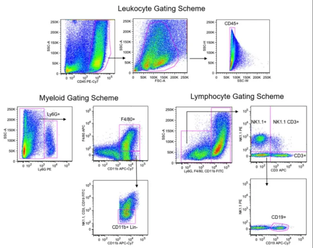
EDTA, 30% BSA) and incubated with anti-CD16/32 prior to adding an antibody cocktail against extracellular antigens. The following antibodies were purchased from Biolegend: anti-CD45 clone 30-F11, anti-Ly6G clone 1A8, anti-F4/80 clone BM8, anti-NK1.1 clone PK136, anti-CD19 clone 6D5, anti-CD11b clone M1/70, anti-CD3 clone 17A2, and Trustain fcX (anti-CD16/32) clone 93. The following isotype controls were also purchased from Biolegend: mouse IgG2a clone MOPC-173, rat IgG2a clone RTK2758, rat IgG2b clone RTK4530. After antibody incubation, cells were washed, fixed, and analyzed using a FACS Aria flow cytometry (BD Biosciences). The number of CD45 cells in each flow cytometry sample was calculated using Bang’s Laboratories Flow Cytometry Absolute Count Standard, which was added prior to data acquisition. FlowJo software (Treestar) was utilized to compensate and analyze data. FMOs with isotype controls were used to determine specific antibody signal. The gating scheme used in the flow cytometry analysis is depicted in Figure 2.3.
2.2.10 Enzyme Linked Immunosorbent Assay (ELISA)
Following euthanasia, epididymal fat pads were harvested at 3, 7, or 14 days after scaffold implant, washed in ice cold PBS, frozen on dry ice, and stored at -80°C until homogenization. Prior to homogenization, tissues were weighed and then homogenized in a defined volume of RIPA buffer containing Halt Protease Inhibitor Cocktail (Thermo Fisher Scientific), PhosStop (Sigma), and phenylmethylsulfonyl fluoride (PMSF) protease inhibitor (Thermo Fisher Scientific) using a benchtop homogenizer. Insoluble materials was removed by centrifugation. Whole tissue homogenate samples were aliquoted and stored at -80°C until use.
All cytokines were measured in whole tissue homogenate using Duoset ELISA kits (R&D Systems); however, several modifications were made to the manufacturer’s instructions to improve sensitivity. A high binding 96-well plate was incubated with capture antibody overnight at 4°C. The plate was then blocked with 10% BSA for 3 hours at room temperature. Samples were incubated on the plate overnight at 4°C. Detection antibody incubation lasted for 3 hours at room temperature before incubation with streptavidin-HRP for 2 hours at room temperature. After incubation with the substrate for 20 minutes, stop solution was added to stop the enzymatic reaction, and absorbance was measured at 450 nm. Background was removed by measuring absorbance at 540 nm. A standard curve was used to determine each cytokine concentration in each sample.
2.2.11 Western Blotting
Whole tissue homogenate was obtained as stated in the previous section. Total protein in the sample was quantified using the Pierce BCA protein assay kit (Thermo Fisher Scientific). 30-50 µg of protein was separated by 8-15% SDS polyacrylamide gel electrophoresis, depending on targeted molecular weight, and transferred to 0.2 µm nitrocellulose membranes. The membranes were blocked with 5% nonfat dry milk in Tris-buffered saline containing Tween-20 (TBST) for 1 hour at room temperature. Primary antibodies for Bax, Mcl-1, and IL-6 were purchased from Cell Signaling Technologies, CD206 was purchased from Abcam, and arginase-1 was purchased from Santa Cruz Biotechnology. Each primary antibody was added to each blot at 1:1000 dilution, with the exception was arginase-1 which was added at 1:100 dilution, in 5% nonfat dry milk in TBST overnight at 4°C. Each membrane was then washed with TBST before incubation with a polyclonal secondary antibody (Abcam) or, in the case of arginase-1, a monoclonal
secondary antibody (Biorad), conjugated to horseradish peroxidase for 1 hour at room temperature. The blots were then developed with SuperSignal enhanced chemiluminescent substrate solution (Pierce). To verify equal loading of samples, blots were incubated under stripping buffer for 20 minutes and washed with TBST. The blot then underwent the same protocol as previously stated to probe for GAPDH (Cell Signaling Technologies). ImageLabs software was used to analyze the relative intensity of each protein.
2.2.12 High Fat Diet Feeding
Mice were allowed to acclimate for 2 weeks prior to being placed on a 60% high fat diet (Research Diets D12492) while a control group was kept on a normal chow diet. The high fat diet was initiated 1 week prior to scaffold implant, and the mice remained on the diet until the end of the study. Five weeks after scaffold implantation, mice were sacrificed under anesthesia, and epididymal fat pads were collected for analysis. Fat pads were homogenized and analyzed for cytokine expression using ELISA.
2.2.13 In Vitro Model of 3T3-L1 Adipocyte Inflammation
On day 9 of differentiation, PLG or PLG+RSV scaffolds were introduced into wells containing 3T3-L1 adipocytes. Prior to addition, all scaffolds were washed in 70% ethanol and then rinsed in sterile PBS. After 72 hours of pretreatment with the scaffold, 10 ng/mL TNF-α was then added to each well and allowed to incubate for an additional 24 hours. Media was collected at this time point for further analysis. Differentiated cells not treated with TNF-α served as a control. Cell media collected at the end of the experiment was analyzed for MCP-1 using a Duoset ELISA kit (R&D Systems).
2.2.14 Statistics
Comparisons between two or more groups over time were conducted with a two-way ANOVA followed by Tukey’s multiple comparisons test. The p-values calculated by the two-way ANOVA which addresses the effect of resveratrol, time, and the interactions between the two are reported as pdrug, ptime, and pinteraction, respectively. Comparisons
between three groups at a single time point were made with a one-way ANOVA followed by Tukey’s multiple comparisons test. Comparisons between two groups at a single time point were made with an unpaired t test. The number of samples analyzed and the statistical analysis performed are detailed in each figure legend. All analyses were completed using GraphPad Prism. In all figures, the error bars denote standard error of the mean (SEM). 2.3 Results
2.3.1 Characterization of Resveratrol Scaffolds
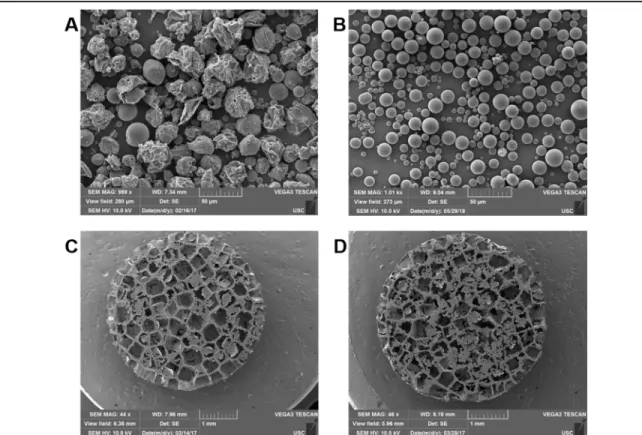
Resveratrol loaded PLG particles (PLG+RSV) were prepared using a single emulsion and solvent evaporation technique. The morphology of these particles and particles containing only the polymer (PLG) is shown in Figure 2.4 A-B. The images show that the morphology of PLG+RSV particles are irregular in shape compared to PLG particles, which are spherical. These particles were then used to fabricate scaffolds using a gas foaming and particulate leaching method. The structure of PLG+RSV and PLG scaffolds are shown in Figure 2.4 C-D. Both scaffolds contain a similar porous structure indicating that particle morphology does not have an impact on scaffold structure. During the gas foaming process, the polymer is saturated with high pressure CO2 (800 psi) which
leads to the nucleation and growth of gas pores in the polymer particles44. The polymer
likely that the gas foaming process is the reason why particle morphology does not impact the final scaffold structure.
The amount of resveratrol in particles and scaffolds was measured by dissolving the polymer in DMSO and measuring the absorbance of the solution at 330 nm. Resveratrol encapsulation within PLG particles is reproducible, with three representative batches containing 54±2, 61±2, and 59±1 µg resveratrol/mg particles (Figure 2.5A). Scaffold fabrication from these particles is also reproducible. After salt leaching, scaffolds made from the particles in Figure 2.5A contained 35±1, 36±3, and 36±3 µg resveratrol/mg scaffold (Figure 2.5B) and weighed 2.29±0.02, 2.48±0.17, and 2.43±0.07 mg (Figure 2.5C). The amount of resveratrol in a scaffold can be calculated by multiplying the loading capacity by the scaffold weight, which was between 80 and 90 µg. Collectively, this data demonstrates we can fabricate resveratrol loaded scaffolds with reproducible drug payloads.
The drug encapsulation efficiency in the particles was 20.3 ± 0.7% (Table 2.1). This encapsulation efficiency was impacted by the mass of PLG particles we were able to recover following the single emulsion/solvent evaporation fabrication procedure. The mass yield for particles was typically 40%. Drug loading capacity dropped from ~60 µg/mg for particles to ~35 µg/mg for scaffolds, giving the scaffolds a drug encapsulation efficiency of 59.6 ± 3.1% (Table 1). Inspection of our fabrication procedure suggests that resveratrol is lost during the salt porogen removal step because scaffolds assayed immediately after gas foaming have a drug encapsulation efficiency of nearly 100% (data not shown).
Resveratrol stability, which is indicated by its absorption at 330 nm92, was studied
later (Figure 2.6 A-B). When particles were analyzed immediately after fabrication, they contained 65 ± 0.5 µg resveratrol/mg particles and when this same batch of particles were analyzed one year later, they contained 61 ± 6 µg resveratrol/mg particles (Figure 2.6A). When scaffolds analyzed directly after fabrication, they contained 37 ± 0.6 µg resveratrol/mg scaffold, and when scaffolds from the same batch were analyzed one year later, they contained 36 ± 0.4 µg resveratrol/mg scaffold (Figure 2.6B). These results indicate that resveratrol activity is not lost after one year when resveratrol particles or scaffolds are kept in dry conditions and protected from light.
To confirm that resveratrol was active after one year of encapsulation in PLG, we investigated the ability of PLG+RSV particles to induce lipolysis in differentiated 3T3-L1 adipocytes by measuring lipid content with an Oil Red O assay. We found that PLG+RSV particles stored for either 2 or 52 weeks significantly decreased lipid content of the adipocytes to a similar extent, while lipid content was not affected by the PLG particles (Figure 2.6C). Representative microscopic images were used to validate the decrease in lipid staining in the PLG+RSV and RSV treated groups (Figure 2.6D). Collectively, these results demonstrate that resveratrol remains bioactive after its encapsulation in PLG and that it remains active for at least one year.
The resveratrol release profile of scaffolds over 14 days was characterized in vitro (Figure 2.7A). At each time point scaffolds were removed from the assay and tested for resveratrol content. The initial amount of resveratrol in the scaffold was taken to be the average amount of resveratrol measured in three scaffolds from the same batch immediately after salt leaching. Resveratrol exhibited a biphasic release pattern (burst followed by a plateau), with ~50% of the drug released in the first 3 days. The amount of
resveratrol released during the plateau phase accounted for ~10% of the initial resveratrol in the scaffold. The release profile indicated that there was ~30% of the resveratrol present within the scaffold after 14 days. Importantly, this burst release of resveratrol could be beneficial in decreasing the acute inflammatory phase which occurs 1-5 days after injury. Minimal scaffold degradation (~5% mass loss) was required for drug release over this time period (Figure 2.7B).
2.3.2 Effect of Resveratrol on Immune Cell Infiltration Following Scaffold Implant To quantify the effect of resveratrol on immune cell infiltration into adipose tissue following scaffold implant, we employed flow cytometry. Total immune cell numbers were measured based on CD45 expression 7 and 14 days after implant (Figure 2.8A). As expected, PLG scaffold implant increased immune cell numbers at day 7. Contrary to our expectations, resveratrol did not decrease immune cell numbers at this time point relative to PLG scaffolds. Surprisingly, immune cell numbers significantly decreased from day 7 to day 14 for both scaffold groups, and we could not detect differences between any of the groups at day 14 by Tukey’s multiple comparison’s test. The data indicate immune cell infiltration following PLG implant into fat largely resolves within 2 weeks without intervention. While not significant, immune cell numbers in the PLG+RSV group tended to be higher at day 7 and lower at day 14 compared to the PLG group.
To better understand these trends, 7 immune cell populations were measured at 7 and 14 days: macrophages (F4/80), neutrophils (Ly6G), monocytes (CD11b), NK cells (NK1.1), T cells (CD3), NKT cells (NK1.1 CD3), and B cells (CD19) (Figure 2.8B-C). For ease of interpretation, immune cell numbers for the scaffold groups are normalized by the naïve controls. At day 7, resveratrol delivery resulted in a significant increase in
neutrophils with a concomitant decrease in T cells, NK cells, and NKT cells (Figure 2.8B). At 14 days, all populations exhibited a trend of decrease, but only monocytes, NK cells, and B cell numbers were significantly lower in resveratrol groups (Figure 2.8C). The data suggest that even though resveratrol dose not decrease the total number of immune cells, it does have an impact on particular immune cell populations.
Flow cytometry revealed that macrophage number was not different between PLG and PLG+RSV at either time point; however, we hypothesized that resveratrol may modify macrophage phenotype. To identify changes in macrophage phenotype, two molecules associated with M2 polarization, CD206 and arginase-193, were measured in scaffold
homogenates 7 days after implant using Western blot (Figure 2.9A).At this time point, CD206 was unchanged in both the PLG and PLG+RSV scaffold groups compared to naïve fat (Figure 2.9B). In contrast, arginase-1 was ~35 fold higher in both PLG and PLG+RSV groups compared to naïve fat (Figure 2.9C). The data indicate that resveratrol delivery does not impact CD206 or arginase-1 expression, but scaffold implant elevates arginase-1 expression above naïve levels.
2.3.3 Effect of Resveratrol on Cytokine Levels Following Scaffold Implant
Cytokines control immune cell infiltration, residence, and survival in the tissue. Thus, we investigated the effect of resveratrol delivery on protein levels of proinflammatory cytokines MCP-1, TNF-α, IL-6, and CXCL1 as well as anti-inflammatory cytokines IL-4, IL-10, and IL-13 in whole tissue homogenates 3, 7, and 14 days after scaffold implant. There was no significant interaction (pinteraction) between drug treatment
and time detected by two-way ANOVA for the analyses performed in Figure 2.10H. MCP-1 exhibited the largest increase following PLG scaffold implant of all cytokines measured,
but was not affected by resveratrol (Figure 2.10A). MCP-1 expression significantly decreased by day 14 as determined by Tukey’s multiple comparisons test. Interestingly, scaffold implant, irrespective of drug payload, did not increase whole tissue levels of TNF-α, IL-6, or CXCL1 compared to naïve levels (Figure 2.10 B-D). We did detect a significant effect of resveratrol on TNF-α (pdrug=0.0147) and CXCL1 (pdrug=0.0168) by two-way
ANOVA (Figure 2.10 H), but drug delivery largely served to bring expression up toward naïve levels. To validate these findings, Western blot was used to detect IL-6 in all groups. We were not able to detect IL-6 in any tissues by this method; however, IL-6 was detected in Raw 264.7 cells after incubation with LPS and Brefeldin A (Figure 2.11).
In terms of anti-inflammatory cytokines, scaffolds, irrespective of drug payload, did not modulate IL-4 expression (Figure 2.10E). On the other hand, there was a significant effect of resveratrol on IL-10 (pdrug=0.0065) and IL-13 (pdrug=0.0023), with resveratrol
increasing expression of both these proteins. In addition, expression of both these molecules increased over time (Figure 2.10F-G). Together, these results suggest that resveratrol’s effect on IL-10 and IL-13 may be responsible for decreases in immune cell numbers at day 14 as measured by flow cytometry (Figure 2.8B-C).
2.3.4Effect of Resveratrol on Bax and Mcl-1 Following Scaffold Implant
Apoptosis is increased in tissues during resolution of inflammation, reflecting the clearance of immune cells that are no longer needed40,94. In addition, resveratrol increases
immune cell apoptosis, especially in neutrophils95–102. Thus, we measured the expression
of Bax, a proapoptotic protein, and Mcl-1, an antiapoptotic protein, in whole tissue protein homogenates 7 and 14 days after implant (Figure 2.12A). Bax and Mcl-1 proteins were present in naïve fat (Figure 2.12A), but only Bax levels increased with scaffold implant
(Figure 2.12A); Mcl-1 levels were largely unaffected (Figure 2.12B). There was no significant interaction between treatment and time detected by two-way ANOVA for Figure 2.12B or Figure 2.12C. There was a trend for resveratrol to increase Bax levels, but it was not significant (pdrug=0.0508); however, over time Bax expression did decrease
(pdrug=0.0040). Collectively, these results suggest that scaffold implant into the fat may
increase rates of apoptosis since proapoptotic Bax increase and antiapoptotic Mcl-1 is relatively unchanged; however, resveratrol’s ability to alter Bax expression, if indeed it does, was difficult to detect.
2.3.5 Ability of Resveratrol to Induce Anti-Inflammatory Responses in Inflammatory Environments
An important application of PLG+RSV scaffolds would be to induce an anti-inflammatory response in inflamed adipose tissue. To investigate the ability of PLG+RSV scaffolds to achieve this, we utilized a common model of adipose tissue inflammation in which mice are fed a high fat diet. Mice received PLG or PLG+RSV scaffolds in both epididymal fat pads 1 week after being fed a high fat diet. The mice were then fed the diet for an additional 5 weeks prior to harvest of the epididymal fat (Figure 2.13A). Cytokine analysis was conducted using ELISA to measure MCP-1, IL-10, and IL-13 in the epididymal fat. High fat diet feeding resulted in an increase in proinflammatory cytokines MCP-1 and a decrease in anti-inflammatory cytokines IL-10 and IL-13 (Figure 2.13B). The results demonstrate that PLG+RSV scaffolds, but not PLG scaffolds, significantly decreased MCP-1 expression in the epididymal fat. Interestingly, PLG+RSV scaffolds did not increase the expression of IL-10 and IL-13.
To investigate resveratrol’s anti-inflammatory effect on adipocytes, we utilized an
in vitro model of adipocyte inflammation. On day 9 after differentiation, cells were treated
with PLG scaffolds or PLG+RSV scaffolds for 72 hours and then treated with 10 ng/mL TNF-α for 24 hours (Figure 2.13C). Media was collected and analyzed for MCP-1 using ELISA. Differentiated cells that were treated with TNF-α secreted ~2.5 fold more MCP-1 compared to untreated differentiated cells (Figure 2.13D). PLG scaffolds had no effect on MCP-1 secretion; however, PLG+RSV scaffolds significantly decreased MCP-1 compared to differentiated cells treated with only TNF-α or pretreated with PLG scaffolds prior to TNF-α treatment.
2.4 Discussion
This report presents the characterization of resveratrol loaded PLG scaffolds and their novel use in modulating the immune response to biomaterial implants in the adipose tissue. Porous PLG scaffolds containing resveratrol were fabricated using a gas foaming/particulate leaching method in which NaCl served as the sacrificial porogen. To our knowledge, this approach is not commonly used for the fabrication of small molecule loaded PLG scaffolds, and our results provide further insight into drug loading and release kinetics of this approach. Our scaffolds contained 37 µg of resveratrol per mg of PLG (Figure 2.5), which is at least 4-fold higher loading than previous studies using biodegradable polyester scaffolds to deliver resveratrol86,91. However, this scaffold
fabrication method was suboptimal because removal of the salt porogen led to a 40% loss of resveratrol loaded in the scaffold. To improve drug loading efficiency of scaffolds, a nonparticulate leaching method to promote scaffold porosity should be investigated. Nonetheless, resveratrol loaded scaffolds fabricated using this method exhibited a burst