368
Fetal Assessment
during Labor
C h a p t e r
DEITRA LEONARD LOWDERMILK
13
• Identify typical signs of nonreassuring fetalheart rate (FHR) patterns.
• Compare FHR monitoring done by intermittent auscultation (IA) with external and internal elec-tronic methods.
• Explain the baseline FHR and evaluate periodic changes.
• Describe nursing measures that can be used to maintain FHR patterns within normal limits.
• Differentiate among the nursing interventions used for managing specific FHR patterns, in-cluding tachycardia and bradycardia; increased and decreased variability; and late and variable decelerations.
• Review the documentation of the monitoring process necessary during labor.
LEARNING OBJECTIVES
acceleration Increase in fetal heart rate (FHR); usu-ally interpreted as a reassuring sign
amnioinfusion Infusion of normal saline warmed to body temperature through an intrauterine catheter into the uterine cavity in an attempt to increase the fluid around the umbilical cord and prevent com-pression during uterine contractions
baseline fetal heart rate Average FHR during a 10-minute period that excludes periodic and episodic changes and periods of marked variability bradycardia Baseline FHR below 110 beats per
minute (beats/min)
deceleration Slowing of FHR attributed to a para-sympathetic response and described in relation to uterine contractions. Types of decelerations include: early deceleration A visually apparent gradual decrease of FHR before the peak of a contrac-tion and return to baseline as the contraccontrac-tion ends; caused by fetal head compression late deceleration A visually apparent gradual
crease of FHR with the lowest point of the de-celeration occurring after the peak of the con-traction and returning to baseline after the contraction ends; caused by uteroplacental in-sufficiency
variable deceleration A visually abrupt de-crease in FHR below the baseline occurring any time during the uterine contracting phase and caused by compression of the umbilical cord. electronic fetal monitoring (EFM) Electronic
sur-veillance of FHR by external and internal methods episodic changes Changes from baseline pat-terns in the FHR that are not associated with uter-ine contractions
hypoxemia Reduction in arterial PO2 resulting in metabolic acidosis by forcing anaerobic glycolysis, pulmonary vasoconstriction, and direct cellular damage
hypoxia Insufficient availability of oxygen to meet the metabolic needs of body tissue
intermittent auscultation Listening to fetal heart sounds at periodic intervals using nonelectronic or ultrasound devices placed on the maternal ab-domen
nonreassuring FHR patterns FHR patterns that in-dicate the fetus is not well oxygenated and re-quires intervention
periodic changes Changes from baseline that oc-cur with uterine contractions
tachycardia Baseline FHR above 160 beats/min tocolysis Inhibition of uterine contractions through
administration of medications; used as an adjunct to other interventions in the management of fetal compromise related to increased uterine activity uteroplacental insufficiency Decline in placental
function (exchange of gases, nutrients, and wastes) leading to fetal hypoxia and acidosis; ev-idenced by late FHR decelerations in response to uterine contractions
Valsalva maneuver Any forced expiratory effort against a closed airway, such as holding one’s breath and tightening the abdominal muscles (e.g., pushing during the second stage of labor) variability Normal irregularity of fetal cardiac
rhythm or fluctuations from the baseline FHR of two cycles or more
T
he ability to assess the fetus by ausculta-tion of the fetal heart was initially de-scribed more than 300 years ago. With the advent of the fetoscope and stethoscope after the turn of the twentieth century, the listener could hear clearly enough to count the fetal heart rate (FHR). When electronic FHR monitoring made its debut for clinical use in the early 1970s, it was anticipated that its use would effect a decrease in cerebral palsy and be more sensitive than stetho-scopic auscultation in predicting and preventing fetal com-promise (Simpson & Knox, 2000). Although neither of these possibilities has been realized, electronic fetal monitoring (EFM) is a useful tool for visualizing FHR patterns on a mon-itor screen or printed tracing.Pregnant women should be informed about the equip-ment and procedures used and the risks, benefits, and lim-itations of intermittent auscultation (IA) and EFM. This chapter discusses the basis for fetal monitoring, the types of monitoring, and nursing assessment and management of nonreassuring fetal status.
Understanding fetal and uteroplacental circulation is im-portant in understanding FHR and uterine activity (UA) monitoring (see Chapter 7).
Fetal Response
Because labor is a period of physiologic stress for the fetus, frequent monitoring of fetal status is part of the nursing care during labor. The fetal oxygen supply must be maintained dur-ing labor to prevent fetal compromise and to promote new-born health after birth. The fetal oxygen supply can decrease in a number of ways:
1. Reduction of blood flow through the maternal vessels as a result of maternal hypertension (chronic hyper-tension or gestational hyperhyper-tension); hypohyper-tension (caused by supine maternal position, hemorrhage, or epidural analgesia or anesthesia); or hypovolemia (caused by hemorrhage)
2. Reduction of the oxygen content in the maternal blood as a result of hemorrhage or severe anemia 3. Alterations in fetal circulation, occurring with
com-pression of the umbilical cord (transient, during uter-ine contractions [UCs]; or prolonged, resulting from BASIS FOR MONITORING
cord prolapse); placental separation or complete abruption; or head compression (head compression causes increased intracranial pressure and vagal nerve stimulation with an accompanying decrease in the FHR)
4. Reduction in blood flow to the intervillous space in the placenta secondary to uterine hypertonus (gener-ally caused by excessive exogenous oxytocin) or sec-ondary to deterioration of the placental vasculature as-sociated with maternal disorders such as hypertension or diabetes mellitus
Fetal well-being during labor can be measured by the re-sponse of the FHR to UCs. In general, reassuring FHR pat-terns are characterized by the following:
•
A baseline FHR in the normal range of 110 to 160 beats per minute (beats/min) with no periodic changes and a moderate baseline variability (see later discussion p. 374)•
Accelerations with fetal movement Uterine ActivityA normal UA pattern in labor is characterized by the fol-lowing:
•
Contractions occurring every 2 to 5 minutes and last-ing less than 90 seconds•
Contractions moderate to strong in intensity, as de-tected by palpation, or intensity is less than 80 mm Hg, as measured by an intrauterine pressure catheter (IUPC)•
Thirty seconds or more elapsing between the end of one contraction and the beginning of the next con-traction•
Between contractions, uterine relaxation should be de-tected by palpation or by an average intrauterine pres-sure of 20 mm Hg or less (Tucker, 2004).Fetal Compromise
The goals of intrapartum FHR monitoring are to identify and differentiate the reassuring patterns from the nonreas-suring patterns, which can be indicative of fetal compromise. Nursing care focuses on interventions promoting adequate fetal oxygenation and interventions for nonreassuring pat-terns if they occur.
Nonreassuring FHR patternsare those associated with fetal hypoxemia, which is a deficiency of oxygen in the
ELECTRONIC RESOURCES
Additional information related to the content in Chapter 13 can be found on the companion website at
http://evolve.elsevier.com/Lowdermilk/Maternity/ • NCLEX Review Questions
• WebLinks
or on the interactive companion CD • NCLEX Review Questions
• Critical Thinking Exercise—Fetal Monitoring • Plan of Care—Electronic Fetal Monitoring
arterial blood. If uncorrected, hypoxemia can deteriorate to severe fetal hypoxia,which is an inadequate supply of oxy-gen at the cellular level. Nonreassuring FHR patterns in-clude the following:
•
Progressive increase or decrease in baseline rate•
Tachycardia of 160 beats/min or more•
Progressive decrease in baseline variability•
Severe variable decelerations (FHR less than 60 beats/min lasting longer than 30 to 60 seconds, with rising baseline, decreasing variability, or slow return to baseline)•
Late decelerations of any magnitude, especially those that are repetitive and uncorrectable•
Absent or undetected FHR variability•
Prolonged deceleration (greater than 60 to 90 seconds)•
Severe bradycardia (less than 70 beats/min)The ideal method of fetal assessment during labor contin-ues to be debated. Results from research studies indicate that both IA of the FHR and electronic FHR monitoring are as-sociated with similar fetal outcomes in low risk intrapartum patients (Feinstein, Sprague, & Trepanier, 2000; Thacker, Stroup, & Chang, 2001). Although IA is a high-touch, low-technology method of assessing fetal status during labor that places fewer restrictions on maternal activity, more than 80% of laboring women in the United States are monitored elec-tronically for at least part of their labor (Albers, 2001). The lack of evidence on the efficacy of EFM should be a factor to consider in decision making about which method of fe-tal assessment is offered to low risk laboring women (Wood, 2003).
Intermittent Auscultation
Intermittent auscultation uses listening to fetal heart sounds at periodic intervals to assess the FHR. IA of the fetal heart can be performed with a Leff scope, a DeLee-Hillis feto-scope, or a Doppler ultrasound device. If a Leff scope is used, the domed side should be opened to the connective tubing to the earpieces. The domed side is then applied to the ma-ternal abdomen. The fetoscope is applied to the listener’s forehead because bone conduction amplifies the fetal heart sounds for counting. The ultrasound device transmits ultrahigh-frequency sound waves reflecting movement of the fetal heart and converts these sounds into an electronic sig-nal that can be counted (Fig. 13-1).
One procedure for performing auscultation is as follows: 1. Perform Leopold maneuvers (see p. 412) by palpating the maternal abdomen to identify fetal presentation and position.
2. Place the listening device over the area of maximal in-tensity (see Fig. 14-6 on p. 413) and clarity of the fe-tal heart sounds to obtain the clearest and loudest sound, which is easiest to count. Apply ultrasound gel to Doppler ultrasound device if used.
MONITORING TECHNIQUES 3. Palpate the abdomen for the absence of UA to be able to count the FHR between contractions.
4. Count the maternal radial pulse while listening to the FHR to differentiate it from the fetal rate.
5. Count the FHR for 30 to 60 seconds between con-tractions to identify the baseline rate. This rate can be assessed only during the absence of UA.
6. Auscultate the FHR during a contraction and for 30 seconds after the end of the contraction to identify any increases or decreases in FHR in response to the con-traction.
By using IA the nurse can assess the baseline FHR, rhythm, and increases and decreases from baseline (Feinstein, 2000). The method and frequency of fetal surveillance dur-ing labor will vary dependdur-ing on maternal-fetal risk factors and the preference of the facility. In the absence of risk fac-tors, one recommended practice is to auscultate the FHR as follows (American Academy of Pediatrics [AAP] & American College of Obstetricians and Gynecologists [ACOG], 2002; Association of Women’s Health, Obstetric and Neonatal Nurses, [AWHONN], 2003):
•
First stageActive phase: every 30 minutes
•
Second stageEvery 15 minutes
If risk factors are present, the FHR is auscultated as follows:
•
First stageActive phase: every 15 minutes
•
Second stageEvery 5 minutes
There is no recommended practice for assessing the FHR in the latent phase of first-stage labor; however, AWHONN (2003) suggests that the FHR be assessed as frequently as ma-ternal vital signs. The FHR also is assessed before and after ambulation, rupture of membranes, and administration of medications and anesthesia, and more frequently when non-reassuring FHR patterns are heard (AWHONN, 2003; Tucker, 2004).
A
B
C
Fig. 13-1 A, Ultrasound fetoscope. B, Ultrasound stetho-scope. C, DeLee-Hillis fetostetho-scope. (Courtesy Michael S. Clement, MD, Mesa, AZ.)
When the FHR is auscultated and docu-mented, it is inappropriate to use the descriptive terms associated with EFM (e.g., moderate variability, variable deceleration) because most of the terms are visual de-scriptions of the patterns produced on the monitor trac-ing. Terms that are numerically defined, however, such as bradycardia and tachycardia, can be used.
Every effort should be made to use the method of fetal assessment the woman desires, if possible. However, aus-cultation of the FHR in accordance with the frequency guidelines just given may be difficult in today’s busy labor and birth units. When used as the primary method of fetal assessment, auscultation requires a 1:1 nurse-to-patient staffing ratio. If acuity and census change so that ausculta-tion standards are no longer met, the nurse must inform the physician or nurse-midwife that continuous EFM will be used until staffing can be arranged to meet the standards.
The woman can become anxious if the examiner cannot readily count the fetal heartbeats. It often takes time for the inexperienced listener to locate the heartbeat and find the area of maximal intensity. To allay the mother’s concerns, she can be told that the nurse is “finding the spot where the sounds are loudest.” If it takes considerable time to locate the fetal heartbeats, the examiner can reassure the mother by offering her an opportunity to listen to them, too. If the examiner cannot locate the fetal heartbeat, assistance should be requested. In some cases ultrasound can be used to help locate the fetal heartbeat. Seeing the FHR on the ultrasound screen will be reassuring to the mother if there was initial dif-ficulty in locating the best area for auscultation.
When using IA, UA is assessed by palpation. The exam-iner should keep his or her hand placed over the fundus be-fore, during, and after contractions. The contraction inten-sity is usually described as mild, moderate, or strong. The contraction duration is measured in seconds, from the be-ginning to the end of the contraction. The frequency of con-tractions is measured in minutes, from the beginning of one contraction to the beginning of the next contraction. The examiner should keep his or her hand on the fundus after the contraction is over to evaluate uterine resting tone or re-laxation between contractions. Resting tone between con-tractions is usually described as soft or relaxed (Goodwin, 2000).
Accurate and complete documentation of fetal status and UA is especially important when IA and palpation are be-ing used because no paper tracbe-ing record of these assessments is provided as with continuous EFM. Labor flow records or computer charting systems that prompt notations of all as-sessments are useful for ensuring such comprehensive doc-umentation.
Electronic Fetal Monitoring
The purpose of electronic FHR monitoring is the ongoing assessment of fetal oxygenation. FHR tracings are analyzed for characteristic patterns that signify specific hypoxic and nonhypoxic events (King & Parer, 2000; Parer & King, 2000).
NURSE ALERT
The two modes of electronic fetal monitoring include the external mode, which uses external transducers placed on the maternal abdomen to assess FHR and UA, and the internal mode, which uses a spiral electrode applied to the fetal pre-senting part to assess the FHR and an IUPC to assess UA and pressure. The differences between the external and in-ternal modes of EFM are summarized in Table 13-1.
External monitoring
Separate transducers are used to monitor the FHR and UCs (Fig. 13-2). The ultrasound transducer works by re-flecting high-frequency sound waves off a moving interface: in this case, the fetal heart and valves; therefore short-term variability and beat-to-beat changes in the FHR cannot be assessed accurately by this method. It is sometimes difficult to reproduce a continuous and precise record of the FHR because of artifacts introduced by fetal and maternal move-ment. The FHR is printed on specially formatted monitor paper. The standard paper speed is 3 cm/min. Once the area of maximal intensity of the FHR has been located, con-ductive gel is applied to the surface of the ultrasound trans-ducer, and the transducer is then positioned over this area. The tocotransducer (tocodynamometer) measures UA transabdominally. The device is placed over the fundus above the umbilicus. UCs or fetal movements depress a pressure-sensitive surface on the side next to the abdomen. The to-cotransducer can measure and record the frequency, regularity, and approximate duration of UCs but not their intensity. This method is especially valuable for measuring UA during the first stage of labor in women with intact membranes or for antepartum testing. Because the tocotransducer of most elec-tronic fetal monitors is designed for assessing UA in the term pregnancy, it may not be sensitive enough to detect preterm
Critical Thinking Exercise
Fetal MonitoringKeri is 18 years old, gravida 1 at term, and has just been admitted to the Labor and Birth Unit. She is assessed to be at low risk for complications at this time. She is ac-companied by her boyfriend and her mother. Keri seems anxious about being in labor, and she tells you that she wants to have her baby monitored on the fetal monitor machine because she thinks that will assure her of hav-ing a good outcome to the birth of her baby. How would you respond to Keri’s statement?
1 Evidence—Is there sufficient evidence to draw con-clusions about what response you should give to Keri? 2 Assumptions—Describe an underlying assumption about the following issues related to continuous EFM in comparison to intermittent auscultation:
a. Low risk versus high risk pregnancies b. Infant outcomes
c. Staffing (nurse/patient ratio)
3 What implications and priorities for responding to Keri can be drawn at this time?
4 Does the evidence objectively support your conclusion? 5 Are there alternative perspectives to your conclusion?
UA. When monitoring the woman in preterm labor, re-member that the fundus may be located below the level of the umbilicus. The nurse may need to rely on the woman to indicate when UA is occurring and to use palpation as an ad-ditional way of assessing contraction frequency.
The external transducer is easily applied by the nurse, but it must be repositioned as the woman or fetus changes
position (see Fig. 13-2, B). The woman is asked to assume a semi-sitting or a lateral position. The equipment is removed periodically to wash the applicator sites and to give back rubs. Use of an external transducer confines the woman to bed. Portable telemetry monitors allow observation of the FHR and UC patterns by means of centrally located elec-tronic display stations. These portable units permit the
TABLE 13-1
External and Internal Modes of Monitoring
EXTERNAL MODE INTERNAL MODE
FETAL HEART RATE
UTERINE ACTIVITY
Modified from Tucker, S. (2004). Pocket guide to fetal monitoring and assessment (5th ed.). St. Louis: Mosby.
Intrauterine pressure catheter (IUPC):This instrument
moni-tors the frequency, duration, and intensity of contractions. The two types of IUPCs are a fluid-filled system and a solid catheter. Both measure intrauterine pressure at the cath-eter tip and convert the pressure into millimcath-eters of mer-cury on the uterine activity panel of the strip chart. Both can be used only when membranes are ruptured and the cervix is sufficiently dilated during the intrapartum period.
Tocotransducer: This instrument monitors frequency and
duration of contractions by means of a pressure-sensing device applied to the maternal abdomen. Used during both the antepartum and the intrapartum periods.
Spiral electrode:This electrode converts the fetal
electrocar-diogram (ECG) as obtained from the presenting part to the fetal heart rate (FHR) via a cardiotachometer. This method can be used only when membranes are ruptured and the cervix is sufficiently dilated during the intrapartum period. Electrode penetrates into fetal presenting part by 1.5 mm and must be attached securely to ensure a good signal.
Ultrasound transducer: High-frequency sound waves
reflect mechanical action of the fetal heart. Noninvasive. Does not require rupture of membranes or cervical dila-tion. Used during both the antepartum and intrapartum periods.
Tocotransducer
(uterine contractions) Ultrasound transducer (FHR)
A B
Fig. 13-2 A, External noninvasive fetal monitoring with tocotransducer and ultrasound trans-ducer. B, Ultrasound transducer is placed below umbilicus, over the area where fetal heart rate is best heard, and tocotransducer is placed on uterine fundus. (B, Courtesy Marjorie Pyle, RNC, Life-circle, Costa Mesa, CA.)
woman to walk around during electronic monitoring. Other monitoring equipment can be used when the woman is sub-merged in water (see Fig. 12-4, C).
Internal monitoring
The technique of continuous internal monitoring allows an accurate appraisal of fetal well-being during labor (Fig. 13-3). For this type of monitoring, the membranes must be ruptured, the cervix sufficiently dilated (2-3 cm), and the pre-senting part low enough to allow placement of the electrode. A small spiral electrode attached to the presenting part shows a continuous FHR on the fetal monitor strip.
Internal monitoring of the FHR may be implemented without internal monitoring of UA. For UA to be moni-tored, a solid or fluid-filled IUPC is introduced into the uterine cavity. A solid catheter has a pressure-sensitive tip that measures changes in intrauterine pressure. A catheter filled with sterile water also can be used. As the catheter is compressed during a contraction, pressure is placed on the pressure transducer or strain gauge; this pressure is then con-verted into a pressure reading in millimeters of mercury. The average pressure during a contraction ranges from 50 to 85 mm Hg. The IUPC can measure the frequency, duration, and intensity of UCs.
The FHR and UA are displayed on the monitor paper, with the FHR in the upper section and UA in the lower sec-tion. Fig. 13-4 contrasts the internal and external modes of electronic monitoring. Note that each small square represents 10 seconds; each larger box of six squares equals 1 minute (when paper is moving through the monitor at 3 cm/min).
Intrauterine pressure transducer (uterine contractions)
Cardiotachometer (FHR)
Electrode Catheter
Fig. 13-3 Diagrammatic representation of internal inva-sive fetal monitoring with intrauterine pressure catheter and spiral electrode in place (membranes ruptured and cervix dilated).
10 seconds 1 minute 61197 61198
Baseline
FHR 140
FHR in bpm UA: intensity not measurable
Penset at 20 Duration Duration Ultr asound T o cotr ansducer Intrauterine resting tone Spir al electrode Intr auter ine pressure catheter UA in mm Hg Variability Intensity FHR in bpm 240 210 180 150 120 90 60 30 100 75 50 25 0 240 210 120 90 60 30 100 75 25 0 Baseline FHR 140 Frequency beginning to beginning 50
Fig. 13-4 Display of fetal heart rate and uterine activity on monitor paper. A, External mode with ultrasound and tocotransducer as signal source. B, Internal mode with spiral electrode and intrauterine catheter as signal source. Frequency of contractions is measured from the beginning of one contraction to the beginning of the next. (From Tucker, S. [2004]. Pocket guide to fetal
mon-itoring and assessment [5th ed.]. St. Louis: Mosby.)
Tachycardiais a baseline FHR greater than 160 beats/ min for a duration of 10 minutes or longer. It can be con-sidered an early sign of fetal hypoxemia, especially when as-sociated with late decelerations and minimal or absent vari-ability. Fetal tachycardia can result from maternal or fetal infection, such as prolonged rupture of membranes with am-nionitis; from maternal hyperthyroidism or fetal anemia; or in response to drugs such as atropine, hydroxyzine (Vistaril), terbutaline, or illicit drugs such as cocaine or methamphet-amines.
Bradycardiais a baseline FHR less than 110 beats/min for a duration of 10 minutes or longer. (Bradycardia should be distinguished from prolonged deceleration patterns, which are periodic changes described later in this chapter.)
180 180 150 120 90 150 120 FHR FHR Undetected
Minimal variability: undetected 5 bpm
180 150 120 90 FHR FHR Moderate variability: 6 to 25 bpm 180 150 120 90 Marked variability: 25 bpm 90
Fig. 13-5 Fetal heart rate variability. A, Absent or unde-tected. B, Minimal. C, Moderate. D, Marked. (Modified from Tucker, S. [2004]. Pocket guide to fetal monitoring and
as-sessment [5th ed.]. St. Louis: Mosby.) Baseline Fetal Heart Rate
The intrinsic rhythmicity of the fetal heart, the central ner-vous system (CNS), and the fetal autonomic nerner-vous system control the FHR. An increase in sympathetic response re-sults in acceleration of the FHR, whereas an augmentation in parasympathetic response produces a slowing of the FHR. Usually a balanced increase of sympathetic and parasym-pathetic response occurs during contractions, with no ob-servable change in the baseline FHR.
Baseline fetal heart rateis the average rate during a 10-minute segment that excludes periodic or episodic changes, periods of marked variability, and segments of the baseline that differ by more than 25 beats/min (National Institute of Child Health and Human Development [NICHD], 1997). The normal range at term is 110 to 160 beats/min.
Variabilityof the FHR can be described as irregular fluc-tuations in the baseline FHR of two cycles per minute or greater (NICHD, 1997). It is a characteristic of baseline FHR and does not include accelerations or decelerations of the FHR. Variability has been described as short term (beat to beat) or long term (rhythmic waves or cycles from baseline). The current definition for research does not distinguish be-tween short-term and long-term variability because in actual practice they are viewed together (NICHD, 1997); however, this definition does identify four ranges of variability as seen in Fig. 13-5. These are based on visualization of the ampli-tude of the FHR in the peak-to-trough segment in beats per minute and include the following:
•
Absent or undetected variability•
Minimal variability (greater than undetected but not more than 5 beats/min)•
Moderate variability (6 to 25 beats/min)•
Marked variability (greater than 25 beats/min) In many facilities, short-term and long-term variability continue to be used to describe the FHR fluctuations. Short-term variability is commonly described as either absent or present while long-term variability may be described using the above categories (Tucker, 2004).Absence of or undetected variability is considered non-reassuring. Diminished variability can result from fetal hy-poxemia and acidosis, as well as from certain drugs that de-press the CNS, including analgesics, narcotics (meperidine [Demerol]), barbiturates (secobarbital [Seconal] and pen-tobarbital [Nembutal]), tranquilizers (diazepam [Valium]), ataractics (promethazine [Phenergan]), and general anes-thetics. In addition, a temporary decrease in variability can occur when the fetus is in a sleep state. These sleep states do not usually last longer than 30 minutes. Table 13-2 contrasts key differences between increased and decreased variability. A sinusoidal pattern—a regular smooth, undulating wave-like pattern—is not included in the current research defini-tion of FHR variability. This uncommon pattern occurs when fetal hypoxia results from Rh isoimmunization or fe-tal anemia (Fig. 13-6).
FETAL HEART RATE PATTERNS
A B C D 200 180 160 140 120 100 80 60 10 8 6 4 2 0 200 180 160 140 120 100 80 60 10 8 6 4 2 0 200 180 160 140 120 100 80 60 10 8 6 4 2 0 200 180 160 140 120 100 80 60 10 8 6 4 2 0 200 180 160 140 120 100 80 60 10 8 6 4 2 0
Fig. 13-6 Sinusoidal fetal heart rate. Note the rhythmic un-dulating pattern. (From: Tucker, S. [2004]. Pocket guide to
It can be considered a later sign of fetal hypoxia and is known to occur before fetal death. Bradycardia can result from placental transfer of drugs such as anesthetics, pro-longed compression of the umbilical cord, maternal pothermia, and maternal hypotension. Maternal supine hy-potension syndrome, caused by the weight and pressure of
the gravid uterus on the inferior vena cava, decreases the re-turn of blood flow to the maternal heart, which then reduces maternal cardiac output and blood pressure. These responses in the mother subsequently result in a decrease in the FHR and fetal bradycardia. Table 13-3 contrasts tachycardia with bradycardia.
TABLE 13-2
Increased and Decreased Variability
INCREASED VARIABILITY DECREASED VARIABILITY
CAUSE
CLINICAL SIGNIFICANCE
NURSING INTERVENTION
From Tucker, S. (2004). Pocket guide to fetal monitoring and assessment (5th ed.). St. Louis: Mosby.
CNS, Central nervous system; FHR, fetal heart rate.
Dependent on cause; intervention not warranted if associ-ated with fetal sleep states or temporarily associassoci-ated with CNS depressants; consider performing external stimulation or scalp stimulation during a vaginal exami-nation to elicit an acceleration of FHR or return to mod-erate variability; consider application of spiral elec-trode; assist health care provider with fetal oxygen saturation monitoring if ordered; prepare for birth if so indicated by the primary health care provider
Priority depends on cause: Observe FHR tracing carefully for any nonreassuring patterns, including decreasing variability and late decelerations; if using external mode of monitoring, consider using internal mode (spiral electrode) for a more accurate tracing. Interven-tion usually not required unless nonreassuring FHR pattern develops
Benign when associated with periodic fetal sleep states, which last 20 to 30 min; if caused by drugs, variability usually increases as drugs are excreted
Decreased variability is not reassuring and is considered a sign of fetal stress unless it has an identifiable tempo-rary (e.g., fetal sleep) or correctable cause
Decreased variability associated with uncorrectable late decelerations indicates presence of fetal acidosis and can result in low Apgar scores
Significance of marked variability not known; increased variability from a previous average variability is earliest FHR sign of mild hypoxemia
Hypoxia or acidosis CNS depressants Analgesics or narcotics Meperidine (Demerol) Alphaprodine (Nisentil) Morphine Pentazocine (Talwin) Barbiturates Secobarbital (Seconal) Pentobarbital (Nembutal) Amobarbital (Amytal) Tranquilizers Diazepam (Valium) Ataractics Promethazine (Phenergan) Propiomazine (Largon) Hydroxyzine (Vistaril) Promazine (Sparine) Parasympatholytics Atropine General anesthetics Prematurity: 24 wk Fetal sleep cycles Congenital abnormalities Fetal cardiac dysrhythmias Early mild hypoxemia
Fetal stimulation by the following: Uterine palpation
Uterine contractions Fetal activity Maternal activity Drugs:
Illicit drugs (e.g., cocaine and methamphetamines) Sympathomyometic (e.g., terbutaline and asthma
Changes in Fetal Heart Rate
Changes in FHR from the baseline are categorized as peri-odic or episperi-odic. Periodic changesare those that occur with UCs. Episodic changesare those that are not associated with UCs. These patterns include accelerations and decelerations (NICHD, 1997).
Accelerations
Accelerationof the FHR is defined as a visually apparent abrupt increase in FHR above the baseline rate. The increase is 15 beats/min or greater and lasts 15 seconds or more, with the return to baseline less than 2 minutes from the beginning of the acceleration. In preterm gestations the definition of an
acceleration is a peak of 10 beats/min or more above base-line for at least 10 seconds. Acceleration of the FHR for more than 10 minutes is considered a change in baseline rate.
Accelerations can be periodic or episodic. Periodic ac-celerations are caused by dominance of the sympathetic nervous response and are usually encountered with breech presentations. Pressure of the contraction applied to the fe-tal buttocks results in accelerations, whereas pressure applied to the head results in decelerations. Accelerations may oc-cur, however, during the second stage of labor in cephalic presentations. Episodic accelerations (Fig. 13-7, B) of the FHR occur during fetal movement and are indications of fe-tal well-being.
TABLE 13-3
Tachycardia and Bradycardia
TACHYCARDIA BRADYCARDIA
DEFINITION
FHR 160 beats/min lasting 10 min FHR 110 beats/min lasting 10 min
CAUSE
CLINICAL SIGNIFICANCE
NURSING INTERVENTION
From Tucker, S. (2004). Pocket guide to fetal monitoring and assessment (5th ed.). St. Louis: Mosby.
ECG, Electrocardiography; FHR, fetal heart rate.
Priority dependent on cause and based on stage of labor, fetal position and station, and fetal status
—intervention not warranted in fetus with heart block diagnosed by ECG
—scalp stimulation may be performed to determine whether the fetus has the ability to compensate phys-iologically for stress (FHR will accelerate)
—all interventions to improve fetal oxygenation (i.e., lateral maternal positioning, hydration, correction of maternal hypotension, maternal oxygenation and dis-continuing oxytocin) may be implemented
—carry out health care provider’s orders based on alle-viating cause
Priority dependent on cause:
—reduce maternal fever with antipyretics as ordered, hydration, and cooling measures
—oxygen at 8-10 L/min by face mask may be of some value
—carry out health care provider’s orders based on alle-viating cause (e.g., assist with fetal pulse oximetry if performed to collect more data about cause)
Bradycardia with moderate variability and absence of pe-riodic changes is not a sign of fetal compromise if FHR remains 80 beats/min; bradycardia caused by hypoxia is a nonreassuring sign when associated with loss of variability and late decelerations
Persistent tachycardia in absence of periodic changes does not appear serious in terms of neonatal outcome (especially true if tachycardia is associated with mater-nal fever); tachycardia is a nonreassuring sign when as-sociated with late decelerations, severe variable decel-erations, or absence of variability
Late fetal hypoxemia or hypoxia
-Adrenergic blocking drugs (propranolol; anesthetics for epidural, spinal, caudal, and pudendal blocks)
Maternal hypotension
Prolonged umbilical cord compression Fetal congenital heart block
Maternal hypothermia
Prolonged maternal hypoglycemia Early fetal hypoxemia
Maternal fever
Parasympatholytic drugs (atropine, hydroxyzine) -Sympathomimetic drugs (ritodrine, isoxsuprine) Intraamniotic infection
Maternal hyperthyroidism Fetal anemia
Fetal heart failure
Fetal cardiac dysrhythmias
EVIDENCE-BASED PRACTICE
Routine Doppler Ultrasound
FINDINGS
• No differences between the two groups were found in an-tenatal admissions or obstetric interventions. One trial found increased perinatal mortality rate in the Doppler group, but when these data were added to the pooled data, the overall difference was not significant. One trial found that the Doppler group was more likely to have re-peat tests. No trials evaluated the ability of second-trimester Doppler ultrasound to predict preeclampsia, in-trauterine growth restriction, or adverse pregnancy outcome. No data were found on acute neonatal prob-lems, long-term neurologic development, or maternal psychologic factors. One trial found that there was an in-crease in birth weight below the 10th percentile in women who had intensive repeated fetal ultrasound and Doppler ultrasound examinations, when compared with women who had only selected Doppler tests.
LIMITATIONS
• Interventions varied among trials: some evaluated bilical artery Doppler alone; others evaluated both um-bilical artery and uteroplacental blood flow. One trial com-pared patients who underwent repeated ultrasound examination plus Doppler with patients who underwent Doppler ultrasound examination only if indicated. Some studies did not allow controls to receive the intervention, and some did allow it. Parameters of measurement dif-fered for the Doppler ultrasound examination. One trial had differing protocols for high risk and low risk women. The homogeneity of the protocols limits generalizability. Many women dropped out of some studies. No trials in-cluded management protocols for abnormal results. CONCLUSIONS
• There is no supporting evidence that routine use of Doppler ultrasound in low risk pregnancy is beneficial to mother or baby. The study showing the intrauterine growth restriction (birth weight less than 10th percentile) suggests that repeated ultrasound examinations may be harmful to the fetus. Doppler ultrasound remains a valu-able tool when indicated for high risk pregnancies. IMPLICATIONS FOR PRACTICE
• Nurses can question the practice of routine Doppler ul-trasound in low risk pregnancy. They can educate patients about the risks and benefits of these routines. As patients become more knowledgeable, they can discuss with their primary health care provider the indications for the test. IMPLICATIONS FOR FURTHER RESEARCH
• Large trials are needed to determine Doppler ultrasound’s ability to predict preeclampsia, intrauterine growth re-striction, and other adverse outcomes in low risk preg-nancies. Outcomes should include maternal psychologic effects, neonatal morbidity, and long-term neurologic de-velopment of the baby. Of particular interest is resolving the issue of the safety of ultrasound.
BACKGROUND
• Prenatal diagnosticians have used a noninvasive tech-nique called Doppler ultrasound since 1977 to visualize the movement of blood in a vessel by detecting the change of frequency of reflected sound. With Doppler ul-trasound the movement of blood in the umbilical artery and the uteroplacental circulation gives information about the quality of perfusion to the fetus. This can screen women with high risk pregnancies for conditions leading to intrauterine growth restriction and gestational hyper-tension perfusion disorders. There is evidence that Doppler ultrasound is a better indicator of fetal well-being than the biophysical profile or electronic fetal mon-itoring.
• Use of screening tests in pregnancy should be preceded by questions about the proven clinical effectiveness of testing: sensitivity (ability to detect a problem), specificity (ability to rule out the problem for truly normal subjects), risks of the testing procedure, and what treatments are reasonably available for those with abnormal results. Test-ing can produce anxiety, inappropriate intervention, and iatrogenic (caused by the caregiver) morbidity and mor-tality. Questions have been raised in the past about the safety of repeated fetal ultrasound examination in gen-eral. Although the procedure is of unquestionable value in high risk pregnancies, these questions regarding the routine use of Doppler ultrasound in low risk pregnancies should be answered, and the answers should be backed up by supportive evidence from randomized, controlled trials.
OBJECTIVES
• The authors of the review sought to assess the safety and efficacy of Doppler ultrasound in low risk pregnancies. The intervention was the use of Doppler ultrasound on women with low risk pregnancies. Maternal outcomes in-cluded fetal monitoring, kick counts, biophysical profile, ultrasound, operative delivery, and psychologic effects. Perinatal outcomes included birth weight, gestational age at birth, preterm birth, respiratory status, Apgar score, ad-mission to special care nursery, morbidity, neural de-velopment at 2 years, and perinatal death.
METHODS Search Strategy
• The authors searched the Cochrane database. Search key-words were not noted.
• Five trials, including 14,338 women, were selected from France, the United Kingdom, and Australia, dated 1992 to 1997.
Statistical Analyses
• Statistical analyses included pooling similar data for metaanalysis and analyzing differences between the Doppler group and controls for each outcome studied. The reviewers accepted results outside the 95% confidence in-terval as significant.
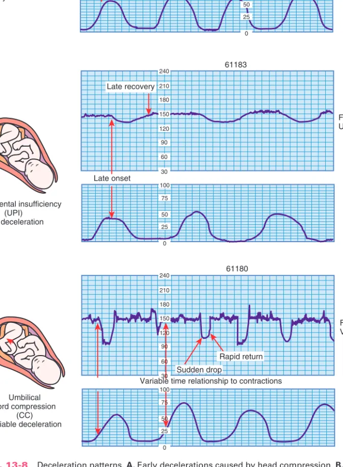
Decelerations
Adeceleration(caused by dominance of parasympathetic response) may be benign or nonreassuring. Three types of decelerations are encountered during labor: early, late, and
variable. FHR decelerations are described by their visual
re-lation to the onset and end of a contraction and by their shape.
Early decelerations. Early deceleration of the FHR is a visually apparent gradual decrease and return to baseline FHR in response to fetal head compression. It is a normal and benign finding (NICHD, 1997). The deceleration gen-erally starts before the peak of the UC and returns to the baseline at the same time as the UC returns to its baseline. Early decelerations may also occur during UCs, during vagi-nal examinations, as a result of fundal pressure, and during placement of the internal mode of fetal monitoring. When present, they usually occur during the first stage of labor when the cervix is dilated 4 to 7 cm. Early decelerations sometimes are seen during the second stage when the woman is pushing.
Because early decelerations are considered to be benign, interventions are not necessary. It is valuable to identify early decelerations so that they can be distinguished from late or variable decelerations, which can be nonreassuring and for which interventions are appropriate. The different charac-teristics of accelerations of the FHR and early decelerations are contrasted in Table 13-4.
Late decelerations. Late deceleration of the FHR is a visually apparent gradual decrease in and return to
base-line FHR associated with UCs (NICHD, 1997). The decel-eration begins after the contraction has started, and the low-est point of the deceleration occurs after the peak of the con-traction. The deceleration usually does not return to baseline until after the contraction is over (Fig. 13-8, B).
Uteroplacental insufficiency causes late decelerations. Persistent and repetitive late decelerations usually indicate the presence of fetal hypoxemia stemming from insufficient placental perfusion. They can be associated with fetal hy-poxemia progressing to hypoxia and acidemia progressing to acidosis. They should be considered an ominous sign when they are uncorrectable, especially if they are associated with decreased variability and tachycardia. Late decelerations caused by the maternal supine hypotension syndrome are usually correctable when the woman turns on her side to dis-place the weight of the gravid uterus off the vena cava. Such lateral positioning allows better return of maternal blood flow to the heart, which in turn increases cardiac output and blood pressure.
Late decelerations caused by uteroplacental insufficiency can result from uterine hyperstimulation with oxytocin, ges-tational hypertension, postdate or postterm pregnancy, am-nionitis, small-for-gestational-age (SGA) fetus, maternal diabetes, placenta previa, abruptio placentae, conduction anesthetics (producing maternal hypotension), maternal car-diac disease, and maternal anemia. The clinical significance and nursing interventions are described in Table 13-5.
Variable decelerations. Variable deceleration is defined as a visual abrupt decrease in FHR below the base-line. The decrease is 15 beats/min or more, lasts at least 15 seconds, and returns to baseline in less than 2 minutes from the time of onset (NICHD, 1997). Variable decelerations oc-cur any time during the uterine contracting phase and are caused by compression of the umbilical cord. Table 13-5 con-trasts late deceleration with variable deceleration.
The pattern of variable decelerations differs from those of early and late decelerations, which closely approximate the shape of the corresponding UC. Instead, variable de-celerations often have a Uor V shape, characterized by a rapid descent to and ascent from the nadir (or depth) of the deceleration (Fig. 13-8, C). Some variable decelerations are preceded and followed by brief accelerations of the FHR, known as “shouldering,” which is an appropriate compen-satory response to compression of the umbilical cord.
Variable decelerations may be related to partial, brief compression of the cord. If encountered in the first stage of labor, they usually can be resolved by changing the mother’s position, such as from one side to the other. Oxy-gen administration by face mask to the mother is some-times helpful. Variable decelerations are most commonly found during the second stage of labor as a result of um-bilical cord compression during fetal descent (Freeman, Garite, & Nageotte, 2003). If repetitive variable decelera-tions occur during the second stage, it is important to dis-courage the woman from pushing with every contraction
240 210 180 150 120 90 60 30 240 210 180 150 120 90 60 30 240 210 180 150 120 90 60 30 100 75 50 25 0 100 75 50 25 0 100 75 50 25 0 4 5 1 7 8 022 4 5 1 7 9 021 4 5 1 8 0 020 3cm/min IOCOext FMP 39%,(22%) 100 80 60 40 20 0 100 80 60 40 20 0 240 210 180 150 120 90 60 30 240 210 180 150 120 90 60 30 MEDS DIL EFF STA ROM pH O2 PULSE TEMP. B/P US1 MEDS DIL EFF STA ROM pH O2 PULSE TEMP. B/P MEDS DIL EFF STA ROM pH O2 PULSE TEMP. B/P
PAGES LEFT PAGES LEFT PAGES LEFT
Fig. 13-7 A, Acceleration of fetal heart rate (FHR) with uter-ine contractions. B, Acceleration of FHR movement. (From Tucker, S. [2004]. Pocket guide to fetal monitoring and
as-sessment [5th ed.]. St. Louis: Mosby.) A
B
240 210 180 150 120 90 60 30 100 75 50 25 0 240 210 180 150 120 90 60 30 100 75 50 25 0 240 210 180 150 120 90 60 30 100 75 50 25 0 61181 Head compression (HC) Early deceleration Uteroplacental insufficiency (UPI) Late deceleration Umbilical cord compression (CC) Variable deceleration 61183 Late onset 61180 FHR Variable shape FHR Uniform shape FHR Uniform shape 61182
Recovery at end of contraction
Late recovery
Variable time relationship to contractions Rapid return Sudden drop
Onset at beginning of contraction
Fig. 13-8 Deceleration patterns. A, Early decelerations caused by head compression. B, Late decelerations caused by uteroplacental insufficiency. C, Variable decelerations caused by cord com-pression. (From Tucker, S. [2004]. Pocket guide to fetal monitoring and assessment [5th ed.]. St. Louis: Mosby.)
A
B
TABLE 13-4
Accelerations and Early Decelerations
ACCELERATION EARLY DECELERATION
Description
Shape Onset
Recovery Less than 2 min from onset
Amplitude Usually 15 beats/min above baseline
Baseline Occurrence
Cause
None required None required
From Tucker, S. (2004). Pocket guide to fetal monitoring and assessment (5th ed.). St. Louis: Mosby.
Nursing intervention
Reassuring pattern not associated with fetal hypoxemia, acidemia, or low Apgar scores Acceleration with fetal movement signifies fetal
well-being, representing fetal alertness or arousal states
Clinical significance
Head compression resulting from the following: —Uterine contractions
—Vaginal examination —Fundal pressure
—Placement of internal mode of monitoring Spontaneous fetal movement
Vaginal examination Reaction to external sounds
Electrode application, scalp stimulation Breech presentation, occiput posterior position Uterine contractions
Fundal pressure Abdominal palpation
Repetitious (occurs with each contraction); usually occurs between 4- and 7-cm dilation and in second stage of labor
Variable; may be repetitive with each contraction
Usually associated with average baseline variability
Usually associated with average baseline vari-ability
Usually proportional to amplitude of contraction; rarely decelerates below 100 beats/min
By end of contraction as uterine pressure returns to its resting tone
Early in contraction phase before peak of contraction
Onset to peak (30 sec; often precedes or occurs simultaneously with uterine contraction)
Uniform shape; mirror image of uterine contraction
May resemble shape of uterine contraction or be spikelike
Transitory decrease of FHR below baseline concurrent with uterine contractions (see Fig. 13-8, A)
Transitory increase of fetal heart rate (FHR) above baseline (see Fig. 13-7)
TABLE 13-5
Late Decelerations and Variable Decelerations
LATE DECELERATION VARIABLE DECELERATION
Description
Shape Onset
Recovery Well after end of contraction Return to baseline is rapid and 2 min from onset, sometimes with transitory acceleration or accel-eration immediately before and after decelera-tion (shouldering or “overshoot”); slow return to baseline with severe variable decelerations Onset of deceleration to the beginning of nadir,
30 sec; decrease in FHR baseline is 15 beats/min, lasting 15 sec; variable times in contracting phase; often preceded by transitory acceleration
Late in contraction phase; after peak of contrac-tion; nadir of deceleration occurs after peak of contraction
Variable; characterized by sudden decrease in FHR in V, U, or Wshape
Uniform; mirror image of uterine contraction; may be deep or shallow
Abrupt decrease in FHR that is variable in dura-tion, intensity, and timing related to onset of contractions (see Fig. 13-8, C)
Transitory gradual decrease in fetal heart rate (FHR) below baseline rate in contracting phase (see Fig. 13-8, B)
TABLE 13-5
Late Decelerations and Variable Decelerations—cont’d
LATE DECELERATION VARIABLE DECELERATION
Deceleration
Baseline
Occurrence Cause
From Tucker, S. (2004). Pocket guide to fetal monitoring and assessment (5th ed.). St. Louis: Mosby.
IV, Intravenous.
The usual priority is:
—Change maternal position (side to side, knee chest)
—if decelerations are severe, proceed with follow-ing measures:
a. Discontinue oxytocin if infusing
b. Administer oxygen at 8-10 L/min with tight face mask
c. Assist with vaginal or speculum examination to assess for cord prolapse
d. Assist with amnioinfusion if ordered e. Assist with fetal oxygen saturation
monitor-ing if ordered
f. Assist with birth (vaginal assisted or ce-sarean) if pattern cannot be corrected The usual priority is:
—Change maternal position (lateral)
—Correct maternal hypotension by elevating legs —Increase rate of maintenance IV solution. —Palpate uterus to assess for hyperstimulation —Discontinue oxytocin if infusing
—Administer oxygen at 8-10 L/min with tight face mask
—Consider internal monitoring for a more accu-rate fetal and uterine assessment
—Fetal scalp or acoustic stimulation
—Assist with fetal oxygen saturation monitoring if ordered
—Assist with birth (cesarean or vaginal assisted) if pattern cannot be corrected
Nursing interventions
Variable decelerations occur in 50% of all labors and usually are transient and correctable
Reassuring variable decelerations last 45 sec and abruptly return to the FHR baseline; normal baseline rate continues; variability does not de-crease
Nonreassuring variable decelerations decrease to
70 beats/min for 60 sec and have a pro-longed return to baseline; baseline rate in-creases, variability is absent
Nonreassuring variable decelerations are associ-ated with fetal acidemia, hypoxemia, and low Apgar scores; severe variable decelerations with average baseline variability just before birth are usually well tolerated
Nonreassuring pattern associated with fetal hy-poxemia, acidemia, and low Apgar scores; con-sidered ominous if persistent and uncorrected, especially when associated with fetal tachycar-dia and loss of variability
Clinical significance
Umbilical cord compression caused by the following:
—Maternal position with cord between fetus and maternal pelvis
—Cord around fetal neck, arm, leg, or other body part
—Short cord —Knot in cord —Prolapsed cord Uteroplacental insufficiency caused by the
following:
—Uterine hyperactivity or hypertonicity —Maternal supine hypotension —Epidural or spinal anesthesia —Placenta previa
—Abruptio placentae —Hypertensive disorders —Postmaturity
—Intrauterine growth restriction —Diabetes mellitus
—Intraamniotic infection
Variable; commonly observed late in labor with fetal descent and pushing
Occurs with each contraction; may be observed at any time during labor
Mild variables usually associated with average baseline variability; moderate and severe vari-ables often associated with decreasing variabil-ity and increasing baseline rate
Often associated with loss of variability and in-creasing baseline rate
Mild: decelerates to any level, 30 sec with abrupt return to baseline
Moderate: decelerates to 70 beats/min for 30 to 60 sec or 70 to 80 bpm for 60 sec
Severe: decelerates to 70 beats/min for 60 sec, with slow return to baseline
Usually proportional to amplitude of contraction; rarely decelerates to 100 beats/min; however, shallow late decelerations have the same signif-icance
Checklist for Fetal Heart Rate and Uterine Activity Assessment (Revised)
Patient’s name Date/time 1. What is the baseline fetal heart rate (FHR)?
Beats/min
Check one of the following as observed on the monitor strip:
Average baseline FHR (110 to 160 beats/min) Tachycardia (160 beats/min)
Bradycardia (110 beats/min) 2. What is the baseline variability?
Absence of variability
Minimal variability (barely detectable up to 5 beats/min)
Moderate variability (6 to 25 beats/min) Marked variability (25 beats/min)
3. Are there any periodic or episodic changes in FHR? Accelerations with fetal movement
Repetitive accelerations with each contraction Early decelerations (head compression) Late decelerations (uteroplacental insuffi-ciency)
Variable decelerations (cord compression) Reassuring (30 to 45 seconds, abrupt return to baseline, normal baseline, moderate variability)
Nonreassuring(60 seconds, slow re-turn to baseline, increasing baseline rate, absence of variability)
Prolonged deceleration (2 minutes to 10 minutes)
4. What is the uterine activity/contraction pattern? Frequency (beginning to beginning of UC) Duration (beginning to end of UC) Abdominal palpation method
Strength (mild, moderate, strong)
Resting time (from end of one contraction to beginning of next one)
Internal monitoring (IUPC) Intensity (mm Hg pressure) Resting tone (mm Hg pressure) COMMENTS:
PANEL NUMBER:
WHAT CAN BE OR SHOULD HAVE BEEN DONE?:
Modified from Tucker, S. (2004). Pocket guide to fetal monitoring and
as-sessment (5th ed.). St. Louis: Mosby. BOX 13-1
so that the fetus has time to recover. Variable decelerations are associated with neonatal depression only when cord compression is severe or prolonged (i.e., tight nuchal cord, short cord, knot in cord, prolapsed cord). Further de-scriptions of the types of variable decelerations, the clini-cal significance, and nursing interventions are given in Table 13-5.
Prolonged decelerations. A prolonged decel-eration is a visually apparent decrease in FHR below the baseline 15 beats/min or more and lasting more than 2 minutes but less than 10 minutes. A deceleration lasting more than 10 minutes is considered a baseline change (NICHD, 1997). Generally the benign causes are pelvic ex-amination, application of a spiral electrode, rapid fetal de-scent, and sustained maternal Valsalva maneuver. Other, less benign, causes are progressive severe variable decelerations, sudden umbilical cord prolapse, hypotension produced by spinal or epidural analgesia or anesthesia, paracervical anes-thesia, tetanic contraction, and maternal hypoxia, which may occur during a seizure. When the deceleration lasts longer than 1 to 2 minutes, a loss of variability with rebound tachycardia usually occurs. Occasionally a period of late de-celerations follows. Prolonged dede-celerations usually are iso-lated events that end spontaneously. However, when a pro-longed deceleration is seen late in the course of severe variable decelerations or during a prolonged series of late decelerations, the prolonged deceleration may occur just be-fore fetal death.
Nurses should notify the physician or nurse-midwife immediately and initiate appropriate treatment for nonreassuring patterns when they see a prolonged deceleration.
The care given to women being monitored by EFM or aus-cultation is the same as that given to the woman having a low risk labor. Care of the woman being monitored by ternal methods may vary. FHR pattern recognition and in-tervention may require a nurse to have additional education and clinical experience.
Assessment and Nursing Diagnoses
The assessment of the woman includes the maternal tem-perature, pulse, respiratory rate, blood pressure, position, comfort, voiding pattern, status of membranes, UC pattern, cervical effacement and dilation, and emotional status. The fetal assessment includes the fetal presentation, fetal posi-tion, FHR, and identification of both reassuring and non-reassuring FHR patterns. A checklist may be used by the nurse to assess the FHR (Box 13-1). All of the assessment information must be documented in the woman’s medical record.
CARE MANAGEMENT
NURSE ALERT
Evaluation of the EFM equipment also must be done to ensure that the equipment is working properly and to allow an accurate assessment of the woman and fetus. A checklist for fetal monitoring equipment can be used to evaluate the equipment functions (Box 13-2).
Nursing diagnoses for the woman who is being monitored electronically for fetal status are based on assessment find-ings. Possible diagnoses include the following:
•
Decreased maternal cardiac output related to—supine hypotension secondary to maternal position
•
Anxiety related to—lack of knowledge concerning fetal monitoring during labor
—restriction of mobility or movement during EFM
•
Impaired fetal gas exchange related to—umbilical cord compression —placental insufficiency
•
Acute pain related to—use of belts to position transducers —maternal position
—vaginal examinations associated with application of maternal or fetal internal monitoring equipment or fetal blood sampling
•
Risk for fetal injury related to—unrecognized hypoxemia, hypoxia, or anoxia —infection secondary to internal monitoring or scalp blood sampling
Expected Outcomes of Care
The primary goals of nursing care are to have a healthy fetal and maternal outcome. The interventions implemented to achieve these outcomes are determined by knowledge of fe-tal status and by standards for care. The planning process in-cludes accommodating the wishes of the woman and family, answering questions, and explaining nursing interventions.
Expected outcomes for the pregnant woman and family and the fetus include the following:
•
The pregnant woman and family will verbalize their understanding of the need for monitoring.•
The pregnant woman and family will recognize and avoid situations that compromise maternal and fetal circulation.•
The fetus will not have any hypoxemic, hypoxic, or anoxic episodes.•
Should fetal compromise occur, it will be identified promptly, and appropriate nursing interventions such as intrauterine resuscitation will be initiated and the physician or nurse-midwife notified.Plan of Care and Interventions
It is the responsibility of the nurse providing care to women in labor to assess FHR patterns, implement independent nursing interventions, document observations and actions according to the established standard of care, and report non-reassuring patterns to the primary care provider (e.g., physi-cian, certified nurse-midwife). See Box 13-3 for a sample pro-tocol for FHR monitoring by IA and EFM during labor.
Although the use of EFM can be reassuring to many par-ents, it can be a source of anxiety to some. Therefore the nurse must be particularly sensitive to and respond appro-priately to the emotional, informational, and comfort needs of the woman in labor and those of her family (Fig. 13-9 and Box 13-4).
Electronic fetal monitoring pattern recognition
Nurses must evaluate many factors to determine whether an FHR pattern is reassuring or nonreassuring. A complete description of FHR tracings includes both qualitative and quantitative descriptions of baseline rate and variability, pres-ence of accelerations, periodic or episodic decelerations, and changes in the FHR pattern over time (NICHD, 1997). Nurses evaluate these factors based on other obstetric Checklist for Fetal Monitoring Equipment
PREPARATION OF MONITOR 1 Is the paper inserted correctly?
2 Are transducer cables plugged into the appropriate outlet of the monitor?
3 Is paper speed set to 3 cm/min?
ULTRASOUND TRANSDUCER
1 Has ultrasound transmission gel been applied to the transducer?
2 Was the fetal heart rate (FHR) tested and noted on the monitor paper?
3 Does a signal light flash or an audible beep occur with each heart beat?
4 Is the belt secure and snug but comfortable for the la-boring woman?
TOCOTRANSDUCER
1 Is the tocotransducer firmly positioned at the site of the least maternal tissue?
2 Has it been applied without gel or paste?
3 Was the uterine activity (UA) reference knob pressed between contractions to adjust the UA baseline to print at the 20 mm Hg line?
4 Is the belt secure and snug but comfortable for the la-boring woman?
SPIRAL ELECTRODE
1 Is the connector attached firmly to the leg plate?
2 Is the spiral electrode attached to the presenting part of the fetus?
3 Is the inner surface of the leg plate covered with elec-trode gel (if necessary)?
4 Is the leg plate properly secured to the woman’s thigh?
INTERNAL CATHETER/STRAIN GAUGE
1 Is the length line on the catheter visible at the introitus?
2 Is it noted on the monitor paper that a UA test or cal-ibration was done?
3 Has the monitor been set to zero according to manu-facturer’s directions?
4 Is the IUPC properly secured to the woman’s thigh?
Modified from Tucker, S. (2004). Pocket guide to fetal monitoring and
as-sessment (5th ed.). St. Louis: Mosby. BOX 13-2
complications, progress in labor, and analgesia or anesthesia. They also must consider the estimated time interval until birth. Interventions are therefore based on clinical judgment of a complex, integrated process (Haggerty & Nuttall, 2000).
Fetal Monitoring Standards
Nurses who care for women during childbirth are legally responsible for correctly interpreting FHR patterns, ini-tiating appropriate nursing interventions based on those patterns, and documenting the outcomes of those interventions. Perinatal nurses are responsible for the timely notification of the physician or nurse-midwife in the event of nonreassuring FHR patterns. Perinatal
LEGAL TIP
nurses also are responsible for initiating the institutional chain of command should differences in opinion arise among health care providers concerning the interpre-tation of the FHR pattern and the intervention required.
Nursing management of nonreassuring pat-terns. The term intrauterine resuscitation is sometimes used to refer to those interventions initiated when a nonreassur-ing FHR pattern is noted; they are directed primarily toward improving uterine and intervillous space blood flow and sec-ondarily toward increasing maternal oxygenation and cardiac output (Parilla, 2002). The following preventive interventions are described in this chapter: avoiding the supine position and encouraging maternal position changes; encouraging Protocol for Fetal Heart Rate Monitoring
MATERNAL AND FETAL ASSESSMENTS
• Obtain a 20-min strip of electronic fetal monitoring (EFM) for all patients admitted to labor unit.
Low Risk Patient
• Auscultate or assess tracing every 30 min in active phase of first stage of labor.
• Auscultate or assess tracing every 15 min in second stage.
High Risk Patient
• Auscultate or assess tracing every 15 min in active phase and every 5 min in second stage.
Auscultation: All Patients
• Count baseline fetal heart rate (FHR) between contractions. • Assess FHR during the contraction and for 30 sec after the
contraction.
• Note increases or decreases of FHR. • Assess FHR before ambulation.
• Interpret FHR data, nursing interventions, and patient re-sponses.
• Notify primary health care provider.
EFM: All Patients
• Assess and interpret baseline FHR, variability of FHR, and presence or absence of decelerations and accelera-tions.
Assessments for All Patients
• Assess uterine activity for frequency and duration, the in-tensity of contractions, and uterine resting tone. • Assess FHR immediately after rupture of membranes,
vaginal examinations, and any invasive procedure.
MATERNAL CARE
• Assist woman to a comfortable position other than supine.
• Change maternal position at least every 2 hr.
EXTERNAL MONITORING Ultrasound Transducer
Function
• Monitors FHR with high-frequency sound waves.
Nursing Care
• Tap transducer before use to ensure sound transmission. • Apply ultrasound transmission gel to transducer, clean abdomen and transducer, and reapply gel every 2h and as needed.
• Massage reddened skin areas gently and reposition belt or adhesive device every 2h and as needed.
• Auscultate FHR with stethoscope or fetoscope if in doubt as to validity of tracing.
• Position and reposition transducer prn to ensure receipt of clear, interpretable FHR data.
Tocotransducer
Function
• Monitors uterine activity via a pressure-sensing device placed on the maternal abdomen.
Nursing Care
• Position and reposition every 2h and as needed on the fundus, where there is the least maternal tissue. • Keep abdominal strap snug but comfortable for the
la-boring woman.
• Adjust knob between contractions to print between 10 and 20 mm Hg on the monitor strip paper.
• Palpate fundus every 30 to 60 min to assess strength of contraction; only frequency and duration of contractions can be assessed with tocotransducer.
• Do not determine woman’s need for analgesia based on uterine activity displayed on monitor strip.
• Gently massage reddened areas under transducer and belt hourly and as needed.
INTERNAL MONITORING Spiral Electrode
Function
• Obtains fetal electrocardiogram (ECG) from presenting part and converts it into FHR.
Nursing Care
• Ensure that the connector to the scalp electrode is ap-propriately attached to leg plate.
• Reapply electrode paste to leg plate if needed. • Observe FHR tracing on monitor strip for variability. BOX 13-3