Title: Cognitive behavioral therapy for depression changes medial prefrontal and ventral
anterior cingulate cortex activity associated with self-referential processing
Shinpei Yoshimuraa,b,*, Yasumasa Okamotoa,b, Keiichi Onodac, Miki Matsunagaa, Go
Okadaa, Yoshihiko Kunisatod, Atsuo Yoshinoa, Kazutaka Uedae, Shin-ichi Suzukid and
ShigetoYamawakia,b
a
Institute of Biomedical and Health Sciences, Hiroshima University, 1-2-3, Kasumi,
Minami-ku, Hiroshima 734-8553, Japan
b
Core Research for Evolutional Science and Technology (CREST)
c
Department of Neurology, Faculty of Medicine, Shimane University, 89-1, Enyacho,
Izumo, 693-8501, Japan
d
Faculty of Human Sciences, Waseda University 2-579-15, Mikajima, Tokorozawa,
Saitama 359-1192, Japan
e Research Center for Advanced Science and Technology, The University of Tokyo,
4-6-1, Komaba, Meguro-ku, Tokyo 153-8904 Japan
*
Corresponding author: Institute of Biomedical and Health Sciences, Hiroshima
University, 1-2-3, Kasumi, Minami-ku, Hiroshima 734-8553, Japan
Keywords
depression; fMRI; cognitive behavioral therapy; self-referential processing; emotion
Abstract
Cognitive behavioral therapy (CBT), an effective treatment for depression, targets
self-referential processing of emotional stimuli. We examined the effects of CBT on
brain functioning during self-referential processing in depressive patients using
functional magnetic resonance imaging (fMRI). Depressive patients (n=23) and healthy
participants (n=15) underwent fMRI scans during a self-referential task using emotional
trait words. The depressive patients had fMRI scans before and after completing a total
of 12 weekly sessions of group CBT for depression, whereas the healthy participants
underwent fMRI scans 12 weeks apart with no intervention. Before undergoing CBT,
the depressive patients showed hyperactivity in the medial prefrontal cortex (MPFC)
during self-referential processing of negative words. Following CBT, MPFC and ventral
anterior cingulate cortex (vACC) activity during self-referential processing among
depressive patients was increased for positive stimuli while it was decreased for
negative stimuli. Improvements in depressive symptoms were negatively correlated
with vACC activity during self-referential processing of negative stimuli. These results
suggest that CBT-related improvements in depressive symptoms are associated with
changes in MPFC and vACC activation during self-referential processing of emotional
Introduction
Depression is an affective disorder characterized by emotional, cognitive, and
behavior dysfunction. Depressive mood, negative cognitive biases, and behavioral
withdrawal interact with each other, and this interaction helps maintain depressive
symptoms (Beevers, 2005). Negative cognitive biases include negative thinking patterns
and memory biases, and in general serve to sustain negative emotional processing. In
turn, negative emotional processing repeatedly reinforces negative cognitive bias,
thereby shaping maladaptive feedback loops (Teasdale, 1985). According to Beck’s
cognitive theory of depression, depressive patients frequently have negative thoughts
about the self (Beck, 2008). This self-referential bias is a key dysfunctional cognition
that maintains and intensifies depression. Self-referential bias is associated with
rumination or maladaptive self-focus (Ingram, 1984; Nolen-Hoeksema et al., 2008;
Pyszczynski & Greenberg, 1987). Several studies provide evidence that rumination and
maladaptive self-focus with regard to stressors or negative emotional events induce
negative mood (Moberly & Watkins, 2008; Mor & Winquist, 2002).
The core brain regions involved in depression-related negative self-referential
bias are the medial prefrontal cortex (MPFC), the ventral anterior cingulate cortex
self-reference processing (Grimm et al., 2009; Johnson et al., 2009; Lemogne et al.,
2009, 2010; 2011; 2012; Northoff, 2007). The vACC and amygdala are associated with
evaluation of emotional stimuli and elicitation of emotional responses (Bush et al.,
2000; Habel et al., 2005; Moran et al., 2006; Siegle et al., 2002). We therefore propose
that the MPFC, vACC, and amygdala might be involved in self-referential processing of
negative emotional stimuli, and as such might constitute a dysfunctional
cognitive-emotional neural circuit that underlies depression.
Cognitive Behavioral Therapy (CBT) seeks to disrupt the vicious cycle of
depressed mood, dysfunctional cognition, and behavioral inhibition that is observed in
depressive disorders. CBT’s targets include negative self-referential cognitions. A few
studies have investigated the effects of CBT on brain functioning in depression. Resting
state PET studies have found that patients who receive CBT show increased activation
in the dorsal ACC and decreased activation in the DLPFC and MPFC, compared with
patients who receive pharmacotherapy (Goldapple et al., 2004; Kennedy et al., 2007).
Using an experimental task to more directly engage affective processing, Fu and
colleagues (2008) reported decreased amygdala activity in depressive patients following
CBT, with dorsal ACC activity being associated with a clinical response. The results of
propose that the abnormal brain activation observed in depressive patients might not
only reflect relatively simple processing of negative emotional stimuli, such as
emotional faces or words, but also more cognitively demanding processing such as
self-referential processing of emotional stimuli (Grimm et al., 2009; Lemogne et al.,
2009, 2010, 2012). However, no studies have examined how CBT might affect brain
functioning associated with negative self-referential cognition. Thus this relationship
and the relationship between CBT and the neural connections between the frontal and
limbic regions remain poorly understood.
Our previous study revealed that depressive patients show high levels of
activation in the MPFC and vACC during self-referential processing (Yoshimura et al.,
2010). We therefore hypothesize that depressive patients (before receiving CBT) would
show greater MPFC and vACC activation relative to healthy participants. Goldapple
and colleagues (2004) reported decreased activation in the MPFC at resting state
following CBT but not after treatment with an antidepressant medication (paroxetine).
Another study also suggested that antidepressants may have no effect on MPFC
overactivation during self-referential processing in depression (Lemogne et al., 2010).
We hypothesize that abnormal MPFC, vACC, and amygdala activation during a
addition, there is some evidence that depression-related neural activity is a sensitive
predictor of CBT treatment outcomes (Fu et al., 2008; Siegle et al., 2006). Therefore, in
the present study we examined the potential association between treatment response to
CBT and neural activity during self-referential processing of emotional stimuli.
Method
Participants
Depressive patients (n=23) were recruited from the outpatients of the
Department of Psychiatry and Neurosciences of the Hiroshima University Hospital.
Inclusion criteria were: (a) diagnosis established by a psychiatrist using the Structured
Clinical Interview for DSM-IV-TR (SCID), and (b) the patient met the criteria for major
depressive disorder according to DSM-IV-TR. Exclusion criteria consisted of current or
previous diagnosis of a bipolar disorder, psychotic spectrum disorder, evidence of
organic brain disorder, current high risk of suicide, substance abuse, mental retardation,
or serious somatic disease. All patients had been taking one or more antidepressant
drugs (i.e., serotonin reuptake inhibitor, serotonin and noradrenalin reuptake inhibitor,
tricyclic antidepressant) for a minimum of 8 weeks without remission of symptoms.
demographic and clinical characteristics of the participants are presented in Table 1.
Nine patients in the current study also participated in our previous study (Yoshimura et
al., 2010).
Healthy control participants (n=15) were recruited from normal populations.
These control participants endorsed no symptoms of depression and had no history of
psychiatric disorder. All patients and control participants had normal vision, and
reported no notable health or eye problems.
The Ethics Committee of Hiroshima University approved the study protocol.
Informed consent was obtained from all participants.
----Table 1 around here----
Evaluation and treatment protocol
The depressive patients participated in 12 weekly, 90-minute sessions of CBT
conducted by a clinical psychologist with groups of five or six participants. The session
topics were as follows: psychoeducation about depression; psychoeducation about
group CBT; self-monitoring of automatic thoughts, behaviors, and mood; understanding
negative self-talk; challenging and restructuring negative thinking about the self;
looking for new ideas and invoking positive thinking; practicing the new ideas and
positive thinking in daily life; evaluating one’s own ideas and thinking during the last
week, and setting up an action plan for the next week; reviewing the outcome of the
program; and finally, relapse prevention.
Patients’ progress was monitored regularly using the Beck Depression
Inventory (BDI). The Hamilton Rating Scale for Depression (HRSD) was used to
evaluate symptom improvement. The Dysfunctional Attitude Scale (DAS), Automatic
Thoughts Questionnaire (ATQ), and Response Styles Questionnaire (RSQ) were used to
measure psychological factors in depressive patients. A more detailed treatment protocol
is described in our previous report (Matsunaga et al., 2010).
The healthy control participants were administered the Beck Depression
Inventory (BDI) at the start and end of the research about12 week interval.
Experimental design
All participants underwent two fMRI scans, about 12 weeks apart, while they
did the judgment tasks. For the depressive patients, the scans occurred before and after
the completion of 12 weeks of CBT. The healthy control participants did not participate
experimental design have been described in our previous study (Yoshimura et al., 2009,
2010).
In the judgment tasks administered during the fMRI scans, the participants
were instructed to make one of four judgments about visually presented words. In the
self-reference condition, participants judged whether or not each trait word described
them. In the other-reference condition, participants judged whether or not each trait
word described the Prime Minister of Japan (Jun-ichiro Koizumi). In the
semantic-processing condition, participants judged whether or not it was difficult to
define each trait word. In the letter-processing condition, participants assessed whether
or not each trait word contained a specific target letter. The semantic-processing and
letter-processing conditions were used to control for extraneous variables such as the
visual and motor processing of stimuli, as well as language processing.
The four judgment conditions each included both positive and negative word
stimuli, resulting in a total of eight conditions overall. For all conditions, participants
made a “yes” or “no” response by pressing a button with the right-hand index or middle
finger, respectively. Button presses were recorded using an MRI-compatible keypad
(Lumitouch, Lightwave Technologies; Richmond, BC, Canada). Participants performed
a block of five trials. At the onset of each condition block, a fixation cross was
displayed for 1000 ms, followed by an instruction cue presented for 3000 ms (e.g.,
“self-reference condition”). Participants then received five trials consisting of a fixation
cross displayed for 1000 ms, followed by an adjective displayed for 3000 ms and the
participant’s response. A fixation point was then displayed for 4000 ms, followed by the
instruction cue for the next block of five trials. Each condition was presented separately.
The duration of each block was 28 s. To control for order effects, blocks within a run
were presented in a pseudo-random order, with no two consecutive blocks featuring the
same instructions. The total time for the self-reference task was 908 s. Both responses
and reaction times were recorded using Presentation software.
fMRI data acquisition
fMRI was performed using a Symphony 1.5 T device (Siemens, Tokyo, Japan).
A time-course series of 227 scans was acquired with T2*- weighted, gradient echo, echo
planar imaging (EPI) sequences. Each volume consisted of 38 slices, with a slice
thickness of 4 mm with no gap, and covered the entire cerebral and cerebellar cortices.
The interval between two successive acquisitions of the same image (TR) was 4000 ms,
the echo time (TE) was 48 ms, and the flip angle was 90°. The field of view (FOV) was
Scan acquisition was synchronized to the onset of each trial. After functional scanning,
structural scans were acquired using a Tl-weighted gradient echo pulse sequence (TR =
12 ms; TE = 3.93 ms; flip angle 25°; FOV 256 mm; voxel dimensions of 1 x 1 x 1 mm),
which facilitated localization.
fMRI data analysis
Image processing and statistical analyses were carried out using Statistical
Parametric Mapping (SPM8) software (Wellcome Department of Cognitive Neurology,
London, UK). The first two and last single volumes of the fMRI run were discarded
because the MR signals were unsteady or empty. All EPI images were spatially
normalized using the Montreal Neurological Institute (MNI) T1 template for group
analysis. Imaging data were corrected for motion and smoothed with a 4 mm full-width,
half-maximum Gaussian filter.
To perform image data analysis, a whole-brain voxel-by-voxel multiple linear
regression model was employed at the individual participant level. The individual
model comprised the covariate of no interest (realignment parameters). We examined
the contrasts between the self-reference conditions and the sum of the two control
conditions (semantic-processing and letter-processing) for both positive and negative
processing, the other-referential condition was excluded from analysis. These contrasts
were submitted to group analysis, using a random effect model. We conducted whole
brain three-way repeated measures ANOVAs as implemented in SPM8 with group
(patients vs. healthy) as a between-subjects factor, and time (time 1 vs. time 2) and
emotional valence (positive vs. negative) as within-subjects factors. To exclude gender
effects, we added participant gender as a nuisance covariate. Activations were reported
p < 0.001 (uncorrected) over the 30-voxel level. All coordinates are reported using the
Montreal Neurological Institute (MNI) coordinate space. Signal intensity from the
three-way interaction active cluster was extracted using the volume of interest (VOI)
tool in SPM8, and multiple comparisons (Bonferroni correction) of three-way
interaction effects were conducted using SPSS.
To examine the brain activation during the self-referential task that predicts
subsequent CBT responses, we performed a correlational analysis between percentage
improvement in HRSD score (= (1-HRSD score at time2/HRSD score at time1)*100)
for each depressive patient after CBT, and activation of brain regions in depressive
patients before CBT was received. In addition, to examine the changes of brain
activation following CBT, which were associated with reduction of rumination (=
correlational analysis between reduction of rumination and activation of brain regions in
depressive patients following CBT.
Results
Effect of CBT (Table 1)
The CBT group treatment was generally effective in reducing depression, as
indicated by changes in BDI scores, F (22, 1) = 23.74, p < 0.001, as well as HRSD
scores, F (22, 1) = 23.60, p < 0.001. There were no significant correlations in initial
HRSD scores before CBT and no percentage changes in HRSD scores as a function of
CBT and BDI score changes for the depression. In addition, CBT contributed to changes
in several psychological scales including the DAS, F (22, 1) = 7.80, p < 0.05, ATQ
(negative scale), F (22, 1) = 12.02, p < 0.005, RSQ (rumination scale), F (22, 1) = 5.77,
p < 0.05, and RSQ (distraction scale), F (22, 1) = 11.85, p < 0.005.
Behavioral data
We conducted a three-way ANOVA for the judgment ratio data in the
self/positive and self/negative conditions with group (patients vs. healthy) as a
between-subjects factor, and time (time 1 vs. time 2) and emotional valence (positive vs.
two depressive patients were incomplete; therefore, only 21 patients were included in
the ANOVA. The three-way interaction was significant (F [1,34]= 5.17, p < 0.05).
Multiple comparisons (a Bonferroni correction) showed significant judgment rate
differences between time 1 and time 2 for the depressive patients in the self/positive
condition (p < 0.05). In the self/negative condition, the difference between time 1 and
time 2 for the depressive patients was marginally significant (p = 0.073).
We conducted a three-way ANOVA for the reaction times in same way as for
the judgment ratios. Only a significant main effect of emotional valence was found (F
[1,34]= 4.71, p < 0.05). Reaction times to negative stimuli were significantly slower
than to positive stimuli (p < 0.05).
fMRI data
A three-way repeated measures ANOVA was conducted to examine possible
differences across group, time, and stimulus valence. There was a significant three-way
interaction effect (Figure1) for the left vACC (MNI coordinates [x, y, z][0, 46, 6],
cluster size = 156 voxels; F = 34.09, df = 1,36), left superior temporal cortex ([-54, -64,
26], cluster size = 70 voxels; F = 23.67, df = 1,36), and the left MPFC ([-6, 44, 44],
cluster size = 33 voxels; F = 20.84, df = 1,36). Post hoc analysis revealed that healthy
(baseline) for the vACC, superior temporal cortex, and MPFC in the self/positive
condition (all p < 0.01). In the self/negative condition, patients showed significant
activation increases relative to healthy participants at time 1 (baseline) for the vACC
and MPFC (all p < 0.05). At time 2, patients showed increased activation in the
self/positive condition compared to healthy participants for the vACC, superior
temporal cortex, and MPFC. In the self/negative condition, healthy participants showed
significant activation increases relative to patients at time 2 for the vACC, superior
temporal cortex, and MPFC (all p < 0.01). In addition, for depressive patients,
activation in these regions significantly increased from time 1 to time 2 for the
self/positive condition (all p < 0.05). Correspondingly, activation in these regions was
significantly decreased from time 1 to time 2 for the self/negative condition (all p <
0.05). On the other hand, for healthy control participants, activation in the MPFC and
vACC was significantly decreased from time 1 to time 2 for the self/positive condition
(all p < 0.05) and increased from time 1 to time 2 for the self/negative condition (all p <
0.01). For depressive patients at time 1, activation in the vACC, superior temporal
cortex, and MPFC in the self/negative condition significantly increased relative to in the
self/positive condition (all p < 0.01). At time 2, depressive patients in the self/negative
condition for the vACC and superior temporal cortex (all p < 0.05). On the other hand,
for healthy control participants at time 1, activation in the vACC and MPFC in the
self/positive condition was significantly increased relative to in the self/negative
condition (all p < 0.05). At time 2, healthy control participants showed significantly
increasing activation in the vACC, superior temporal cortex, and MPFC in the
self/negative condition relative to that in the self/positive condition (all p < 0.01).
There were no other significant interactions.
-Figure 1 around here-
There was a significant negative correlation between percent changes in HRSD
scores and vACC activation in the self/negative condition, before CBT was received (r
= -0.60, p < 0.01; r2 = 0.36, p < 0.005). This negative correlation indicates that patients
with greater clinical improvement following CBT had lower activity in the vACC
(Figure 2A).
In addition, there was a significant positive correlation between percent
changes in the rumination score and vACC activation changes in the self/negative
condition following CBT (r = 0.49, p < 0.05; r2 = 0.24, p < 0.05). This positive
correlation indicates that patients with greater improvement in rumination following
-Figure 2 around here-
Discussion
Self-referential processing of negative stimuli, as one form of negative
rumination, is thought to be an important cognitive feature of depression
(Nolen-Hoeksema et al., 2008). Excessive negative self-rumination results in further
deterioration of depressive mood (Nolen-Hoeksema et al., 1993; Watkins & Moulds,
2005). CBT seeks to improve depressed mood by intervention at the level of
cognitions and behaviors. In the present study of depressive patients, we examined how
CBT affects brain functioning that underlies self-referential processing of negative
stimuli. Compared to control participants, depressive patients showed increased
activation in the MPFC during the self/negative condition. Following group CBT,
depressive patients showed decreased activation in the MPFC and vACC during
self-referential processing of negative words, and increased activation in these regions
during the processing of positive words. Correlational analysis revealed an association
between treatment response and pre-treatment vACC activity during self/negative
conditions. These findings represent the first report of CBT-related changes in vACC
Previous studies have reported that the MPFC might play an important role in
self-referential processing (Fossati et al., 2003, 2004; Kelley et al., 2002; Moran et al.,
2006; Schmitz et al., 2006). In addition, our previous work revealed that the MPFC is
associated with self-referential or other-referential processing of emotional stimuli
(Yoshimura et al., 2009). The vACC was also engaged, particularly during
self-referential processing of negative emotional stimuli. In addition, our previous work
(Yoshimura et al., 2010) with depressive patients revealed that MPFC and vACC
activations during self-referential processing of negative emotional stimuli are
correlated with symptoms of depression. This relationship between the MPFC and the
vACC might underlie a “vicious cycle” of self-referential and negative emotional
processing (Northoff, 2007).
The present study identified decreased activation in the MPFC and vACC
following CBT, which suggests one mechanism whereby CBT might attenuate
depressive cognitive-emotional processing. In addition, CBT was associated with
increased MPFC and vACC activity during self-referential processing of positive
stimuli. Ritchey et al. (2011) reported increased ventral MPFC activity during the
processing of positive stimuli following CBT. However, this CBT effect on brain
positive as well as neutral and negative stimuli.
The present results suggest that the increases in more balanced, positive
thinking and pleasant activity that follow successful CBT promote a more positive
self-concept, such that the MPFC and vACC become more responsive to positive
stimuli during self-referential processing. Previous functional brain imaging studies of
CBT have reported that CBT alters resting state activation of the MPFC, ACC, and
DLPFC (Goldapple et al., 2004; Seminowicz et al., 2004). These data imply that CBT
works to effect top-down modulation from the cortical to the limbic region (Goldapple
et al., 2004). Our findings appear to support top-down modulation, given the direct
MPFC and vACC activation changes that we observed.
CBT for depression includes at least two essential components. One is
identifying and correcting inaccurate thoughts associated with negative emotion
(cognitive restructuring). Another is helping patients to engage in more pleasurable
activities, such as exercise or listening to music (behavioral activation). Recent brain
imaging studies of emotion regulation as a component of CBT report that the lateral
prefrontal cortex and MPFC are associated with emotion regulation, and that these
regions inhibit the amygdala (Etkin et al., 2006; Ochsner et al., 2002; Ochsner & Gross,
that maintain depression, such as rumination, overgeneralization, and self-focused
attention. These dysfunctional cognitive processes might be associated with amygdala
or ventral/rostral ACC activation. Some research reports that improvements in
depressive symptoms are associated with attenuated activation in these regions.
However, the present study did not find decreased amygdala activity during
self-referential processing of negative stimuli. These results perhaps indicate that
decreased negative emotional processing occurs concurrently with decreased
self-referential processing.
On the other hand, Dichter and colleagues (2009) suggested that behavioral
interventions for depression (behavioral activation therapy) might enhance brain reward
system functioning, which includes regions such as the striatum involved in reward
selection. We can assume that cognitive and behavioral interventions improve
depressive symptoms to a similar degree, but perhaps via different mechanisms. The
present intervention included both types of intervention and we could not relate these
separate components to specific patterns of brain activation. Future research could
distinguish between CBT components, and examine the effect of each component on
brain functioning associated with changes in cognition and behavior.
activation in the self/negative condition. The negative correlation between treatment
response and vACC activation in the self/negative condition suggests that depressive
patients with relatively low sensitivity to negative emotion benefitted most from CBT or
other treatment. In the study of Siegle et al. (2006), depressive patients received CBT
and participated in an fMRI scan during a self-referential task. They reported that
treatment improvement was correlated with vACC activity before receiving CBT, as
was also found in our present study. They suggested that CBT is useful for depressive
patients who cannot engage in sufficient emotional regulation.
Although our present results are consistent with the results of Siegle et al.
(2006), we have a different interpretation. Because CBT decreased vACC activation
during the self/negative condition, the vACC activation observed here was likely
associated with self-referential processing in the form of rumination, rather than
emotional regulation. In addition, recent research has reported an association between
rumination and vACC function in the context of both self-referential processing and
depression (Berman et al., 2011; Cooney et al., 2010; Heatherton, 2011; Ray et al.,
2005). These researchers suggest that vACC activity might reflect attention toward
one’s internal emotional state in depression.
cognitive tasks using emotional stimuli often activate the vACC in depression (Fu et al.,
2008; Keedwell et al., 2005; Matthews et al., 2008) One possible explanation about
vACC functioning in depression may be that vACC activation reflects emotional
processing which is emphasized by ruminative processing which focuses on negative
aspects of self. Indeed, rumination is thought to be associated with less responsiveness
to both antidepressant drugs and CBT (Ciesla & Roberts, 2002; Schmaling et al., 2002),
because depressive patients who have severe rumination might have difficulty learning
CBT components such as monitoring mood, emotion regulation, and engaging positive
activity. Patients who engage in limited rumination may typically respond quite well to
standard CBT that is focused on challenging the content of negative cognitions. We
consider that this interpretation is supported by the correlation between the reduction of
rumination and activation changes in the vACC during the self/negative condition
following CBT. However, vACC functioning in depression is a complicated issue,
which needs further consideration.
Limitations
The present study has limitations. First, patient symptom severity was
relatively mild. The present findings remain to be generalized to patients with more
received pharmacotherapy throughout their participation in CBT, such that their
treatment histories were not controlled. Thus, we could not rule out pharmacological
treatment effects on the brain activity that we observed in the present study. It might be
possible that changes of brain activation and clinical symptoms were due to
pharmacotherapy or spontaneous rather than produced solely by CBT. Third, test-retest
reliability of the task used in present study was poor. The fMRI indicated that healthy
participants showed increasing activity in the vACC and MPFC in the self/negative
condition, and decreasing activity in these regions from time 1 to time 2. Some other
research studies have reported variability of signal change by repeated fMRI
measurement(Fu et al., 2008; Jensen et al., 2012; Johnstone et al., 2005). We consider
that if the signal changes that we found in both depressive and healthy participants were
spontaneous, then the direction of signal change might be same. But the signal changes
from time 1 to time 2 between depressive and healthy participants were in opposite
directions. In addition, there were no significant correlations between brain activity and
BDI or behavioral data in the healthy participants. It is difficult to speculate about the
reason for these relative activation changes from time 1 to time 2, but activation
changes from time 1 to time 2 in depressive patients were explained by receiving CBT
In summary, we found that CBT influences MPFC and vACC functioning
related to self-referential processing of negative emotional stimuli. It is therefore
possible that the effect of CBT on depressive symptoms is mediated by MPFC and
vACC activity associated with self-referential processing. In addition, the present study
suggests the possibility that vACC functioning could be a useful index for treatment
response to CBT for depression.
Acknowledgments
This study was supported by the following: a Grant-in-Aid for Scientific Research and
“Integrates research on neuropsychiatric disorders”, the Strategic Research Program for
Brain Sciences by the Ministry of Education, Culture, Sports, Science and Technology
of Japan; Research on Psychiatric and Neurological Diseases and Mental Health,
Ministry of Health, Labour and Welfare; Core Research for Evolutional Science and
Technology (CREST), Japan Science and Technology Agency; Japan Society for the
Promotion of Science (JSPS) Fellows.
Financial Disclosures
References
Beck, A. T. (2008). The evolution of the cognitive model of depression and its neurobiological correlates. The American journal of psychiatry, 165(8), 969–77. doi:10.1176/appi.ajp.2008.08050721
Beevers, C. G. (2005). Cognitive vulnerability to depression: a dual process model. Clin Psychol Rev, 25(7), 975–1002. doi:S0272-7358(05)00026-7 [pii]
10.1016/j.cpr.2005.03.003
Berman, M. G., Peltier, S., Nee, D. E., Kross, E., Deldin, P. J., & Jonides, J. (2011). Depression, rumination and the default network. Social cognitive and affective neuroscience, 6(5), 548–55. doi:10.1093/scan/nsq080
Bush, G., Luu, P., & Posner, M. I. (2000). Cognitive and emotional influences in anterior cingulate cortex. Trends Cogn Sci, 4(6), 215–222. Retrieved from
http://www.ncbi.nlm.nih.gov/entrez/query.fcgi?cmd=Retrieve&db=PubMed&dopt =Citation&list_uids=10827444
Ciesla, J. A., & Roberts, J. E. (2002). Self-directed thought and response to treatment for depression: A preliminary investigation. Journal of Cognitive Psychotherapy, 16(4), 435–453.
Cooney, R. E., Joormann, J., Eugène, F., Dennis, E. L., & Gotlib, I. H. (2010). Neural correlates of rumination in depression. Cognitive, Affective, & Behavioral Neuroscience, 10(4), 470–478. Retrieved from
http://www.ncbi.nlm.nih.gov/pubmed/21098808
Dichter, G. S., Felder, J. N., Petty, C., Bizzell, J., Ernst, M., & Smoski, M. J. (2009). The effects of psychotherapy on neural responses to rewards in major depression. Biol Psychiatry, 66(9), 886–897. doi:S0006-3223(09)00826-9 [pii]
10.1016/j.biopsych.2009.06.021
Etkin, A., Egner, T., Peraza, D. M., Kandel, E. R., & Hirsch, J. (2006). Resolving emotional conflict: a role for the rostral anterior cingulate cortex in modulating activity in the amygdala. Neuron, 51(6), 871–882. doi:S0896-6273(06)00626-X [pii] 10.1016/j.neuron.2006.07.029
Fossati, P, Hevenor, S. J., Lepage, M., Graham, S. J., Grady, C., Keightley, M. L., Craik, F., et al. (2004). Distributed self in episodic memory: neural correlates of
successful retrieval of self-encoded positive and negative personality traits. Neuroimage, 22(4), 1596–1604. Retrieved from
http://www.ncbi.nlm.nih.gov/entrez/query.fcgi?cmd=Retrieve&db=PubMed&dopt =Citation&list_uids=15275916
Fossati, Philippe, Hevenor, S. J., Graham, S. J., Grady, C., Keightley, M. L., Craik, F., & Mayberg, H. (2003). In search of the emotional self: an fMRI study using positive and negative emotional words. The American journal of psychiatry, 160(11), 1938–45. Retrieved from http://www.ncbi.nlm.nih.gov/pubmed/14594739
Fu, C. H., Williams, S. C., Cleare, A. J., Scott, J., Mitterschiffthaler, M. T., Walsh, N. D., Donaldson, C., et al. (2008). Neural responses to sad facial expressions in major depression following cognitive behavioral therapy. Biol Psychiatry, 64(6), 505–512. doi:S0006-3223(08)00565-9 [pii] 10.1016/j.biopsych.2008.04.033
Goldapple, K., Segal, Z., Garson, C., Lau, M., Bieling, P., Kennedy, S., & Mayberg, H. (2004). Modulation of cortical-limbic pathways in major depression:
treatment-specific effects of cognitive behavior therapy. Arch Gen Psychiatry, 61(1), 34–41. doi:10.1001/archpsyc.61.1.34 61/1/34 [pii]
Grimm, S., Ernst, J., Boesiger, P., Schuepbach, D., Hell, D., Boeker, H., & Northoff, G. (2009). Increased self-focus in major depressive disorder is related to neural abnormalities in subcortical-cortical midline structures. Hum Brain Mapp, 30(8), 2617–2627. doi:10.1002/hbm.20693
Habel, U., Klein, M., Kellermann, T., Shah, N. J., & Schneider, F. (2005). Same or different? Neural correlates of happy and sad mood in healthy males. Neuroimage, 26(1), 206–214. Retrieved from
http://www.ncbi.nlm.nih.gov/entrez/query.fcgi?cmd=Retrieve&db=PubMed&dopt =Citation&list_uids=15862220
Heatherton, T. (2011). Neuroscience of Self and Self-Regulation. Annual review of psychology, 363–390. doi:10.1146/annurev.psych.121208.131616.Neuroscience Ingram, R. E. (1984). Toward an information-processing analysis of depression.
Jensen, K. B., Kosek, E., Wicksell, R., Kemani, M., Olsson, G., Merle, J. V, Kadetoff, D., et al. (2012). Treatment with Cognitive Behavioral Therapy increases
pain-evoked activation of the prefrontal cortex in patients suffering from chronic pain. Pain. doi:10.1016/j.pain.2012.04.010
Johnson, M. K., Nolen-Hoeksema, S., Mitchell, K. J., & Levin, Y. (2009). Medial cortex activity, self-reflection and depression. Social cognitive and affective neuroscience, 4(4), 313–27. doi:10.1093/scan/nsp022
Johnstone, T., Somerville, L. H., Alexander, A. L., Oakes, T. R., Davidson, R. J., Kalin, N. H., & Whalen, P. J. (2005). Stability of amygdala BOLD response to fearful faces over multiple scan sessions. NeuroImage, 25(4), 1112–23.
doi:10.1016/j.neuroimage.2004.12.016
Keedwell, P. a, Andrew, C., Williams, S. C. R., Brammer, M. J., & Phillips, M. L. (2005). A double dissociation of ventromedial prefrontal cortical responses to sad and happy stimuli in depressed and healthy individuals. Biological psychiatry, 58(6), 495–503. doi:10.1016/j.biopsych.2005.04.035
Kelley, W. M., Macrae, C. N., Wyland, C. L., Caglar, S., Inati, S., & Heatherton, T. F. (2002). Finding the self? An event-related fMRI study. J Cogn Neurosci, 14(5), 785–794. Retrieved from
http://www.ncbi.nlm.nih.gov/entrez/query.fcgi?cmd=Retrieve&db=PubMed&dopt =Citation&list_uids=12167262
Kennedy, S. H., Konarski, J. Z., Segal, Z. V, Lau, M. A., Bieling, P. J., McIntyre, R. S., & Mayberg, H. S. (2007). Differences in brain glucose metabolism between responders to CBT and venlafaxine in a 16-week randomized controlled trial. Am J Psychiatry, 164(5), 778–788. doi:164/5/778 [pii] 10.1176/appi.ajp.164.5.778 Lemogne, C, le Bastard, G., Mayberg, H., Volle, E., Bergouignan, L., Lehericy, S.,
Allilaire, J. F., et al. (2009). In search of the depressive self: extended medial prefrontal network during self-referential processing in major depression. Soc Cogn Affect Neurosci, 4(3), 305–312. doi:nsp008 [pii] 10.1093/scan/nsp008 Lemogne, C, Mayberg, H., Bergouignan, L., Volle, E., Delaveau, P., Lehericy, S.,
over the course of depression: a pilot study. J Affect Disord, 124(1-2), 196–201. doi:S0165-0327(09)00496-0 [pii] 10.1016/j.jad.2009.11.003
Lemogne, Cédric, Delaveau, P., Freton, M., Guionnet, S., & Fossati, P. (2012). Medial prefrontal cortex and the self in major depression. Journal of affective disorders, 136(1-2), e1–e11. doi:10.1016/j.jad.2010.11.034
Lemogne, Cédric, Gorwood, P., Bergouignan, L., Pélissolo, A., Lehéricy, S., & Fossati, P. (2011). Negative affectivity, self-referential processing and the cortical midline structures. Social cognitive and affective neuroscience, 6(4), 426–433. Retrieved from http://www.ncbi.nlm.nih.gov/pubmed/20519253
Matsunaga, M., Okamoto, Y., Suzuki, S., Kinoshita, A., Yoshimura, S., Yoshino, A., Kunisato, Y., et al. (2010). Psychosocial functioning in patients with
Treatment-Resistant Depression after group cognitive behavioral therapy. BMC Psychiatry, 10, 22. doi:1471-244X-10-22 [pii] 10.1186/1471-244X-10-22 Matthews, S. C., Strigo, I. a, Simmons, A. N., Yang, T. T., & Paulus, M. P. (2008).
Decreased functional coupling of the amygdala and supragenual cingulate is related to increased depression in unmedicated individuals with current major depressive disorder. Journal of affective disorders, 111(1), 13–20.
doi:10.1016/j.jad.2008.05.022
Moberly, N. J., & Watkins, E. R. (2008). Ruminative self-focus and negative affect: an experience sampling study. J Abnorm Psychol, 117(2), 314–323.
doi:2008-05639-006 [pii] 10.1037/0021-843X.117.2.314
Mor, N., & Winquist, J. (2002). Self-focused attention and negative affect: a meta-analysis. Psychol Bull, 128(4), 638–662. Retrieved from
http://www.ncbi.nlm.nih.gov/entrez/query.fcgi?cmd=Retrieve&db=PubMed&dopt =Citation&list_uids=12081086
Moran, J. M., Macrae, C. N., Heatherton, T. F., Wyland, C. L., & Kelley, W. M. (2006). Neuroanatomical evidence for distinct cognitive and affective components of self. J Cogn Neurosci, 18(9), 1586–1594. Retrieved from
http://www.ncbi.nlm.nih.gov/entrez/query.fcgi?cmd=Retrieve&db=PubMed&dopt =Citation&list_uids=16989558
Nolen-Hoeksema, S., Morrow, J., & Fredrickson, B. L. (1993). Response styles and the duration of episodes of depressed mood. J Abnorm Psychol, 102(1), 20–28. Retrieved from
http://www.ncbi.nlm.nih.gov/entrez/query.fcgi?cmd=Retrieve&db=PubMed&dopt =Citation&list_uids=8436695
Nolen-Hoeksema, S., Wisco, B. E., & Lyubomirsky, S. (2008). Rethinking rumination. Perspectives on Psychological Science, 3(5), 400–424.
Northoff, G. (2007). Psychopathology and pathophysiology of the self in depression - neuropsychiatric hypothesis. J Affect Disord, 104(1-3), 1–14.
doi:S0165-0327(07)00084-5 [pii] 10.1016/j.jad.2007.02.012
Ochsner, K. N., Bunge, S. A., Gross, J. J., & Gabrieli, J. D. (2002). Rethinking feelings: an FMRI study of the cognitive regulation of emotion. J Cogn Neurosci, 14(8), 1215–1229. doi:10.1162/089892902760807212
Ochsner, K. N., & Gross, J. J. (2005). The cognitive control of emotion. Trends Cogn Sci, 9(5), 242–249. doi:S1364-6613(05)00090-2 [pii] 10.1016/j.tics.2005.03.010 Pyszczynski, T., & Greenberg, J. (1987). Self-regulatory perseveration and the
depressive self-focusing style: a self-awareness theory of reactive depression. Psychol Bull, 102(1), 122–138. Retrieved from
http://www.ncbi.nlm.nih.gov/entrez/query.fcgi?cmd=Retrieve&db=PubMed&dopt =Citation&list_uids=3615702
Ray, R. D., Ochsner, K. N., Cooper, J. C., Robertson, E. R., Gabrieli, J. D., & Gross, J. J. (2005). Individual differences in trait rumination and the neural systems
supporting cognitive reappraisal. Cogn Affect Behav Neurosci, 5(2), 156–168. Retrieved from
http://www.ncbi.nlm.nih.gov/entrez/query.fcgi?cmd=Retrieve&db=PubMed&dopt =Citation&list_uids=16180622
Ritchey, M., Dolcos, F., Eddington, K. M., Strauman, T. J., & Cabeza, R. (2011). Neural correlates of emotional processing in depression: changes with cognitive behavioral therapy and predictors of treatment response. Journal of psychiatric research, 45(5), 577–87. doi:10.1016/j.jpsychires.2010.09.007
Schmaling, K. B., Dimidjian, S., Katon, W., & Sullivan, M. (2002). Response styles among patients with minor depression and dysthymia in primary care. J Abnorm Psychol, 111(2), 350–356. Retrieved from
http://www.ncbi.nlm.nih.gov/entrez/query.fcgi?cmd=Retrieve&db=PubMed&dopt =Citation&list_uids=12003456
Schmitz, T. W., Rowley, H. A., Kawahara, T. N., & Johnson, S. C. (2006). Neural correlates of self-evaluative accuracy after traumatic brain injury.
Neuropsychologia, 44(5), 762–773. doi:S0028-3932(05)00264-2 [pii] 10.1016/j.neuropsychologia.2005.07.012
Seminowicz, D. A., Mayberg, H. S., McIntosh, A. R., Goldapple, K., Kennedy, S., Segal, Z., & Rafi-Tari, S. (2004). Limbic-frontal circuitry in major depression: a path modeling metanalysis. Neuroimage, 22(1), 409–418.
doi:10.1016/j.neuroimage.2004.01.015 S1053811904000497 [pii]
Siegle, G. J., Carter, C. S., & Thase, M. E. (2006). Use of FMRI to predict recovery from unipolar depression with cognitive behavior therapy. Am J Psychiatry, 163(4), 735–738. doi:163/4/735 [pii] 10.1176/appi.ajp.163.4.735
Siegle, G. J., Steinhauer, S. R., Thase, M. E., Stenger, V. A., & Carter, C. S. (2002). Can’t shake that feeling: event-related fMRI assessment of sustained amygdala activity in response to emotional information in depressed individuals. Biol Psychiatry, 51(9), 693–707. Retrieved from
http://www.ncbi.nlm.nih.gov/entrez/query.fcgi?cmd=Retrieve&db=PubMed&dopt =Citation&list_uids=11983183
Teasdale, J. D. (1985). Psychological treatments for depression: how do they work? Behav Res Ther, 23(2), 157–165. doi:0005-7967(85)90024-5 [pii]
Wager, T. D., Davidson, M. L., Hughes, B. L., Lindquist, M. a, & Ochsner, K. N. (2008). Prefrontal-subcortical pathways mediating successful emotion regulation. Neuron, 59(6), 1037–50. doi:10.1016/j.neuron.2008.09.006
Watkins, E., & Moulds, M. (2005). Distinct modes of ruminative self-focus: impact of abstract versus concrete rumination on problem solving in depression. Emotion, 5(3), 319–328. doi:2005-11380-007 [pii] 10.1037/1528-3542.5.3.319
Yoshimura, S., Okamoto, Y., Onoda, K., Matsunaga, M., Ueda, K., Suzuki, S., & Shigetoyamawaki. (2010). Rostral anterior cingulate cortex activity mediates the relationship between the depressive symptoms and the medial prefrontal cortex activity. J Affect Disord, 122(1-2), 76–85. doi:S0165-0327(09)00277-8 [pii] 10.1016/j.jad.2009.06.017
Yoshimura, S., Ueda, K., Suzuki, S., Onoda, K., Okamoto, Y., & Yamawaki, S. (2009). Self-referential processing of negative stimuli within the ventral anterior cingulate gyrus and right amygdala. Brain Cogn, 69(1), 218–225.
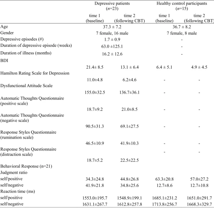
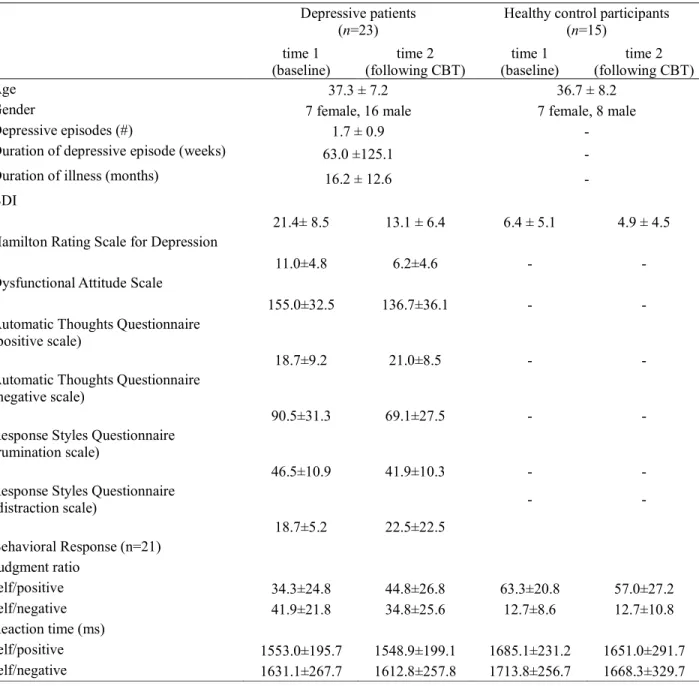
Table 1. Demographic and clinical characteristics of the sample
Depressive patients (n=23)
Healthy control participants (n=15) time 1 (baseline) time 2 (following CBT) time 1 (baseline) time 2 (following CBT) Age 37.3 ± 7.2 36.7 ± 8.2
Gender 7 female, 16 male 7 female, 8 male
Depressive episodes (#) 1.7 ± 0.9 -
Duration of depressive episode (weeks) 63.0 ±125.1 -
Duration of illness (months) 16.2 ± 12.6 -
BDI
21.4± 8.5 13.1 ± 6.4 6.4 ± 5.1 4.9 ± 4.5
Hamilton Rating Scale for Depression
11.0±4.8 6.2±4.6 - -
Dysfunctional Attitude Scale
155.0±32.5 136.7±36.1 - -
Automatic Thoughts Questionnaire (positive scale)
18.7±9.2 21.0±8.5 - -
Automatic Thoughts Questionnaire (negative scale)
90.5±31.3 69.1±27.5 - -
Response Styles Questionnaire (rumination scale)
46.5±10.9 41.9±10.3 - -
Response Styles Questionnaire
(distraction scale) - - 18.7±5.2 22.5±22.5 Behavioral Response (n=21) Judgment ratio self/positive 34.3±24.8 44.8±26.8 63.3±20.8 57.0±27.2 self/negative 41.9±21.8 34.8±25.6 12.7±8.6 12.7±10.8 Reaction time (ms) self/positive 1553.0±195.7 1548.9±199.1 1685.1±231.2 1651.0±291.7 self/negative 1631.1±267.7 1612.8±257.8 1713.8±256.7 1668.3±329.7
Figure legend
Figure1
(A): Picture and graph displays activation in the MPFC (medial prefrontal cortex: green
region) and parameter estimates for each condition in the three-way interaction.
(B): Picture and graph displays activation in the vACC (ventral anterior cingulate
cortex: red region) and parameter estimates for each condition in the three-way
interaction. Both clusters of activities were overlaid on T-1 weighted anatomical brain
images.
Figure 2
Left scatter plot and associated correlation coefficient illustrate the relationship between
vACC parameter estimates and percent symptom improvement in the self/negative
condition (A). Right scatter plot and associated correlation coefficient illustrate the
relationship between changes of vACC parameter estimates and percent changes of
rumination score on the Rumination Styles Questionnaire in the self/negative condition
Table 1. Demographic, clinical characteristics data of the sample
727
Depressive patients (n=23)
Healthy control participants (n=15) time 1 (baseline) time 2 (following CBT) time 1 (baseline) time 2 (following CBT) Age 37.3 ± 7.2 36.7 ± 8.2
Gender 7 female, 16 male 7 female, 8 male
Depressive episodes (#) 1.7 ± 0.9 -
Duration of depressive episode (weeks) 63.0 ±125.1 -
Duration of illness (months) 16.2 ± 12.6 -
BDI
21.4± 8.5 13.1 ± 6.4 6.4 ± 5.1 4.9 ± 4.5
Hamilton Rating Scale for Depression
11.0±4.8 6.2±4.6 - -
Dysfunctional Attitude Scale
155.0±32.5 136.7±36.1 - -
Automatic Thoughts Questionnaire (positive scale)
18.7±9.2 21.0±8.5 - -
Automatic Thoughts Questionnaire (negative scale)
90.5±31.3 69.1±27.5 - -
Response Styles Questionnaire (rumination scale)
46.5±10.9 41.9±10.3 - -
Response Styles Questionnaire
(distraction scale) - - 18.7±5.2 22.5±22.5 Behavioral Response (n=21) Judgment ratio self/positive 34.3±24.8 44.8±26.8 63.3±20.8 57.0±27.2 self/negative 41.9±21.8 34.8±25.6 12.7±8.6 12.7±10.8 Reaction time (ms) self/positive 1553.0±195.7 1548.9±199.1 1685.1±231.2 1651.0±291.7 self/negative 1631.1±267.7 1612.8±257.8 1713.8±256.7 1668.3±329.7
Left scatter plot and associated correlation coefficient illustrate the relationship between vACC parameter estimates and percent symptom improvement in the self/negative condition (A). Right scatter plot and associated correlation coefficient illustrate the relationship between changes of vACC parameter estimates
and percent changes of rumination score on the Rumination Styles Questionnaire in the self/negative condition following CBT (B).