0095-1137/10/$12.00 doi:10.1128/JCM.01098-10
Copyright © 2010, American Society for Microbiology. All Rights Reserved.
Recognition of Potentially Novel Human Disease-Associated
Pathogens by Implementation of Systematic 16S rRNA
Gene Sequencing in the Diagnostic Laboratory
䌤
†
Peter M. Keller,
1‡ Silvana K. Rampini,
2‡ Andrea C. Bu
¨chler,
3Gerhard Eich,
4Roger M. Wanner,
5Roberto F. Speck,
3Erik C. Bo
¨ttger,
1and Guido V. Bloemberg
1*
University of Zurich, Institute of Medical Microbiology, Gloriastrasse 30/32, CH-8006 Zurich, Switzerland1; Division of Internal Medicine,
University Hospital Zurich, Raemistrasse 100, CH-8091 Zurich, Switzerland2; Division of Infectious Diseases and Hospital Epidemiology,
University of Zurich, University Hospital of Zurich, Raemistrasse 100, CH-8091 Zurich, Switzerland3; Division of
Infectious Diseases and Hospital Hygiene, Department of Internal Medicine, Stadtspital Triemli,
CH-8063 Zurich, Switzerland4; and Department of Internal Medicine,
Kantonsspital Winterthur, CH-8401 Winterthur, Switzerland5
Received 1 June 2010/Returned for modification 19 June 2010/Accepted 2 July 2010
Clinical isolates that are difficult to identify by conventional means form a valuable source of novel human pathogens. We report on a 5-year study based on systematic 16S rRNA gene sequence analysis. We found 60 previously unknown 16S rRNA sequences corresponding to potentially novel bacterial taxa. For 30 of 60 isolates, clinical relevance was evaluated; 18 of the 30 isolates analyzed were considered to be associated with human disease.
16S rRNA gene sequence analysis is broadly considered the “gold standard” in bacterial identification (6, 29). In daily clin-ical diagnostics, accurate bacterial identification is essential in judging whether a bacterial isolate is to be considered the causative agent of an infectious disease or merely a colonizer. In our study, we aimed to characterize the bacterial diversity encountered in a diagnostic laboratory by revealing potentially novel, clinically relevant species, according to the current spe-cies definition by the Clinical and Laboratory Standards Insti-tute (22).
Routine 16S rRNA gene sequencing is implemented in our laboratory and is a fixed part of our diagnostic algorithms for identification of bacterial isolates (1, 2, 32). We retrospectively reanalyzed 16S rRNA gene sequences collected during 2004 to 2008 to identify potentially novel bacterial taxa of clinical rel-evance. The Institute of Medical Microbiology (IMM) serves the 850-bed University Hospital of Zurich and surrounding smaller hospitals. Bacterial isolates from blood, cerebrospinal fluid, wounds, joint aspirates, respiratory samples, genitouri-nary swabs, feces, and urine were recovered by culture on appropriate media according to standard procedures (19). Iso-lates that could not be identified by phenotypic methods un-derwent sequencing. 16S rRNA gene analysis was performed as previously described (1). Homology analyses were per-formed using the SmartGene Integrated Database Network System (IDNS) (24) and NCBI GenBank databases. For the first screening of our large data collection, we selected isolates
with sequence homology of⬍99.0% to members of described
taxa, regarding these as potentially novel species; isolates with
sequence homology of⬍95% were regarded as representatives
of a novel genus (2). The boundary for novel families was
⬍87.5% homology and, for novel orders,⬍78.4% 16S rRNA
sequence homology (30). After the first screening, we used
more stringent cutoff values (⬍97.5% for species) for taxa with
significant interspecies 16S rRNA divergence; i.e., members of
thePaenibacillaceaefamily and theClostridialesorder (6, 25).
During the 5-year study period, 1,663 cultured isolates were subjected to 16S rRNA gene sequence analysis (Table 1). Of those, 60 isolates (0.4‰; see Table S1 in the supplemental
material) had a 16S rRNA gene homology of⬍99% to
mem-bers of accepted taxa on the date of the first interpretation. A total of 11 of the 60 sequences with a 16S rRNA homology of
⬍99% in the first-time analysis could be allocated to a species
established during the study term as a novel species by others:
Acinetobacter septicus (16, 20),Brevibacterium ravenspurgense
(17),Corynebacterium freiburgense(12),Corynebacterium
mas-siliense(n⫽2) (18),C. mastitidis(10, 18),C. pyruviciproducens
(26), C. ureicelerivorans (11, 31), Neisseria zoodegmatis (28),
Paenibacillus barengoltzii (21), and the reclassified
Campy-* Corresponding author. Mailing address: University of Zurich, In-stitute of Medical Microbiology, Gloriastrasse 30/32, CH-8006 Zurich, Switzerland. Phone: 41 44 634 2887. Fax: 41 44 634 4906. E-mail: [email protected].
† Supplemental material for this article may be found at http://jcm .asm.org/.
‡ P.M.K. and S.K.R. contributed equally to the work.
[image:1.585.301.540.607.724.2]䌤Published ahead of print on 14 July 2010.
TABLE 1. Clinical bacterial isolates with 16S rRNA gene homology⬍99% (n⫽60)
Taxonomic group
No. of isolates with indicated 16S rRNA homology
⬍99% to⬎95% ⬍95%
Enteric Gram-negative rods 0 0
Fastidious Gram-negative rods 1 4
Gram-negative cocci 1 0
Gram-negative nonfermenters 7 1
Gram-positive cocci 12 2
Gram-positive rods 26 6
Total 47 13
3397
on May 16, 2020 by guest
http://jcm.asm.org/
on May 16, 2020 by guest
http://jcm.asm.org/
lobacter ureolyticus(previously known as Bacteroides
ureolyti-cus) (27).
We calculated dendrograms to assess phylogenetic relation-ships (Fig. 1). Potentially novel streptococcal species clustered with known pathogens. For example, within the streptococci, one isolate (GenBank accession no. GU797873) shared 98.8%
se-quence homology withS. infantis; another isolate (GU797840)
shared 97.8% homology withS. pneumoniae. Within the
Ba-cillales order (Fig. 1B), eight novel Paenibacillus sequence
types were recovered: three of them (GU797882, GU797868,
and GU797869) were distantly related (⬍95% sequence
ho-mology) toPaenibacillus chinjuensis-P. validus, and two isolates
(GU797838 and GU797854) were related to P. timonensis
(97.3% and 94.5% sequence homology). Several bacterial
fam-ilies are represented in theClostridialesorder. The novel
se-quences recovered in the Clostridialesorder all belonged to
different families (Fig. 1C). We found two representatives of
the Fusobacterialesorder (Fig. 1C): one isolate (GU797848)
was related toFusobacterium russii(sequence homology 98.8%),
and one isolate (GU797890) was most closely related to
Lepto-trichia buccalis(98.9% sequence homology). Eleven novel
se-quences belonged to the Pseudomonadales order, and 3 of
those (GU797845, GU797842, and GU797892) represented
potential novelAcinetobacterspp. (Fig. 1D). The
Actinomyce-talesorder (Fig. 1E) comprises 25 potentially novel taxa (data
for 24 taxa are given in the figure). Six corynebacterial isolates,
i.e., Corynebacterium freiburgense (GU797839),
Corynebacte-rium massiliense (GU797864 and GU797833), C. mastitidis
(GU797866),C. ureicelerivorans(GU797878), andC.
pyruvicip-roducens (GU797881), clustered with type strain sequences
that were established as novel species during the study period.
[image:3.585.49.473.70.467.2]We recovered three Nocardia spp. (GU797846, GU797858,
FIG. 1. Phylogeny of 55 of 60 cultured isolates recovered from clinical specimens with homology of⬍99% to 16S rRNA gene sequences of members of published taxa. The dendrograms were calculated using CLUSTAL V alignment and a matrix of Jukes-Cantor distances determined by the neighbor-joining method using DNASTAR Lasergene MegAlign 7.0 software. Taxonomic order adherence of 55 taxons identified in this study: (A)Lactobacillales; (B)Bacillales; (C)FusobacterialesandClostridiales; (D)Pseudomonadales; (E)Actinomycetales. Study isolates are shown in bold. Species in regular type were selected as (published) type strains of the different taxa. We usedEscherichia coliK-12rrnA[NCBI GenBank accession no. EG30084] andMycobacterium tuberculosisH37Rvrrs[NC_000962] as outgroups.
on May 16, 2020 by guest
http://jcm.asm.org/
and GU797874); two of them (GU797846 and GU797858)
belonged to the Nocardia asteroides complex and one
(GU797874) was related toNocardia flavorosea. Four
poten-tially novel Actinobaculum spp. (GU797861, GU797867,
GU797872, and GU797883/GU797308) were attributed to the same taxonomic clade according to 16S rRNA sequence
phy-logeny results, with Actinobaculum schaalii as the nearest
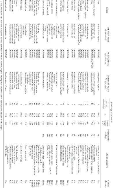
neighboring species (95.7 to 98.5% sequence homology). To assess the clinical relevance of the microbiological findings, we selected 30 isolates for which sufficient clinical data were available and performed reviews of patient charts (Table 2). We reviewed the patient charts for clinical signs and symptoms of infection, inflammation parameters such as fever, leukocyte count, C-reactive protein (CRP) and procalcitonin levels, ra-diological and laboratory findings (serology and bacterial cul-ture results), previous infections or bacterial isolates, anti-biotic treatment, and clinical diagnosis (see Table S2 in the supplemental material). We established a clinical score in-corporating the different parameters mentioned above to determine the likelihood (codified as “yes,” “likely,” “un-likely,” “no”) of an infectious disease in each case and to assess the association of the bacterial isolate found with dis-ease.
Clinical relevance was attributed to 18 isolates. In 10 cases, patient history, laboratory findings, and clinical course follow-ing antibiotic therapy guided by the isolate’s drug susceptibility testing results were compatible with a pathogenic role for the isolated microorganism. In eight cases, we concluded that the bacterial isolate was likely to have been the cause of an infec-tion. The putative novel disease-associated species mostly
be-longed to the Actinomycetales order of Gram-positive rods.
The recently described Acinetobacter septicus (GU797892)
(16), twoActinobaculumspp. (GU797883 and GU797872), and
aGardnerellasp. (GU797857) were each identified as present
in samples from patients with urinary tract infections. In one
case, we recovered a potentially novelActinobaculumsequence
type (GU797872) from several blood cultures of a patient suffering from urosepsis, underlining the pathogenic
poten-tial of species of the Actinobaculum genus in ascending
urinary tract infections. A potentially novelAcinetobactersp.
(GU797845), most closely related toAcinetobacter
calcoaceti-cus, was identified in the peritoneal dialysate of a 22-year-old
patient with kidney failure. He exhibited infective monobacte-rial peritonitis as a complication of continuous ambulatory
peritoneal dialysis (CAPD). A previously unknown
Actinomy-cessp. (GU797891) was isolated from a 49-year old female
patient suffering from severe, acute, suppurative parotitis.
Neisseria zoodegmatis (previouslyNeisseria CDC EF-4 group
[28]) was found in an isolate from a patient with a wound and a history of a cat bite (GU797849). A novel sequence type of
Paenibacillus barengoltzii(21) was cultured from a central venous
catheter in the jugular vein of a patient with a burn injury
(GU797838). A potentially novelStreptococcussp. (GU797859),
belonging to theStreptococcus anginosusgroup, was found in
an isolate from a septic patient suffering from cholangitis. A
potentially novelStreptococcussp. related toStreptococcus
ora-lis (GU797840) was cultured from a hip joint aspirate of a
patient with hip prosthesis infection. Bacterial isolates that were found to be irrelevant to a patient’s clinical disease entity
(12/30) were considered to represent skin flora (e.g.,
Paeniba-cillusspp. orRuminococcusspp.) or to belong to
environmen-tal bacteria (e.g.,Aquabacteriumspp. orKineosphaeraspp.).
Broad-range 16S rRNA gene amplification readily allows the detection of members of as-yet-unknown bacterial taxa (4, 5, 9). Major hypervariable regions are present in the first 500 bp of the roughly 1,600 bp comprising the 16S rRNA gene downstream of the conserved primer target sites (5, 9, 29). Thus, analysis of this part of the gene sequence allows the recognition of potentially novel taxa based on previously
es-tablished cutoff values of⬍99% homology for new species and
⬍95% homology for new genera (1–3, 7). While such a general
cutoff is appropriate for overall first analysis of large data sets, we note that the boundaries for species definition by 16S rRNA sequence homology may be different for different phyla
(13, 25). A less stringent cutoff value (i.e.,⬍99.6% homology)
could have been used to delimit different species in bacterial
groups such as theStreptococcus mitisgroup or nonfermenters
(13). Conversely, for species belonging to thePaenibacillaceae
family and theClostridialesorder, a more stringent cutoff value
(i.e., 97.5% homology) is more appropriate (6) and was there-fore applied after the first selection performed with the 99% cutoff value.
In 2008, the fraction of bacterial isolates submitted for mo-lecular identification was 0.8%. Previous investigations re-ported rates of 0.5% to 1% for a similar study setup (7) and a rate of 14% for isolates restricted to aerobic Gram-positive rods (2). Gram-positive rods and Gram-negative cocci are overrepresented in the group of sequenced isolates in our comparative analysis of phenotypic and 16S rRNA-based iden-tification methods. Of the 1,663 (3.7%) sequences determined during the study period, 60 were judged to be representatives of potentially novel species or novel genera. A recent review (29), summarizing 16S rRNA gene-based studies published from 2001 to 2007, calculated that 215 unique sequences re-covered during this period from human specimens represented potentially novel species. Of the 215, 29 belonged to novel genera. During our study, the number of 16S rRNA sequences deposited in the NCBI nucleotide database increased by a factor of 15. The SmartGene IDNS 16S rRNA database, which is a curated database derived from the NCBI repository, in-creased in size by a factor of 4. Despite this increase in the number of sequences deposited, the recovery of sequences
with⬍99% homology to members of established taxa in our
data set during 2004 to 2008 remained relatively constant at between 2.4% and 5.1%. This finding may reflect the fact that many of the sequences deposited in public databases were the outcome of large-scale ecological or environmental (meta-genomic) sequencing projects and did not include sequences of clinical laboratory isolates.
Bacterial taxonomic classification has advanced differently in various taxonomic groups (25): Phenotypic methods readily allow species determination below the resolution of 16S rRNA-based sequence analysis in studies of enteric
Gram-negative bacteria (14, 15). In contrast, theActinomycetales
or-der is a rich but poorly investigated group (2). For example,
within theCorynebacteriumgenus, 18 novel species were validly
described from 2004 to 2009. A total of 5 of these, namely,
Corynebacterium freiburgense(12), Corynebacterium
pyruvicip-roducens(26),C. mastitidis(10, 18),C. massiliense(18), andC.
ureicelerivorans(11, 31), were also identified in our study.
on May 16, 2020 by guest
http://jcm.asm.org/
TABLE 2. Subset analysis of microbial and clinical data of 30 patients a Source 16S rRNA-based identification NCBI GenBank accession no. Match with highest % homology Homology of 1st match Polymicrobial etiology Clinical diagnosis Clinical relevance No. of mismatches % Match length (bp) Urine Acinetobacter septicus GU797892 Acinetobacter septicus 0 100.0 531 No Urinary tract infection (urothelial carcinoma) Yes Peritoneal dialysate Acinetobacter sp. GU797845 Acinetobacter calcoaceticus 10 98.3 574 No Peritonitis (CAPD) Yes Urine Actinobaculum sp. GU797883, GU797308 Actinobaculum schaalii 26 96.6 768 No Ascending urinary tract infection (pigtail catheter) Yes Blood culture Actinobaculum sp. GU797872 Actinobaculum schaalii 10 98.5 641 No Urosepsis, urothelial carcinoma Yes Parotid gland aspirate Actinomyces sp. GU797891 Actinomyces naeslundii 15 97.4 573 Yes Parotid inflammation Yes Urine Gardnerella sp. GU797857 Gardnerella vaginalis 6 98.9 527 No Urinary tract infection Yes Tissue (hand) Neisseria zoodegmatis GU797849 Neisseria zoodegmatis 3 99.4 525 Yes Cat bite Yes Central venous catheter Paenibacillus barengoltzii GU797838 Paenibacillus barengoltzii 3 99.4 505 No Intravascular catheter-associated infection Yes Blood culture Streptococcus sp. GU797859 Streptococcus constellatus 8 98.6 571 No Cholangitis, sepsis Yes Femur bone Streptococcus sp. GU797840 Streptococcus oralis 9 97.8 403 No Hip prosthesis infection with soft tissue abscess Yes Pleural aspirate Actinomyces sp. GU797884 Actinomyces odontolyticus 17 96.6 496 Yes Anastomosis insuf ficiency (pneumonectomy) Likely Blood culture Burkholderiales order GU797888 Sutterella stercoricanis 54 89.8 530 No Small intestine ischemia Likely Corneal tissue Corynebacterium mastitidis GU797866 Corynebacterium mastitidis 0 100.0 463 Yes Chronic blepharitis Likely Intravenous catheter Corynebacterium massiliense GU797833 Corynebacterium massiliense 1 99.8 556 No Sepsis (unclear focus of infection) Likely Spongiosa tissue Mogibacterium sp. GU797879 Mogibacterium timidum 7 98.7 538 Yes Maxillary bone necrosis Likely Sputum Nocardia sp. GU797874 Nocardia flavorosea 8 98.4 494 Yes Upper lobe pneumonia (COPD) b Likely Deep wound swab Peptostreptococcaceae family GU797889 Anaerococcus octavius 28 94.7 530 Yes Axillary abscess Likely Superficial wound Pseudomonas sp. GU797855 Pseudomonas fulva 10 98.1 526 Yes Ulceration, digit II of right foot (diabetes mellitus) Likely Contact lens Kocuria sp. GU797852 Kocuria marina 26 96.1 668 Contact lens-associated ceratitis Unlikely Hip joint aspirate Paenibacillaceae family GU797869 Paenibacillus chinjuensis 34 92.9 480 Rheumatoid arthritis Unlikely Knee joint aspirate Paenibacillaceae family GU797870 Paenibacillus pocheonensis 35 93.8 563 Intravenous drug abuse, hepatitis C Unlikely Blood culture Propioniferax sp. GU797880 Propioniferax innocua 27 96.4 720 Aplastic anemia Unlikely Blood culture Aquabacterium sp. GU797863 Aquabacterium citratiphilum 22 95.8 520 Fever, AML c No Blood culture Campylobacter ureolyticus GU797876 Campylobacter ureolyticus 1 99.8 519 Fever, neutropenia No Bone biopsy Corynebacterium pyruviciproducens GU797881 Corynebacterium pyruviciproducens 0 100.0 745 Open bone fracture No Sputum Kineosphaera sp. GU797835 Kineosphaera limosa 24 95.6 549 Chronic bronchitis No urine Brevibacteriaceae family GU797885 Leucobacter tardus 57 92.2 734 Urinary tract infection No Bursa aspirate Paenibacillaceae family GU797882 Paenibacillus validus 32 93.5 493 Trochanteric bursitis No Blood culture Paenibacillaceae family GU797875 Paenibacillus contaminans 51 90.7 551 HIV infection, Pneumocystis jirovecii pneumonia No Blood culture Ruminococcus sp. GU797893 Ruminococcus obeum 22 95.9 539 HIV infection No a The likelihood of a relevant infectious disease associated with the microbiological findings was estimated after retrospective patient chart analy sis. b COPD, chronic obstructive pulmonary disease. c AML, acute myelogenous leukemia.
on May 16, 2020 by guest
http://jcm.asm.org/
[image:5.585.97.488.72.725.2]When we calculated phylogenetic trees based on partial 16S rRNA sequences (Fig. 1), we found that differentiation was numerically strong (as measured by nucleotide substitutions of
base pairs) in theActinomycetales, Clostridiales,
Fusobacteria-les, andPseudomonalesorders whereas it was less profound in
theLactobacillalesorder and, more specifically, in the
Strepto-coccaceae family. Regarding potentially novel Streptococcus
spp., further molecular analysis of additional loci (by, e.g.,
sodA,rpoB, and recAsequence homology) would be required
to determine exact phylogenetic relationships (8, 23). In summary, out of 1,663 bacterial isolates subjected to 16S rRNA sequencing during a 5-year period, we recovered 60 clinical bacterial isolates that were indicative of the presence of putative novel bacterial species. Of these 60 isolates, 9 were established as novel pathogens in the literature during the period of the study. A total of 18 (60%) isolates showed clinical relevance in a subset analysis of 30 of the 60 isolates. Isolates with clinical implications are mostly representatives of genera
that comprise known pathogens (i.e.,Streptococcusspp.,
Acti-nobaculum spp., Actinomyces spp., and Neisseria spp.). Our
findings underline the importance of 16S rRNA gene sequenc-ing in routine identification algorithms designed to recognize novel pathogens in the diagnostic laboratory.
We thank the laboratory technicians for their dedicated help. The study was supported by the University of Zurich.
REFERENCES
1.Bosshard, P. P., S. Abels, M. Altwegg, E. C. Bo¨ttger, and R. Zbinden.2004.
Comparison of conventional and molecular methods for identification of aerobic catalase-negative gram-positive cocci in the clinical laboratory. J. Clin. Microbiol.42:2065–2073.
2.Bosshard, P. P., S. Abels, R. Zbinden, E. C. Bo¨ttger, and M. Altwegg.2003.
Ribosomal DNA sequencing for identification of aerobic gram-positive rods in the clinical laboratory (an 18-month evaluation). J. Clin. Microbiol.41:
4134–4140.
3.Bosshard, P. P., R. Zbinden, S. Abels, B. Bo¨ddinghaus, M. Altwegg, and
E. C. Bo¨ttger.2006. 16S rRNA gene sequencing versus the API 20 NE system
and the VITEK 2 ID-GNB card for identification of nonfermenting Gram-negative bacteria in the clinical laboratory. J. Clin. Microbiol.44:1359–1366.
4.Bo¨ttger, E. C.1996. Approaches for identification of microorganisms. ASM
News62:247–250.
5.Bo¨ttger, E. C.1989. Rapid determination of bacterial ribosomal RNA
se-quences by direct sequencing of enzymatically amplified DNA. FEMS Mi-crobiol. Lett.53:171–176.
6.Clarridge, J. E., III.2004. Impact of 16S rRNA gene sequence analysis for
identification of bacteria on clinical microbiology and infectious diseases. Clin. Microbiol. Rev.17:840–862.
7.Drancourt, M., P. Berger, and D. Raoult.2004. Systematic 16S rRNA gene
sequencing of atypical clinical isolates identified 27 new bacterial species associated with humans. J. Clin. Microbiol.42:2197–2202.
8.Drancourt, M., V. Roux, P. E. Fournier, and D. Raoult.2004.rpoBgene
sequence-based identification of aerobic Gram-positive cocci of the gen-eraStreptococcus,Enterococcus,Gemella,Abiotrophia, andGranulicatella. J. Clin. Microbiol.42:497–504.
9.Edwards, U., T. Rogall, H. Blocker, M. Emde, and E. C. Bo¨ttger.1989.
Isolation and direct complete nucleotide determination of entire genes. Characterization of a gene coding for 16S ribosomal RNA. Nucleic Acids Res.17:7843–7853.
10.Fernandez-Garayzabal, J. F., M. D. Collins, R. A. Hutson, E. Fernandez, R.
Monasterio, J. Marco, and L. Dominguez.1997.Corynebacterium mastitidis
sp. nov., isolated from milk of sheep with subclinical mastitis. Int. J. Syst. Bacteriol.47:1082–1085.
11.Ferna´ndez-Natal, M. I., J. A. Saez-Nieto, S. Valdezate, R. H.
Rodriguez-Pollan, S. Lapena, F. Cachon, and F. Soriano.2009. Isolation of
Corynebac-terium ureicelerivoransfrom normally sterile sites in humans. Eur. J. Clin. Microbiol. Infect. Dis.28:677–681.
12.Funke, G., R. Frodl, K. A. Bernard, and R. Englert.2009.Corynebacterium
freiburgensesp. nov., isolated from a wound obtained from a dog bite. Int. J. Syst. Evol. Microbiol.59:2054–2057.
13.Janda, J. M., and S. L. Abbott.2007. 16S rRNA gene sequencing for
bac-terial identification in the diagnostic laboratory: pluses, perils, and pitfalls. J. Clin. Microbiol.45:2761–2764.
14.Janda, J. M., and S. L. Abbott.2006. The familyEnterobacteriaceae:
taxo-nomic considerations, p. 7–14.InJ. M. Janda (ed.), The enterobacteriaceae, 2nd ed. ASM Press, Washington, DC.
15.Johnson, J. R.2000. Shigella andEscherichia coliat the crossroads:
Machiavel-lian masqueraders or taxonomic treachery? J. Med. Microbiol.49:583–585.
16.Kilic, A., H. Li, A. Mellmann, A. C. Basustaoglu, M. Kul, Z. Senses, H.
Aydogan, C. W. Stratton, D. Harmsen, and Y. W. Tang.2008.Acinetobacter
septicussp. nov. association with a nosocomial outbreak of bacteremia in a neonatal intensive care unit. J. Clin. Microbiol.46:902–908.
17.Mages, I. S., R. Frodl, K. A. Bernard, and G. Funke.2008. Identities of
Arthrobacterspp. andArthrobacter-like bacteria encountered in human clin-ical specimens. J. Clin. Microbiol.46:2980–2986.
18.Merhej, V., E. Falsen, D. Raoult, and V. Roux. 2009. Corynebacterium
timonensesp. nov. andCorynebacterium massiliensesp. nov., isolated from human blood and human articular hip fluid. Int. J. Syst. Evol. Microbiol.
59:1953–1959.
19.Murray, P. R., and E. J. Baron.2007. Manual of clinical microbiology, 9th
ed. ASM Press, Washington, DC.
20.Nemec, A., M. Musilek, M. Vaneechoute, E. Falsen, and L. Dijkshoorn.2008.
Lack of evidence for “Acinetobacter septicus” as a species different from
Acinetobacter ursingii? J. Clin. Microbiol.46:2826–2827.
21.Osman, S., M. Satomi, and K. Venkateswaran.2006.Paenibacillus
pasa-denensis sp. nov. andPaenibacillus barengoltziisp. nov., isolated from a spacecraft assembly facility. Int. J. Syst. Evol. Microbiol.56:1509–1514.
22.Petti, C. A., P. P. Bosshard, M. E. Brandt, J. E. Clarridge III, T. V.
Feld-blyum, P. Foxall, M. R. Furtado, N. Pace, and G. W. Procop.2006.
Inter-pretive criteria for identification of bacteria and fungi by DNA target se-quencing: approved guideline, vol. MM18-A. Clinical and Laboratory Standards Institute (CLSI), Wayne, PA.
23.Poyart, C., G. Quesne, S. Coulon, P. Berche, and P. Trieu-Cuot.1998.
Identi-fication of streptococci to species level by sequencing the gene encoding the manganese-dependent superoxide dismutase. J. Clin. Microbiol.36:41–47.
24.Simmon, K. E., A. C. Croft, and C. A. Petti.2006. Application of SmartGene
IDNS software to partial 16S rRNA gene sequences for a diverse group of bacteria in a clinical laboratory. J. Clin. Microbiol.44:4400–4406.
25.Stackebrandt, E.2006. Defining taxonomic ranks, p. 29–57.InM. Dworkin
and S. Falkow (ed.), The prokaryotes: a handbook on the biology of bacteria, 3rd ed. Springer, New York, NY.
26.Tong, J., C. Liu, P. Summanen, H. Xu, and S. M. Finegold.2010.
Coryne-bacterium pyruviciproducenssp. nov., a pyruvic acid producer. Int. J. Syst. Evol. Microbiol.60:1135–1140.
27.Vandamme, P., L. Debruyne, E. De Brandt, and E. Falsen.2 October 2009,
posting date. Reclassification of Bacteroides ureolyticusas Campylobacter ureolyticuscomb. nov. Int. J. Syst. Evol. Microbiol. [Epub ahead of print.]
28.Vandamme, P., B. Holmes, H. Bercovier, and T. Coenye.2006. Classification
of Centers for Disease Control Group Eugonic Fermenter (EF)-4a and EF-4b asNeisseria animalorissp. nov. andNeisseria zoodegmatissp. nov., respectively. Int. J. Syst. Evol. Microbiol.56:1801–1805.
29.Woo, P. C., S. K. Lau, J. L. Teng, H. Tse, and K. Y. Yuen.2008. Then and
now: use of 16S rDNA gene sequencing for bacterial identification and discovery of novel bacteria in clinical microbiology laboratories. Clin. Mi-crobiol. Infect.14:908–934.
30.Yarza, P., M. Richter, J. Peplies, J. Euzeby, R. Amann, K. H. Schleifer, W.
Ludwig, F. O. Glockner, and R. Rossello-Mora.2008. The All-Species Living
Tree project: a 16S rRNA-based phylogenetic tree of all sequenced type strains. Syst. Appl. Microbiol.31:241–250.
31.Yassin, A. F.2007.Corynebacterium ureicelerivoranssp. nov., a lipophilic
bacte-rium isolated from blood culture. Int. J. Syst. Evol. Microbiol.57:1200–1203.
32.Zbinden, A., E. C. Bottger, P. P. Bosshard, and R. Zbinden.2007. Evaluation
of the colorimetric VITEK 2 card for identification of gram-negative non-fermentative rods: comparison to 16S rRNA gene sequencing. J. Clin. Mi-crobiol.45:2270–2273.