Dilution Method for Susceptibility Testing of
Campylobacter
spp.
Mirva Lehtopolku,a,bPirkko Kotilainen,a,bPauli Puukka,cUlla-Maija Nakari,dAnja Siitonen,dErkki Eerola,ePentti Huovinen,a,eand Antti J. Hakanena,b
Antimicrobial Resistance Unit,aPopulation Studies Unit,cand Bacteriology Unit,dNational Institute for Health and Welfare (THL), Turku and Helsinki, Finland; Department of Medicine, Turku University Hospital and University of Turku, Turku, Finlandb; and Department of Medical Microbiology and Immunology, University of Turku, Turku, Finlande
The agar dilution method has been standardized by the CLSI for the susceptibility testing ofCampylobacterspecies, and accord-ing to these standards, the disk diffusion method should be used only in screenaccord-ing for macrolide and ciprofloxacin resistance. Nevertheless, the disk diffusion test is currently widely used, since it is easy to perform in clinical microbiology laboratories. In this study, the disk diffusion method was compared to the agar dilution method by analyzing thein vitroactivities of seven anti-microbial agents against 174Campylobacterstrains collected in Finland between 2003 and 2008. Recommendations of the CLSI were followed using Mueller-Hinton agar plates with 5% of sheep blood. For each strain, the disk diffusion tests were performed two to four times. Of the 33 erythromycin-resistant strains (MIC,>16g/ml), 24 (73%) constantly showed a 6-mm erythromy-cin inhibition zone (i.e., no inhibition), while for seven strains the inhibition zone varied from 6 to 44 mm in repeated measure-ments. Among the 141 erythromycin-susceptible strains (MIC,<16g/ml), erythromycin inhibition zones varied between 6 and 61 mm. Of the 87 ciprofloxacin-resistant strains, 47 (54%) showed 6-mm inhibition zones, while 40 strains showed inhibition zones between 6 and 60 mm. Significant differences between the repetitions were observed in the disk diffusion for all antimicro-bial agents and all strains except for the macrolide-resistant strains regarding the macrolides. For 17 (10%) strains, the variation in repeated measurements was substantial. These results show that the disk diffusion method may not be a reliable tool for the susceptibility testing ofCampylobacterspp. Further studies are needed to assess whether the disk diffusion test could be im-proved or whether all susceptibilities of campylobacters should be tested using an MIC-based method.
C
ampylobacters are one of the most common causes of bacterial gastroenteritis in humans (2). Though bacterial gastroenteritis is usually a mild and self-limiting disease, antimicrobial therapy is needed in severe and prolonged cases, in immunocompromised pa-tients, and in pregnant women as well as in very young and very old patients (2). Currently, macrolides and fluoroquinolones are consid-ered to be the first- and second-choice alternatives for the antimicro-bial treatment of campylobacteriosis. In recent years, however, resis-tance against both of these antimicrobial groups has complicated the empirical treatment of bacterial gastroenteritis (1). Therefore, reli-able routine susceptibility testing is required to provide an adequate antimicrobial treatment for severely ill patients with campylobacte-riosis. Moreover, correct measures are needed to monitor the Cam-pylobacterresistance situation worldwide.Several laboratory methods have been applied for the suscep-tibility testing ofCampylobacterspecies. The agar dilution and broth dilution methods have been standardized by the Clinical and Laboratory Standards Institute (CLSI) (3, 4, 5, 6). The Etest is also an MIC-based method which has been widely used although not standardized. The disk diffusion method has been standard-ized by the CLSI, but according to those standards, it should be used only as a screening method for resistance to erythromycin and ciprofloxacin: a disk diffusion zone of 6 mm (growth up to the edge of a 6-mm disk) indicates resistance, while any inhibition zone would require an MIC determination of susceptibility (4).
A number of previous studies have compared the disk diffu-sion method with the other susceptibility testing methods for
Campylobacterspp. In some of those studies, the results have been in line, though in some other studies, inconsistencies between the methods have been observed (8, 9, 15, 17–20). Nevertheless, the
disk diffusion test is currently widely used, since it is easy to per-form in clinical microbiology laboratories.
The aim of the present study was to compare the performance of the disk diffusion method with the agar dilution method in susceptibility testing of campylobacters to seven different antimi-crobial agents. In addition, the repeatability of the disk diffusion method was evaluated in determining thein vitroefficacy of these same antimicrobial agents towardCampylobacterspp. The main interest was focused on erythromycin and ciprofloxacin suscepti-bilities, since they are clinically the two most important antimi-crobial agents in the treatment of campylobacteriosis. Five addi-tional antimicrobial agents were chosen based on possible clinical relevance and in order to find out whether there are differences within antimicrobial groups.
(This work was presented in part at the 20th European Congress of Clinical Microbiology and Infectious Diseases [ECCMID], Vi-enna, Austria, 2010.)
MATERIALS AND METHODS
Campylobacterisolates.The study collection consisted of 174 Campylo-bacterstrains isolated from stool specimens from Finnish patients be-tween 2003 and 2008. Of these strains, 23 were collected locally in the area
Received30 May 2011Returned for modification22 July 2011 Accepted30 October 2011
Published ahead of print9 November 2011
Address correspondence to Mirva Lehtopolku, [email protected].
Copyright © 2012, American Society for Microbiology. All Rights Reserved.
doi:10.1128/JCM.01090-11
on May 16, 2020 by guest
http://jcm.asm.org/
of the Turku University Hospital. The rest of the strains were collected from 10 different hospital districts around Finland; their susceptibilities have been reported in our earlier paper (14). The cultivation of the stool samples and the preliminary identification of the samples were carried out by standard microbiological methods. The hippurate hydrolysis test was used for determination of theC. jejunistrains. All hippurate-positive iso-lates were determined asCampylobacter jejuni, and all hippurate-negative isolates were confirmed by PCR as eitherC. jejuniorC. coli(16). The final study collection was composed of 151C. jejunistrains and 23C. coli strains.Campylobacter jejunitype strain DSM 4688 (same as ATCC 33560 and NCTC 11351),C. colitype strain DSM 4689 (ATCC 33559 and NCTC 11366),Staphylococcus aureusATCC 29213, andEscherichia coliATCC 25922 were used as controls in susceptibility testing.
Agar dilution and the Etest method.The MICs of theCampylobacter strains were determined by the standard agar plate dilution method for six antimicrobials (3, 12). The 90-mm plates were incubated in a microaerobic atmosphere (CampyPak; BBL) at 35⫾1°C for 48 h. The MIC determinations were done twice for each strain. The antimicrobial agents evaluated were clindamycin, erythromycin, nalidixic acid, and tetracycline (Sigma, Stein-heim, Germany) and azithromycin and ciprofloxacin (Fluka, Buchs, Switzer-land). The MICs of the isolates to tigecycline were determined by the Etest (Biodisk AB, Solna, Sweden) as described previously (14).
Disk diffusion method.The following antimicrobial disks were used: clindamycin 2g, erythromycin 15g, nalidixic acid 30g, tetracycline 30g, azithromycin 15g, ciprofloxacin 5g, and tigecycline 15g (Oxoid, United Kingdom). The disk diffusion tests were made according to the following instructions. Plates were dried at 35°C for 1.5 to 2 h. Inocula prepared in sterile NaCl at a density adjusted to a 0.5 McFarland turbidity standard were delivered onto Mueller-Hinton agar plates sup-plemented with 5% defibrinated sheep blood filled with an amount of agar giving a uniform depth of 4⫾0.5 mm. The disks were added and incu-bated in a microaerobic atmosphere at 35⫾1°C for 48 h. Sterile blotting paper was used between the lid and plate to inhibit excess moisture during the incubation. Three plates were made for each strain for the testing of susceptibility toward the different antimicrobials from the same inocu-lum at one measurement time. A maximum of three disks at the same time were applied onto one agar plate to make sure of a proper reading of even large inhibition zones. Tigecycline was always placed alone onto one agar plate. All disk diffusion tests were done according to the same instructions for each strain, three to four times for erythromycin and two to four times for the other antimicrobials. No inhibition zone indicated a disk diffusion zone of 6 mm, i.e., growth up to the edge of a 6-mm disk.
Data analysis. The susceptibility data were analyzed using the WHONET5.4 computer program (http://www.whonet.org). Statistical analyses were made using SPSS 17.0 and SAS for Windows 9.1 programs. To describe the variability within the two or four disk diffusion test results, coefficients of variation (%) and maximum differences (mm) between the tests were determined for all strains. For the susceptible strains, repeated-measures analysis of variance was used to compare the repetitions. Due to the skewed distributions for the resistant strains, pairwise differences between the repetitions were tested by nonparametric Wilcoxon rank sum test. The Bon-ferroni method was used to adjust for the multiple comparisons. APvalue of
⬍0.05 was considered statistically significant.
RESULTS
As determined by the agar dilution method, a total of 33 Campy-lobacterstrains were erythromycin resistant (MICⱖ16g/ml) and 141 strains were susceptible (MIC⬍16g/ml) (Table 1). A total of 87 strains were ciprofloxacin resistant (MICⱖ4g/ml) and 87 strains were susceptible (MIC⬍4g/ml). According to the Etest, all 174 strains were susceptible to tigecycline. The MIC determinations were made twice for each strain obtaining identi-cal susceptibility results.
For the erythromycin-resistant strains, the total number of the measurements for the erythromycin disk was 129. Of the 33
erythromycin-resistant strains, 24 (73%) showed an erythromy-cin inhibition zone of 6 mm (i.e., no inhibition zone) in all re-peated measurements (total number, 115). Seven (21%) erythromycin-resistant strains with MICs ofⱖ128g/ml showed inhibition zones between 6 and 44 mm; two (6.1%) of the erythromycin-resistant strains, with MICs of 16 and 64g/ml, respectively, showed inhibition zones between 22 and 42 mm in all repeated measurements. For the 141 erythromycin-susceptible isolates, a total of 477 measurements were performed. For these susceptible strains, the inhibition zones for erythromycin varied from 6 to 61 mm. Ten (2.1%) measurements were equal to or less than 20 mm, and two (0.42%) were 6 mm.
For the ciprofloxacin-resistant strains, the total number of the measurements for the ciprofloxacin disk was 312. Of the 87 ciprofloxacin-resistant strains, 47 (54%) showed a ciprofloxacin inhibition zone of 6 mm in all repeated measurements (total num-ber, 231), while for 40 (46%) strains, the inhibition zone varied from 6 to 60 mm. In 11 (12%) measurements, the inhibition zone was over 10 mm. For six ciprofloxacin-resistant strains with MICs between 4 and 32g/ml, the inhibition zones were equal to or over 20 mm in all repeated measurements. For the 87 ciprofloxacin-susceptible isolates, a total of 267 measurements were performed. For these susceptible strains, the inhibition zones for ciprofloxa-cin varied between 6 and 66 mm. A 6-mm inhibition zone was observed in nine (3.4%) measurements. The results for azithro-mycin, clindaazithro-mycin, nalidixic acid, tetracycline, and tigecycline are shown in Table 1.
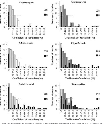
For all measurements of azithromycin, ciprofloxacin, clinda-mycin, erythroclinda-mycin, nalidixic acid, and tetracycline, the coeffi-cients of variation (CV) were determined for the resistant and susceptible strains (Fig. 1; Table 1). For all compounds, strains were found to have a substantial variation between the repetitions of the disk diffusion test. For the erythromycin disk diffusion tests, the CV variation was less than 5% for 53 strains and greater than or equal to 15% for 36 strains. For ciprofloxacin, the variation was small for 64 strains and substantial for 52 strains. Seventeen strains showed substantial variations for both erythromycin and cipro-floxacin. For all of the strains susceptible to these antimicrobial agents, the mean of maximum difference between two different measurements was over 8 mm (Table 1). Even for the resistant strains, the mean values of maximum difference over 4 mm were found for ciprofloxacin, nalidixic acid, and tetracycline. The mean values of coefficient of variation for all antimicrobial agents were over 10%. When the different repetition times were evaluated, significant differences were observed for all antimicrobial agents and for all strains except for the macrolide-resistant strains re-garding erythromycin and azithromycin (Table 1).
DISCUSSION
The aim of the present study was to evaluate the adequacy of the disk diffusion method in comparison with the agar dilution method for determining the efficacy of important antimicrobial compounds to-wardCampylobacterspp. In so doing, significant differences were found for the majority of the antimicrobial agents analyzed when the disk diffusion tests were repeated and the results obtained at different measurement times compared. As many as 17 (10%) of the 174 strains showed a substantial variation in repeated measurements for erythromycin and ciprofloxacin. However, no significant differences in repeatability were observed in the group of macrolide-resistant strains regarding erythromycin and azithromycin. The reasons for
on May 16, 2020 by guest
http://jcm.asm.org/
TABLE 1 Correlation between MICs and inhibition zone values a Antimicrobial agent Agar plate dilution Disk diffusion b No. of strains
Resistance breakpoint (
g/ml)
Susceptibility according to
MIC determinations MIC range ( g/ml) No. of total measurements
Inhibition zone variation (mm)
No. of measurements with no inhibition zone Mean value for max difference (mm) Mean value for coefficient of variation (%) P c Erythromycin 33 ⱖ 16 Resistant 16– ⬎ 128 129 6–44 115 (89%) 2.79 10.04 No significant pairwise differences 141 Susceptible 0.5–8 477 6–61 2 (0.42%) 8.91 11.5 ⬍ 0.001 Azithromycin 31 ⱖ 4 Resistant 64– ⬎ 128 121 6–57 110 (91%) 3.55 10.68 No significant pairwise differences 143 Susceptible ⱕ 0.062–1 480 6–64 4 (0.83%) 10.66 12.28 ⬍ 0.001 Clindamycin 33 ⱖ 8 Resistant 8–128 127 6–58 103 (81%) 6.21 18.32 ⬍ 0.05 for 3 of 6 pairwise differences 141 Susceptible 0.125–4 479 6–60 7 (1.5%) 10.54 14.91 0.026 Ciprofloxacin 87 ⱖ 4 Resistant 4– ⬎ 32 312 6–60 231 (74%) 5.49 19.24 ⬍ 0.01 for all pairwise differences 87 Susceptible 0.06–1 267 6–66 9 (3.4%) 10.38 11.07 0.013 Nalidixic acid 86 ⱖ 32 Resistant 32–256 310 6–44 239 (77%) 4.42 16.86 ⬍ 0.01 for all pairwise differences 87 Susceptible 2–16 234 6–56 9 (3.8%) 8.72 13.19 ⬍ 0.001 Tetracycline 65 ⱖ 16 Resistant 16– ⬎ 128 230 6–62 101 (44%) 6.91 22.2 ⬍ 0.01 for all pairwise differences 107 Susceptible 0.1–4 332 6–70 2 (0.60%) 10.05 10.08 0.014 Tigecycline ⱖ 0.5 Resistant 174 Susceptible ⱕ 0.008–0.064 609 6–80 1 (0.16%) 10.8 9.93 0.81 aThe maximum difference between different measurement times and the mean value for coefficient of variation were determined for five antimicrobial age nts. Also, P values were determined for different measurement times. bDisk diffusion tests were repeated for each strain 2 to 4 times. cSignificance of the difference between repetitions. Due to very skewed distributions in the resistant strains, differences between the repetitions were tested by nonparametric Wilcoxon rank sum test. In the susceptible strains, repeated measures were tested by analysis of variance.
on May 16, 2020 by guest
http://jcm.asm.org/
the better performance regarding the macrolide-resistant strains are still unclear. One explanation for the result could be that the number of those strains was rather small. Thus, further studies are needed to evaluate this finding with a greater number of macrolide-resistant
Campylobacterstrains.
Of the 129 measurements made for the strains classified as erythromycin resistant by the MICs, 89% showed no inhibition zones (i.e., an inhibition zone of 6 mm), while in 11% of them, the inhibition zones exceeded 6 mm, indicating a need for a more accurate susceptibility determination to demonstrate erythromy-cin resistance. For ciprofloxaerythromy-cin, the situation was even more wor-risome: for the ciprofloxacin-resistant strains, only 74% of the results showed no inhibition zones in 312 measurements. It is noteworthy that even 66% of the measurements for tetracycline would have required MIC-based determinations of susceptibility, if the rule of the 6-mm inhibition zone as an indication of
resis-tance would have been followed also for this antimicrobial. On the other hand, 0.42% and 3.4% of all measurements for the erythromycin-susceptible and ciprofloxacin-susceptible strains, respectively, did not show any inhibition zones. Thus, following the CLSI guidelines, a small number of susceptibleCampylobacter
strains would have been falsely classified as resistant. Apart from these false-resistant strains, our findings support the CLSI stan-dardization that the disk diffusion method should be used as a screening method only for resistance to erythromycin and cipro-floxacin in Campylobacter spp. and that any inhibition zone around the disk demands an MIC-based determination of suscep-tibility (3, 4, 5, 6, 13).
These results indicate that there is a need for a standardized pro-tocol for susceptibility testing in clinical microbiology laboratories, as well as determining clear resistance breakpoints and interpretive cri-teria forCampylobacterspp. The falsely diagnosed resistant strains
FIG 1Coefficients of variation for all repeated measurements of six antimicrobial agents studied were determined for 174Campylobacter jejuniand
Campylo-bacter colistrains. Measurements were repeated two to four times for each strain. R means a resistant strain and S a susceptible strain.
on May 16, 2020 by guest
http://jcm.asm.org/
[image:4.585.113.475.65.502.2]may lead to an excessive use of more toxic and possibly even less effective antimicrobial treatment for patients with campylobacterio-sis (19). The most serious threat is that recampylobacterio-sistant strains falsely diag-nosed as susceptible may lead to ineffective antimicrobial treatment even in invasive and life-threatening infections. Infections with resis-tant strains have been reported in association with a 5-fold increase of the risk of invasive illness or death (7). Especially for that reason, it is of importance to be able to correctly distinguish the resistant strains. Moreover, adequate worldwide monitoring ofCampylobacter resis-tance is impossible if the resisresis-tance rates are falsely reported due to unreliable susceptibility testing.
Several previous papers have focused on the efficacy and accuracy of the disk diffusion method and the Etest method compared to the agar plate dilution or broth microdilution methods, with somewhat contradictory results (8, 9, 15, 17, 19, 20). In these studies, the disk diffusion tests were performed only once for each strain. Gaudreau et al. (8, 9) have found the disk diffusion method to be a reliable, easy and inexpensive method for the testing of the susceptibility ofC. jejunito erythromycin, ciprofloxacin, and tetracycline. Corroborat-ing the present study, the results of, e.g., van der Beek et al. are differ-ent (19). They reinvestigated 48 erythromycin-resistantC. jejuniand
C. colistrains retrospectively to reevaluate erythromycin resistance, and only 11 to 14% of theC. jejunistrains and 67% of theC. coli
strains were erythromycin resistant in the second analysis. In that study, the initial susceptibility testing was performed in most cases by the disk diffusion method and the reinvestigation was carried out using broth microdilution. The authors conclude that routine deter-mination of the erythromycin resistance inC. jejuniandC. colishows unacceptable interlaboratory variation. Nonstandardized suscepti-bility testing methods may be involved in the differences of the sus-ceptibility results obtained by the disk diffusion method, including (i) various protocols of the methods used, (ii) long incubation time for campylobacters, (iii) inaccuracy of the measurements between differ-ent times or between differdiffer-ent persons measuring the inhibition zone, and (iv) different methods for achieving microaerobic conditions during the incubation. In their paper, van der Beek et al. (19) specu-lated on the possibility that differences could also be caused by insta-bility of the erythromycin resistance. In the present study, no rising trend during the repetitions was observed in the inhibition zone vari-ation between the different measurement times. Therefore, the insta-bility of the erythromycin resistance does not seem to be the factor underlying the variation in our strains.
In conclusion, our results show that the disk diffusion method may not be a reliable tool for susceptibility testing of Campylobac-terspp. This is a major concern due to the wide use of the disk diffusion method in routine clinical laboratories as well as in some research laboratories. Accurate determination ofCampylobacter
susceptibility and resistance is of vital importance to ensure an adequate antimicrobial therapy for patients with severe forms of the disease and, also, to efficiently monitor the antimicrobial re-sistance situation ofCampylobacterspp. worldwide. Further stud-ies are needed to assess whether the disk diffusion test method could be improved or whether all susceptibility testing of campy-lobacters should be done using an MIC-based method.
ACKNOWLEDGMENTS
We thank Erkki Nieminen for technical assistance and Tarja Boman, Katri Kylä-Mattila, Minna Lamppu, Tarja Laustola, and Tuula Randell for labora-tory assistance.
This study was financially supported by grants from the Maud Kuistila Memorial Foundation, Finnish Cultural Foundation, and the Turku Uni-versity Central Hospital Research Fund.
REFERENCES
1.Aarestrup FM, Engberg J.2001. Antimicrobial resistance of thermophilic
Campylobacter.Vet. Res.32:311–321.
2.Allos BM. 2001.Campylobacter jejuniinfections: update on emerging
issues and trends. Clin. Infect. Dis.32:1201–1206.
3. CLSI. 2009. Performance standards for antimicrobial susceptibility
testing; 19th informational supplement M100 –S19. Clinical and Labora-tory Standards Institute, Wayne, PA.
4.CLSI.2006. Methods for antimicrobial dilution and disk susceptibility
testing of infrequently isolated or fastidious bacteria; approved guide-line M45-A. Clinical and Laboratory Standards Institute, Wayne, PA.
5.CLSI.2008. Performance standards for disk and dilution susceptibility
testing from bacteria isolated from animals 31-A3. Clinical and Labora-tory Standards Institute, Wayne, PA.
6.CLSI.2009. Methods for dilution antimicrobial susceptibility test for
bacteria that grow aerobically; approved standard— 8th edition. CLSI document M07–A8. Clinical and Laboratory Standards Institute, Wayne, PA.
7.Coker AO, et al.2002. Human campylobacteriosis in developing
coun-tries. Emerg. Infect. Dis.8:237–244.
8.Gaudreau C, et al.2008. Comparison of disk diffusion and agar dilution
methods for erythromycin, ciprofloxacin, and tetracycline susceptibility testing ofCampylobacter coliand for tetracycline susceptibility testing of Campylobacter jejunisubsp.jejuni. Antimicrob. Agents Chemother.52: 4475– 4477.
9.Gaudreau C, et al.2007. Comparison of disk diffusion and agar dilution
methods for erythromycin and ciprofloxacin susceptibility testing of Campylobacter jejunisubsp.jejuni. Antimicrob. Agents Chemother.51: 1524 –1526.
10. Reference deleted. 11. Reference deleted.
12. Hakanen AJ, et al.2003. Multidrug resistance inCampylobacter jejuni
strains collected from Finnish patients during 1995-2000. J. Antimicrob. Chemother.52:1035–1039.
13. King A. 2001. Recommendations for susceptibility tests on fastidious
organisms and those requiring special handling. J. Antimicrob. Che-mother.48(Suppl. 1):77– 80.
14. Lehtopolku M, et al. 2010. Antimicrobial susceptibilities of
multidrug-resistantCampylobacter jejuniandC. colistrains: in vitro activities of 20 antimicrobial agents. Antimicrob. Agents Chemother.
54:1232–1236.
15. McGill K, et al. 2009. Comparison of disc diffusion and epsilometer
(E-test) testing techniques to determine antimicrobial susceptibility of Campylobacterisolates of food and human clinical origin. J. Microbiol. Methods79:238 –241.
16. Nakari UM, et al.2008. Correct identification and discrimination
be-tweenCampylobacter jejuniandC. coliby a standardized hippurate test and species-specific polymerase chain reaction. Eur. J. Clin. Microbiol. Infect. Dis.27:513–518.
17. Schönberg-Norio D, et al.2006. Activities of telithromycin,
erythromy-cin, fluoroquinolones, and doxycycline againstCampylobacterstrains iso-lated from Finnish subjects. Antimicrob. Agents Chemother. 50: 1086 –1088.
18. Valdivieso-Garcia A, et al.2009. Cost analysis and antimicrobial
suscep-tibility testing comparing the E test and the agar dilution method in Cam-pylobacter jejuniandCampylobacter coli.Diagn. Microbiol. Infect. Dis.
65:168 –174.
19. van der Beek MT, et al.2010. Inaccuracy of routine susceptibility tests for
detection of erythromycin resistance ofCampylobacter jejuniand Campy-lobacter coli.Clin. Microbiol. Infect.16:51–56.
20. Varela NP, et al.2008. Comparison of agar dilution and E-test for
anti-microbial susceptibility testing ofCampylobacter coliisolates recovered from 80 Ontario swine farms. Can. J. Vet. Res.72:168 –174.