Typing of
Mycoplasma pneumoniae
Rebecca J. Brown,a,bMatthew T. G. Holden,cO. Brad Spiller,aVictoria J. Chalkerb
Cardiff University, School of Medicine, Department of Child Health, University Hospital of Wales, Cardiff, United Kingdoma; Bacteriology Reference Department, Public Health England, London, United Kingdomb; University of St. Andrews, School of Medicine, Medical & Biological Sciences, North Haugh, St. Andrews, United Kingdomc
Mycoplasma pneumoniae
is a major human respiratory pathogen causing both upper and lower respiratory disease in humans
of all ages, and it can also result in other serious extrapulmonary sequelae. A multilocus sequence typing (MLST) scheme for
M.
pneumoniae
was developed based on the sequences of eight housekeeping genes (
ppa
,
pgm
,
gyrB
,
gmk
,
glyA
,
atpA
,
arcC
, and
adk
)
and applied to 55
M. pneumoniae
clinical isolates and the two type strains M129 and FH. A total of 12 sequence types (STs)
re-sulted for 57
M. pneumoniae
isolates tested, with a discriminatory index of 0.21 STs per isolate. The MLST loci used in this
scheme were shown to be stable in 10 strains following 10 sequential subculture passages. Phylogenetic analysis of concatenated
sequences of the eight loci indicated two distinct genetic clusters that were directly linked to multilocus variable-number tandem
repeat analysis (MLVA) type. Genetic MLST clustering was confirmed by genomic sequence analysis, indicating that the MLST
scheme developed in this study is representative of the genome. Furthermore, this MLST scheme was shown to be more
discrimi-natory than both MLVA and P1 typing for the
M. pneumoniae
isolates examined, providing a method for further and more
de-tailed analysis of observed epidemic peaks of
M. pneumoniae
infection. This scheme is supported by a public Web-based
data-base (http://pubmlst.org/mpneumoniae).
M
ycoplasma pneumoniae
is a common cause of
community-acquired pneumonia (CAP) transmitted by aerosol or close
contact (1).
M. pneumoniae
may cause other serious
extrapulmo-nary sequelae, such as encephalitis (2). The pathogen is found in
all age groups, with a higher prevalence in children age 5 to 14
years (3,
4). Admissions to a United Kingdom hospital in patients
with CAP that were attributed to
M. pneumoniae
were estimated at
18% in 1982 and 4% in 1999 (5). Major increases and decreases in
M. pneumoniae
infection have occurred periodically in the United
Kingdom; historically, epidemics have occurred at approximately
4-year intervals and have lasted 12 to 15 months, concurrent with
sporadic infection at a lower level and seasonal peaks from
De-cember to February (4,
6). However, globally, peaks of infection
have been observed in either summer or autumn, with no obvious
explanation for this seasonal variation (7–10).
Typing of clinical isolates by molecular methods is of
impor-tance for the understanding of the epidemiology of
M.
pneu-moniae
infection and for an analysis of endemic outbreaks. It is
generally considered that molecular typing of
M. pneumoniae
is
hampered by the fact that the pathogen is a genetically
homolo-gous species (11). Initial molecular typing targeted the gene
en-coding the major surface protein (P1) of
M. pneumoniae
.
PCR-restriction fragment length polymorphism (PCR-RFLP) analysis
of the P1 gene, which encodes a major adhesion, is the most
com-mon genotyping method. This enables the separation of isolates
into two types, 1 and 2 (11–13). More recent studies utilize the
repetitive regions, RepMp2/3 and RepMp4, which can be found in
the P1 gene, for molecular typing and have resulted in the
identi-fication of an additional subtype and three variants of these
subtypes (14,
15). Multilocus variable-number tandem-repeat
(VNTR) analysis (MLVA) has also been used, which is based on
the variation in the copy number of tandem repeated sequences
(VNTRs) found at different loci across the genome. The variation
in the copy number of these tandem repeats (TRs) depends on the
isolate tested. Initially, 265 strains were grouped into 26 MLVA
types based on five VNTR loci (Mpn1 and Mpn13 to Mpn16), and
an additional 18 novel types have since been reported (16–18).
However, the Mpn1 locus is unstable in both clinical strains and in
laboratory passages, and most of the novel types came from
vari-ations in Mpn1; therefore, there is international consensus that
this locus should be removed from the typing scheme (19).
Multilocus sequence typing (MLST) was previously attempted
for the molecular typing of
M. pneumoniae
; however, due to the
homogeneity of the
M. pneumoniae
species, very little
polymor-phisms were found in the housekeeping genes examined, and it
was previously concluded that MLST with housekeeping and
structural genes was not useful for molecular typing (22). Only
three housekeeping genes were thoroughly examined for
poly-morphisms across 30 isolates of either P1 type 1, 2, or a variant
strain. The other genes selected for analysis were examined against
a single representative strain from each subtype. In this study, an
MLST scheme was developed with a high discriminatory ability to
differentiate
M. pneumoniae
isolates based on sequence
polymor-Received14 May 2015Returned for modification9 June 2015
Accepted14 July 2015
Accepted manuscript posted online22 July 2015
CitationBrown RJ, Holden MTG, Spiller OB, Chalker VJ. 2015. Development of a
multilocus sequence typing scheme for molecular typing ofMycoplasma
pneumoniae. J Clin Microbiol 53:3195–3203.doi:10.1128/JCM.01301-15.
Editor:N. A. Ledeboer
Address correspondence to Victoria J. Chalker, [email protected]. O.B.S. and V.J.C. are co-senior authors.
Supplemental material for this article may be found athttp://dx.doi.org/10.1128
/JCM.01301-15.
Copyright © 2015, American Society for Microbiology. All Rights Reserved.
doi:10.1128/JCM.01301-15
on May 16, 2020 by guest
http://jcm.asm.org/
phisms within eight housekeeping genes, improving on all
pub-lished typing methods for
M. pneumoniae
.
MATERIALS AND METHODS
M. pneumoniaestrains, culture conditions, and sample preparation.
The strains analyzed in this study are listed inTable 1. Fifty-fiveM. pneu-moniaestrains were submitted to Public Health England, United King-dom, for clinical diagnostic purposes, and the twoM. pneumoniaetype strains, FH (NCTC 10119, ATCC 15531) and M129 (ATCC 29342), were obtained from National Collection of Type Cultures (NCTC) (held by Public Health England). All strains were triple cloned onMycoplasmaagar (Mycoplasma Experience, Surrey, United Kingdom) and confirmed to be M. pneumoniae by amplification of thep1gene (23).
All strains were subsequently cultured inMycoplasmaliquid me-dium (MLM) (Mycoplasma Experience, Surrey, United Kingdom). For genomic sequencing, strains were grown in 100 ml of broth culture, and genomic DNA was extracted using the GenElute bacterial genomic DNA kit (Sigma, Dorset, United Kingdom). PCR amplification was performed on bacterial DNA from a 500-l 4-day culture that was released by boiling lysis (95°C for 10 min) following centrifugation at 17,000⫻gfor 10 min, removal of all MLM, and resuspension in 50l of sterile water.
Multilocus sequence typing.Housekeeping genes considered to be conserved in other bacterial species under a low rate of selective pressure were chosen for analysis (Table 2). The locus sequences were selected using the available genome sequences ofM. pneumoniaeFH and M129 (FH GenBank accession no.NC_017504.1, and M129 GenBank accession
no.NC_000912.1) and the available whole-genome sequences of 35
clin-ical isolates. Ten genes were included for initial analysis: RecA protein (recA), inorganic phosphatase (ppa), phosphoglycerate mutase (pgm), DNA gyrase subunit B (gyrB), guanylate kinase (gmk), serine hydroxym-ethyltransferase (glyA), elongation factor P (efp), ATP synthase subunit␣ (atpA), carbamate kinase (arcC), and adenylate kinase (adk); however, recAandefpwere excluded from the resulting MLST scheme. The locus regions for PCR amplification were selected based on areas of the protein-coding sequences (CDS) containing nucleotide polymorphisms.
PCR utilizing the primers listed inTable 3was used to amplify the target genes from a further 20M. pneumoniaeclinical isolates. Amplifica-tion of each of the locus sequences was performed in a DNA thermocycler (Techne Prime, Stone, United Kingdom) in 50-l reaction mixtures con-taining 1⫻GoTaq Flexi buffer (Promega, Southampton, United King-dom), 1.5 mM MgCl2, 0.2 mM deoxynucleoside triphosphates, 0.5
pmol/l each primer, 1.56 units of GoTaq DNA polymerase (Promega), and 2.5l of template DNA. PCRs consisted of an initial denaturation step of 3 min at 94°C, followed by 35 cycles of 60 s at 94°C, 60 s at 60°C, and 60 s at 72°C. A final extension step was maintained for 10 min at 72°C. The primer sequences and PCR product sizes are shown inTable 3. The PCR products were analyzed on 1.5% agarose gels with ethidium bromide vi-sualization. All PCRs were performed in duplicate.
PCR amplicons were purified using a Qiagen miniprep kit (Qiagen, Inc., Hilden, Germany), as per the manufacturer’s instructions, and se-quenced using the amplification primers, which was performed by MWG Eurofins (Ebersberg, Germany). The sequences obtained from each cor-responding forward and reverse primer were assembled and trimmed for double-stranded high-quality sequences. All the sequences obtained for each locus were aligned using Clustal W (Vector NTI, Paisley, United Kingdom), and different allelic types (ATs) (sequences with at least a 1-nucleotide difference) were assigned sequential numbers. The combi-nation of the eight alleles determined the allelic profile of a strain, and each unique allelic profile was designated a unique sequence type (ST). Open reading frame amino acid sequences were identified using the ExPASy translation tool (Mycoplasma setting [web.expasy.org/translate/]) for each AT. The deduced amino acid sequences were aligned using Clustal W (Vector NTI) for each locus, and synonymous changes were identified.
MLVA and P1 typing.MLVA types were determined as described by Dégrange et al. (16), excluding the VNTR locus Mpn1 and using
interna-tional nomenclature consensus (19). P1 types were determined as de-scribed by Dumke et al. (15).
Genomic sequencing.Genomic sequence data for 35 isolates were obtained using the Illumina Nextera XT sample prep kit (Illumina, Cam-bridge, United Kingdom) and sequenced on an Illumina HiSeq 2500 plat-form with TruSeq rapid SBS kits (200 cycles; Illumina) and cBOT for cluster generation (Illumina). Fastq reads were trimmed using Trimmo-matic 0.32, with the following parameters: leading, 30; trailing, 30; slid-ingwindow, 10:30; and minlen, 50 (20). The Illumina reads were assem-bled to the M129 type strain (GenBank accession no.NC_000912.1) using SPAdes version 2.5.0 (21) and mapped to M129 using Geneious version 8.0.4. Sequencing yielded at least one contig of between 99,047 bp and 324,397 bp with homology to the M129 type strain (GenBank accession
no.NC_000912.1), passing quality and coverage checks. Identification as
M. pneumoniaefrom the genomic data was confirmed with 16S rRNA sequence analysis. The Illumina reads for all isolates were mapped against the reference chromosome of M129 (EMBL accession no.U00089) using SMALT (http://www.sanger.ac.uk/resources/software/smalt/) to identify single-nucleotide polymorphisms (SNPs), as previously described (39). Regions of recombination in the whole chromosomes of the isolates were analyzed using Genealogies Unbiased By recomBinations In Nucleotide Sequences (GUBBINS) (40).
Phylogenetic analysis.The locus sequences corresponding to each strain were concatenated head-to-tail for diversity analysis. Sequence analyses and tree construction were performed using MEGA 6.0. Neigh-bor-joining trees were constructed for each individual locus and concat-enated sequences using Kimura’s two-parameter model (24,25). Maxi-mum-likelihood trees were constructed for each individual locus using the Jukes-Cantor model of sequence evolution (26). Maximum-likeli-hood trees were constructed from concatenated sequences of the eight MLST loci using the generalized time-reversible (GTR) model of sequence evolution, with uniform rates of variation (27). Bootstrap analyses with 1,000 replicates were performed for every phylogenetic tree (28). Related-ness between STs was analyzed based on allelic profiles using eBURST version3. Maximum-likelihood trees were constructed from genomic se-quences after the removal of areas of recombination. In total, 1,854 SNP sites were identified in comparison to the M129 reference chromosome. Three regions were predicted to contain SNP sites that had arisen by recombination, and these contained 28 SNP sites.
RESULTS
MLST of
M. pneumoniae
.
An initial examination of 10 gene
tar-gets in the two type strains M129 and FH and the genomic
se-quences of 35
M. pneumoniae
clinical isolates identified variation,
in the form of SNP differences, in 8 out of the 10 genes. The
recA
and
efp
genes were 100% conserved in all sequences analyzed and
were therefore excluded from the MLST scheme. Genomic
se-quence analysis and additional PCR and sequencing of all eight
targets in a further 20 clinical isolates resolved a total of 12 STs.
The discriminatory typing ability for
M. pneumoniae
was 0.21 ST
per isolate. The number of SNPs observed within each individual
locus and the percentage of polymorphic sites are indicated in
Table 3, with
pgm
having the highest number of SNPs (10 SNPs)
and the highest percentage of polymorphic sites corrected for
se-quence length (0.93%). The number of alleles per locus ranged
from two (
ppa
,
gyrB
,
gmk
, and
arc
) to four (
atpA
) (Table 3).
Ex-amination of the Hunter-Gaston diversity index (DI) (which
ranges from 0.0 for no diversity to 1.0 for complete diversity)
indicated moderate diversity between the STs (DI, 0.784; 95%
confidence interval [CI], 0.716 to 0.852), with the greatest
indi-vidual diversity shown in
pgm
(DI, 0.620; 95% CI, 0.566 to 0.674)
and the lowest diversity shown in
arcC
(DI, 0.069; 95% CI, 0.000
to 0.158).
on May 16, 2020 by guest
http://jcm.asm.org/
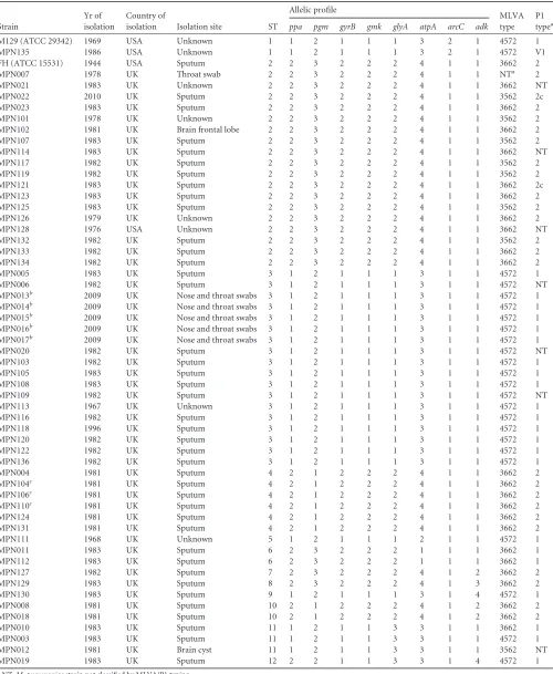
TABLE 1Description ofM. pneumoniaestrains used in this study, their sequence type, allelic profile, and MLVA and P1 types
Strain
Yr of isolation
Country of
isolation Isolation site ST
Allelic profile
MLVA type
P1 typea ppa pgm gyrB gmk glyA atpA arcC adk
M129 (ATCC 29342) 1969 USA Unknown 1 1 2 1 1 1 3 2 1 4572 1
MPN135 1986 USA Unknown 1 1 2 1 1 1 3 2 1 4572 V1
FH (ATCC 15531) 1944 USA Sputum 2 2 3 2 2 2 4 1 1 3662 2
MPN007 1978 UK Throat swab 2 2 3 2 2 2 4 1 1 NTa 2
MPN021 1983 UK Unknown 2 2 3 2 2 2 4 1 1 3662 NT
MPN022 2010 UK Sputum 2 2 3 2 2 2 4 1 1 3562 2c
MPN023 1983 UK Sputum 2 2 3 2 2 2 4 1 1 3662 2
MPN101 1978 UK Unknown 2 2 3 2 2 2 4 1 1 3562 2
MPN102 1981 UK Brain frontal lobe 2 2 3 2 2 2 4 1 1 3662 2
MPN107 1983 UK Sputum 2 2 3 2 2 2 4 1 1 3562 2
MPN114 1983 UK Sputum 2 2 3 2 2 2 4 1 1 3662 NT
MPN117 1982 UK Sputum 2 2 3 2 2 2 4 1 1 3562 2
MPN119 1982 UK Sputum 2 2 3 2 2 2 4 1 1 3562 2
MPN121 1983 UK Sputum 2 2 3 2 2 2 4 1 1 3662 2c
MPN123 1983 UK Sputum 2 2 3 2 2 2 4 1 1 3662 2
MPN125 1983 UK Sputum 2 2 3 2 2 2 4 1 1 3562 2
MPN126 1979 UK Unknown 2 2 3 2 2 2 4 1 1 3662 2
MPN128 1976 USA Unknown 2 2 3 2 2 2 4 1 1 3662 NT
MPN132 1982 UK Sputum 2 2 3 2 2 2 4 1 1 3562 2
MPN133 1982 UK Sputum 2 2 3 2 2 2 4 1 1 3662 2
MPN134 1982 UK Sputum 2 2 3 2 2 2 4 1 1 3662 2
MPN005 1983 UK Sputum 3 1 2 1 1 1 3 1 1 4572 1
MPN006 1982 UK Sputum 3 1 2 1 1 1 3 1 1 4572 NT
MPN013b 2009 UK Nose and throat swabs 3 1 2 1 1 1 3 1 1 4572 1
MPN014b 2009 UK Nose and throat swabs 3 1 2 1 1 1 3 1 1 4572 1
MPN015b 2009 UK Nose and throat swabs 3 1 2 1 1 1 3 1 1 4572 1
MPN016b 2009 UK Nose and throat swabs 3 1 2 1 1 1 3 1 1 4572 1
MPN017b 2009 UK Nose and throat swabs 3 1 2 1 1 1 3 1 1 4572 1
MPN020 1982 UK Sputum 3 1 2 1 1 1 3 1 1 4572 NT
MPN103 1982 UK Sputum 3 1 2 1 1 1 3 1 1 4572 1
MPN105 1983 UK Sputum 3 1 2 1 1 1 3 1 1 4572 1
MPN108 1983 UK Sputum 3 1 2 1 1 1 3 1 1 4572 1
MPN109 1982 UK Sputum 3 1 2 1 1 1 3 1 1 4572 NT
MPN113 1967 UK Unknown 3 1 2 1 1 1 3 1 1 4572 1
MPN116 1982 UK Sputum 3 1 2 1 1 1 3 1 1 4572 1
MPN118 1996 UK Sputum 3 1 2 1 1 1 3 1 1 4572 1
MPN120 1982 UK Sputum 3 1 2 1 1 1 3 1 1 4572 1
MPN122 1982 UK Sputum 3 1 2 1 1 1 3 1 1 4572 1
MPN136 1982 UK Sputum 3 1 2 1 1 1 3 1 1 4572 1
MPN004 1981 UK Sputum 4 2 1 2 2 2 4 1 1 3662 2
MPN104c 1981 UK Sputum 4 2 1 2 2 2 4 1 1 3662 2
MPN106c 1981 UK Sputum 4 2 1 2 2 2 4 1 1 3662 2
MPN110c 1981 UK Sputum 4 2 1 2 2 2 4 1 1 3662 2
MPN124 1981 UK Sputum 4 2 1 2 2 2 4 1 1 3662 2
MPN131 1981 UK Sputum 4 2 1 2 2 2 4 1 1 3662 2
MPN111 1968 UK Unknown 5 1 2 1 1 1 2 1 1 4572 1
MPN011 1983 UK Sputum 6 2 3 2 2 2 1 1 1 3662 1
MPN112 1983 UK Sputum 6 2 3 2 2 2 1 1 1 3662 1
MPN127 1982 UK Sputum 7 2 3 2 2 2 4 1 2 3662 2
MPN129 1983 UK Sputum 8 2 3 2 2 2 4 1 3 3662 2
MPN130 1983 UK Sputum 9 1 2 1 1 1 3 1 4 4572 1
MPN008 1981 UK Sputum 10 2 1 2 2 2 4 1 2 3662 2
MPN018 1981 UK Sputum 10 2 1 2 2 2 4 1 2 3662 2
MPN010 1983 UK Sputum 11 1 2 1 1 3 3 1 1 3662 1
MPN003 1983 UK Sputum 11 1 2 1 1 3 3 1 1 4572 1
MPN012 1981 UK Brain cyst 11 1 2 1 1 3 3 1 1 3562 NT
MPN019 1983 UK Sputum 12 2 2 1 1 3 3 1 4 4572 1
aNT,M. pneumoniaestrain not classified by MLVA/P1 typing. b
Strains isolated from the same patient. cStrains isolated from the same patient.
on May 16, 2020 by guest
http://jcm.asm.org/
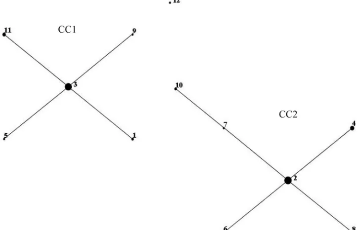
Neighbor-joining and maximum-likelihood trees constructed
from concatenated sequences of the eight loci for the 57
M.
pneu-moniae
isolates (Fig. 1) illustrated two genetically distinct clusters,
which were confirmed by eBURST examination of relatedness
(Fig. 2). The two clusters, designated clonal complex 1 (CC1) and
CC2, contained ST1, ST3, ST5, ST9, and ST11, and ST2, ST4, ST6,
ST7, ST8, and ST10, respectively. ST12 located distal to the two
main clusters; however, phylogenetic analysis revealed closer
po-sitioning to CC1. Neighbor-joining and maximum-likelihood
trees were constructed for the eight loci individually (data not
shown), and the topology of both neighbor-joining and
maxi-mum-likelihood trees was consistent for all loci and concatenated
sequences.
Five homogenous strains (
M. pneumoniae
MPN13 to MPN17)
originating from nose and throat swabs from the same patient
with Stevens-Johnson syndrome had identical STs (ST3).
Addi-tionally, two clinical isolates (
M. pneumoniae
MPN104 and
MPN106) originating from separate sputum samples from a
pa-tient with bronchopneumonia taken 4 days apart also had
identi-cal STs (ST4). This indicates a single clonal population responsible
for infection in these cases.
The possibility of synonymous sequence changes (indicating a
pressure to conserve amino acid sequence and protein structure)
was investigated by comparing the predicted translated sequences
for each locus. Analysis of the deduced amino acid sequences of
the eight loci for the 57 strains indicated that both synonymous
and nonsynonymous SNPs occurred, of which approximately
44% resulted in an amino acid change. Nonsynonymous SNPs are
highlighted in Fig. SA2 in the supplemental material. The amino
acid sequences for ArcC, Gmk, and GyrB yielded homologous
sequences for all ATs, numbering at two ATs for each locus. In
comparison, Pgm analysis resulted in the largest number of
non-TABLE 2MLST loci used in established bacterial MLST schemes also present inM. pneumoniaeBacterial species
Presence of MLST locusa
recA ppa pgm gyrB gmk glyA efp atpA arcC adk
Bacillus cereus ✓
Chlamydia trachomatis ✓
Campylobacter jejuni ✓ ✓
Escherichia coli ✓ ✓ ✓
Enterococcus faecium ✓ ✓
Haemophilus influenzae ✓ ✓
Helicobacter pylori ✓ ✓ ✓
Moraxella catarrhalis ✓ ✓ ✓
Neisseria meningitidis ✓ ✓
Staphylococcus aureus ✓ ✓
Staphylococcus epidermidis ✓
Streptococcus suis ✓
Vibrio vulnificus ✓
Yersinia pseudotuberculosis ✓
[image:4.585.39.545.78.249.2]aMLST loci were chosen based on the frequency of use in other bacterial MLST schemes (http://www.mlst.net/) and the presence of the gene in the published M129 and FH whole genomes.
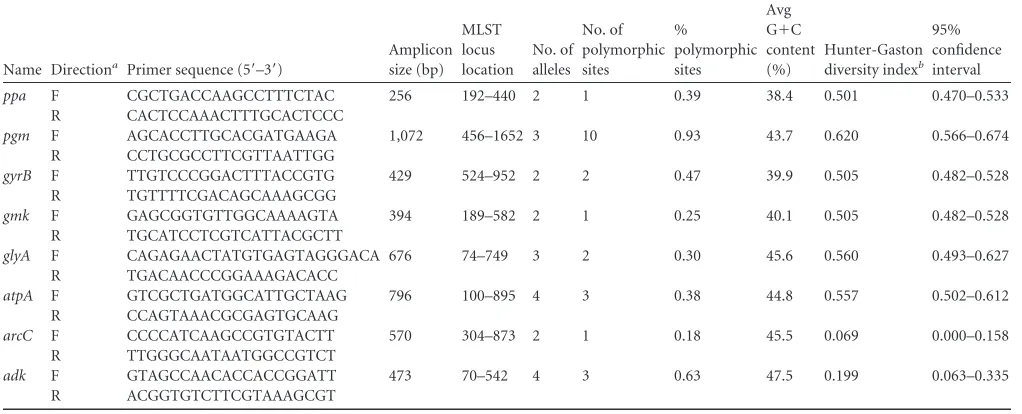
TABLE 3Primer pairs developed in this study and variability of the different loci
Name Directiona Primer sequence (5=–3=)
Amplicon size (bp)
MLST locus location
No. of alleles
No. of polymorphic sites
%
polymorphic sites
Avg G⫹C content (%)
Hunter-Gaston diversity indexb
95% confidence interval
ppa F CGCTGACCAAGCCTTTCTAC 256 192–440 2 1 0.39 38.4 0.501 0.470–0.533
R CACTCCAAACTTTGCACTCCC
pgm F AGCACCTTGCACGATGAAGA 1,072 456–1652 3 10 0.93 43.7 0.620 0.566–0.674 R CCTGCGCCTTCGTTAATTGG
gyrB F TTGTCCCGGACTTTACCGTG 429 524–952 2 2 0.47 39.9 0.505 0.482–0.528
R TGTTTTCGACAGCAAAGCGG
gmk F GAGCGGTGTTGGCAAAAGTA 394 189–582 2 1 0.25 40.1 0.505 0.482–0.528
R TGCATCCTCGTCATTACGCTT
glyA F CAGAGAACTATGTGAGTAGGGACA 676 74–749 3 2 0.30 45.6 0.560 0.493–0.627 R TGACAACCCGGAAAGACACC
atpA F GTCGCTGATGGCATTGCTAAG 796 100–895 4 3 0.38 44.8 0.557 0.502–0.612
R CCAGTAAACGCGAGTGCAAG
arcC F CCCCATCAAGCCGTGTACTT 570 304–873 2 1 0.18 45.5 0.069 0.000–0.158
R TTGGGCAATAATGGCCGTCT
adk F GTAGCCAACACCACCGGATT 473 70–542 4 3 0.63 47.5 0.199 0.063–0.335
R ACGGTGTCTTCGTAAAGCGT
a
F, forward; R, reverse.
bHunter-Gaston diversity index (DI) ranges from 0.0 for no diversity to 1.0 for complete diversity.
on May 16, 2020 by guest
http://jcm.asm.org/
[image:4.585.39.550.501.708.2]synonymous changes in the amino acid sequence, with four
changes in the sequence between three ATs.
The MLST scheme was applied to the published complete
ge-nome sequences of
M. pneumoniae
available from NCBI for
strains 309 (GenBank accession no.
NC_016807.1), M129-B7
(GenBank accession no.
CP003913.2), and M29 (GenBank
acces-sion no.
NZ_CP008895.1) and to assembled contigs available
from NCBI for strains PO1 (GCA_000319655.1), PI 1428 (GCA_
000319675.1), and 19294 (GCA_000387745.1). These strains were
determined to belong to ST2, ST1, ST3, ST2, ST1, and ST7,
respec-tively.
The stability of each MLST locus was assessed in 10
M.
pneu-moniae
isolates. Isolates were retyped following short-term
pas-sage (10 sequential subculture paspas-sages) in liquid medium. All loci
were found to be completely stable, with no SNPs in comparison
to the original isolate.
FIG 1Phylogenetic trees were constructed based on concatenated sequences of eight housekeeping loci for 12 unique STs using maximum-likelihood (A) and neighbor-joining (B) methods. Bootstrap support values of⬎70% are shown. STs are indicated by differential shading.
FIG 2eBURST version 3 was used to analyze the 12 unique STs resolved for all 57M. pneumoniaeisolates. Two main clonal complexes (CC) were defined. The size of each dot is proportional to the number of isolates included in the analysis for each ST.
on May 16, 2020 by guest
http://jcm.asm.org/
[image:5.585.95.491.67.307.2] [image:5.585.112.475.464.697.2]Genomic sequence analysis.
Three regions of SNPs were
pre-dicted to have arisen by homologous recombination in the
chro-mosomes of the 35 clinical
M. pneumoniae
isolates (Fig. 3), one of
which distinguished genomic clade 1 (GC1) from GC2, and the
other two occurred within GC1. Area 1 was predicted to occur in
all strains in GC1, area 2 in 10 strains, and a single strain, MPN113,
had a single additional predicted area of recombination, area 3.
Following removal of the predicted areas of recombination, two
distinct genetic clades were identified, GC1 and GC2 (Fig. 3).
Ex-cellent parity was found using this method and concatenated
MLST sequences, with all strains colocating to the corresponding
CC and GC.
Comparison to other typing methods.
There was no obvious
link between the ST and the year when the strains were collected,
the patient’s age, and the sample origin; however, limited numbers
of strains were available per year, and for some years, there were
no strains. Indeed, multiple STs can be observed in a single year.
Furthermore, ST was related to P1 type (Table 1), with the two
most common STs, ST2 and ST3, containing strains that were P1
type 2 and P1 type 1, respectively. Additionally, this MLST scheme
was also comparable to MLVA typing. The two major clusters
observed, CC1 and CC2, were directly linked to MLVA type: CC1
contained MLVA type 4572, whereas CC2 contained MLVA types
3662 and 3562. Each ST contained only one MLVA type, with the
exception of ST2, which contained both 3662 and 3562, and ST11,
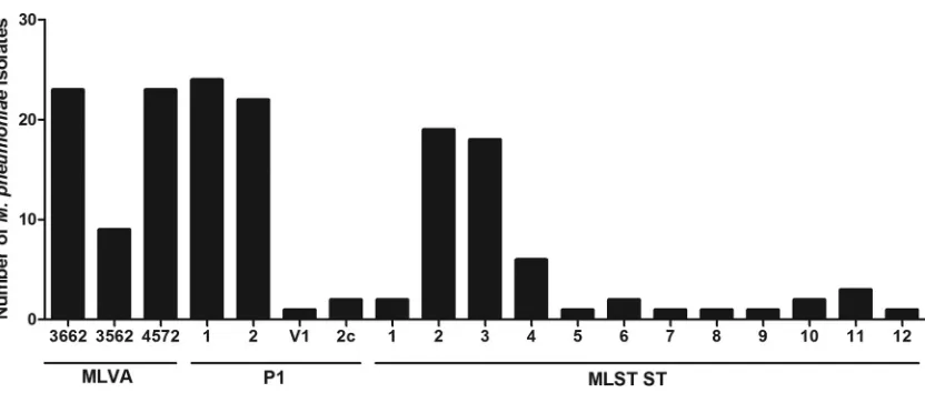
which contained 4572, 3662, and 3562 (Table 1). Distributions of
MLVA type, P1 type, and ST can be observed in
Fig. 4, indicating
that P1 type 1, MLST ST2, and MLVA types 3662 and 4572
oc-curred most frequently in the isolates tested.
In the isolates tested in this scheme, MLST was deemed to be
more discriminatory than both MLVA typing and P1 typing,
re-sulting in 0.21, 0.05, and 0.07 types per isolate, respectively. This
was confirmed by examination of the Hunter-Gaston DI, which
indicated higher discriminatory ability for the MLST scheme (DI,
0.784; 95% CI, 0.716 to 0.852) than that of the current MLVA
scheme (DI, 0.633; 95% CI, 0.583 to 0.683) and P1 typing (DI,
0.567; 95% CI, 0.510 to 0.625).
Online database.
An
M. pneumoniae
MLST online database
was created for both MLST alleles, profile definitions, and isolate
data (29) (http://pubmlst.org/mpneumoniae).
0 200000 400000 600000 800000
210
M129 MPN135 MPN113 MPN111 MPN108 MPN118 MPN103 MPN120 MPN136 MPN130 MPN116 MPN105 MPN109 MPN122 MPN101 MPN106 MPN124 MPN110 MPN131 MPN104 MPN126 MPN128 MPN102 MPN129 MPN134 MPN133 MPN132 MPN121 MPN117 MPN107 MPN125 MPN119 MPN112 MPN114 MPN123 MPN127
GC1
GC2
FIG 3Prediction of recombination in theM. pneumoniaeisolate chromosomes. Regions of variation in the genomes of the 35 clinicalM. pneumoniae isolates and the type strain M129, which are predicted to have arisen by homologous recombination, are shown on the right. Red blocks indicate recombination predicted to have occurred on internal nodes, and blue indicates taxon-specific recombination. Isolates are ordered according to the phylogenetic tree displayed on the left. The track along the top of the figure displays the M129 chromosome and annotation, in which protein-coding sequences (CDS) are indicated in light blue.
on May 16, 2020 by guest
http://jcm.asm.org/
[image:6.585.39.560.64.417.2]DISCUSSION
MLST has been used to genotype several species of bacteria,
in-cluding several
Mycoplasma
species:
Mycoplasma agalactiae
,
My-coplasma bovis
, and
Mycoplasma hyorhinis
(30–32). This study has
described the successful development of a novel
M. pneumoniae
MLST scheme to allow the characterization of clinical isolates.
This scheme was successfully used to discriminate 55 clinical
iso-lates of
M. pneumoniae
from British patients (with the exception
of two U.S. isolates) within the reference laboratory collection,
from respiratory and extrapulmonary sites, and the two type
strains M129 and FH. Eight housekeeping genes were identified as
suitable targets for the scheme, and these were used to genotype
M. pneumoniae
isolates by either PCR followed by sequencing or
whole-genome sequence analysis.
gyrB
contains a quinolone
resis-tance-determining region (QRDR) with documented
in vitro
mu-tations at amino acid positions 443, 464, and 483. The clinical use
of quinolones may increase selective pressure
in vivo
, resulting in a
high mutation rate (33). However, the
gyrB
locus sequence
ampli-fied in this MLST scheme is in a different region of the gene from
the QRDR and is therefore considered a suitable MLST target. The
stability of the eight loci was evaluated
in vitro
and was confirmed
before and after 10 repeated passages of 10 strains in liquid
me-dium. However, stability over a larger number of passages in
liq-uid medium and an evaluation of stability using
in vitro
tissue
culture were not assessed.
The discriminatory power of this MLST scheme with the eight
loci was 0.784 for the collection of 57 isolates. In comparison, the
Hunter-Gaston DI of the P1 typing method for the 57 isolates was
0.567, and the DI of the MLVA scheme was 0.633; therefore, this
MLST scheme was more discriminatory for the isolates tested.
However, it has been shown that the established MLVA method is
more discriminatory than P1 typing (16), which was confirmed in
this study. The allelic diversity of each of the MLST loci varied
significantly at each locus, with the
pgm
,
glyA
,
atpA
,
gyrB
,
gmk
, and
ppa
loci being more discriminatory than the
adk
and
arcC
loci.
The association of this set of markers with varied Hunter-Gaston
DIs makes this MLST, in theory, more optimal for
epidemiologi-cal studies than other existing methodologies.
Analysis of
M. pneumoniae
infection at the individual patient
level was possible using this scheme. Multiple clinical isolates were
available from two of 50 patients: five were from a patient with
Stevens-Johnson syndrome (MPN013 to MPN017), and two were
from a patient with bronchopneumonia, taken 4 days apart. In
both cases, the ST, MLVA type, and P1 type remained the same,
indicating that a single clonal isolate was responsible for infection.
Recurring infection or reinfection of
M. pneumoniae
can be
deter-mined using this scheme. Recurring infection would have the
same ST as the original infection, whereas reinfection with
M.
pneumoniae
would likely be a different ST. Genetic MLST
insta-bility in isolates might occur; however, in this study, this was not
seen over 10 passages.
The eBURST analysis illustrates the relationship of STs on the
basis of the number of MLST loci that differ between two STs.
Analysis of this population modeling indicates that the two
clus-ters, CC1 and CC2, differed by more than one locus, but within
each cluster, the STs did not differ by more than one locus. Within
a cluster, this highlights the homogenous nature of the
M.
pneu-moniae
species; however, a definitive split can be observed
be-tween the two clusters in both ST and MLVA type. A possible
divergent clade with ST12 from CC1 is also apparent; however,
more isolates need to be typed by this method to confirm this
observation. Few typing methods have been able to detect
signif-icant differences between strains, including one previous attempt
to subtype
M. pneumoniae
by MLST with housekeeping and
struc-tural genes (12,
15,
22). The previous MLST was determined to be
not sufficiently discriminatory to be used for epidemiological
pur-poses. However, the MLST scheme developed in this study was
able to discriminate between
M. pneumoniae
isolates and resulted
in two genetically distinct clusters, indicating significant
differ-ences between strains.
A comparison between genomic sequence analysis after the
removal of predicted areas of recombination and phylogenetic
analysis of concatenated MLST sequences showed similar
topol-ogy and the same distinct genetic clustering. This indicates that
this MLST scheme is representative of the genome and confirms
that
M. pneumoniae
can be subdivided into two distinct genetic
lineages.
Typing of clinical
M. pneumoniae
isolates is becoming
increas-ingly important due to the global increase in
M. pneumoniae
in-fections and the increase in macrolide-resistant strains (34,
35). This scheme provides a more discriminatory method than
both the MLVA and P1 typing methods currently in use,
allow-FIG 4Distribution of MLVA, P1 type, and ST for 57M. pneumoniaeisolates (each group defined by lines below).on May 16, 2020 by guest
http://jcm.asm.org/
[image:7.585.85.501.62.239.2]ing further and more detailed analysis of observed epidemic
peaks of
M. pneumoniae
infection. Community outbreaks of
pneumonia caused by
M. pneumoniae
have been described
world-wide (36–38), and it would be interesting to evaluate this MLST
scheme in such epidemic situations. The level of discrimination of
this typing method and its usefulness in epidemic analysis should
be confirmed by comparing outbreak-related strains to a set of
control strains that were isolated from a similar time period and
geographical area but that are not epidemiologically related. More
severe or adverse infections with
M. pneumoniae
are seen in some
patients. The reason for this is not clear; however, it can be
postu-lated that this is due to specific microbe pathogenicity (identified
through genetic markers) or variance in host susceptibility. This
method could assist in determining if this is a strain-specific
phe-nomenon. One advantage of MLST is that it is PCR based and does
not require the growth of bacteria, which can be a lengthy process
for
M. pneumoniae
. However, there is a large amount of
sequenc-ing required for this method, which can be laborious and
expen-sive; therefore, its adaptation for widespread use directly on
clin-ical specimens would be beneficial.
In conclusion, this study presents a robust MLST scheme that
has proven discriminatory for
M. pneumoniae
, providing isolate
characterization and a higher level of discrimination than MLVA
and P1 typing methods. In addition, phylogenetic analysis of both
STs and whole-genome sequence data revealed two genetically
distinct clusters. Crucially, this scheme for
M. pneumoniae
is also
supported by a public Web-based database (http://pubmlst.org
/mpneumoniae).
ACKNOWLEDGMENTS
This work was funded by Public Health England. These studies were sup-ported by funding initiatives by the National Institute for Social Care and Health Research (NISCHR) (research support from the Welsh Govern-ment) via the registered research group Microbial and Infection Transla-tional Research Group (MITReG) and Children and Young Persons Re-search Network (CYPRN).
We declare no conflicts of interest.
REFERENCES
1.Waites K, Talkington D.2004.Mycoplasma pneumoniaeand its role as a human pathogen. Clin Microbiol Rev17:697–728.http://dx.doi.org/10
.1128/CMR.17.4.697-728.2004.
2.Meyer Sauteur P, Jacobs B, Spuesens E, Jacobs E, Nadal D, Vink C, van Rossum AMC.2014. Antibody responses toMycoplasma pneumoniae: role in pathogenesis and diagnosis of encephalitis? PLoS Pathog10:
e1003983.http://dx.doi.org/10.1371/journal.ppat.1003983.
3.Polkowska A, Harjunpää A, Toikkanen S, Lappalainen M, Vuento R, Vuorinen T, Kauppinen J, Flinck H, Lyytikäinen O.2012. Increased incidence ofMycoplasma pneumoniaeinfection in Finland, 2010 –2011. Euro Surveill 17:pii⫽20072. http://www.eurosurveillance.org/View
Article.aspx?ArticleId⫽20074.
4.Chalker VJ, Stocki T, Mentasti M, Fleming D, Sadler C, Ellis J, Ber-mingham A, Harrison TG.2011.Mycoplasma pneumoniaeinfection in primary care investigated by real-time PCR in England and Wales. Eur J Clin Microbiol Infect Dis30:915–921.http://dx.doi.org/10.1007/s10096
-011-1176-3.
5.Howard LS, Sillis M, Pasteur MC, Kamath AV, Harrison BD. 2005. Microbiological profile of community-acquired pneumonia in adults over the last 20 years. J Infect50:107–113.http://dx.doi.org/10.1016/j.jinf.2004 .05.003.
6.Chalker V, Stocki T, Litt D, Bermingham A, Watson J, Fleming D, Harrison T.2012. Increased detection ofMycoplasma pneumoniae in-fection in children in England and Wales, October 2011 to January 2012. Euro Surveill 17:pii⫽20081. http://www.eurosurveillance.org
/ViewArticle.aspx?ArticleId⫽20081.
7.Jacobs E.2012.Mycoplasma pneumoniae: now in the focus of clini-cians and epidemiologists. Euro Surveill 17:pii⫽20084. http://www
.eurosurveillance.org/ViewArticle.aspx?ArticleId⫽20084.
8.Liu J, Ai H, Xiong Y, Li F, Wen Z, Liu W, Li T, Qin K, Wu J, Liu Y.
2015. Prevalence and correlation of infectious agents in hospitalized chil-dren with acute respiratory tract infections in central China. PLoS One
10:e0119170.http://dx.doi.org/10.1371/journal.pone.0119170. 9.Rastawicki W, Kaluzewski S, Jagielski M, Gierczyski R.1998.
Epidemi-ology ofMycoplasma pneumoniaeinfections in Poland: 28 years of surveil-lance in Warsaw 1970 –1997. Euro Surveill 3:99 –100. http://www
.eurosurveillance.org/ViewArticle.aspx?ArticleId⫽95.
10.Tjhie JH, Dorigo-Zetsma JW, Roosendaal R, Van Den Brule AJ, Bestebroer TM, Bartelds AI, Vandenbroucke-Grauls CM.2000. Chla-mydia pneumoniaeandMycoplasma pneumoniaein children with acute respiratory infection in general practices in The Netherlands. Scand J In-fect Dis32:13–17.http://dx.doi.org/10.1080/00365540050164155. 11. Cousin-Allery A, Charron A, de Barbeyrac B, Fremy G, Skov Jensen
J, Renaudin H, Bebear C. 2000. Molecular typing of Mycoplasma pneumoniaestrains by PCR-based methods and pulsed-field gel elec-trophoresis. Application to French and Danish isolates. Epidemiol In-fect124:103–111.
12. Dorigo-Zetsma JW, Dankert J, Zaat SAJ.2000. Genotyping of Myco-plasma pneumoniaeclinical isolates reveals eight P1 subtypes within two genomic groups. J Clin Microbiol38:965–970.
13. Sasaki T, Kenri T, Okazaki N, Iseki M, Yamashita R, Shintani M, Sasaki Y, Yayoshi M.1996. Epidemiological study ofMycoplasma pneumoniae infections in Japan based on PCR-restriction fragment length polymor-phism of the P1 cytadhesin gene. J Clin Microbiol34:447– 449. 14. Dumke R, Von Baum H, Luck PC, Jacobs E.2010. Subtypes and variants
ofMycoplasma pneumoniae: local and temporal changes in Germany 2003–2006 and absence of a correlation between the genotype in the re-spiratory tract and the occurrence of genotype-specific antibodies in the sera of infected patients. Epidemiol Infect138:1829 –1837.http://dx.doi
.org/10.1017/S0950268810000622.
15. Dumke R, Lück PC, Noppen C, Schaefer C, von Baum H, Marre R, Jacobs E.2006. Culture-independent molecular subtyping ofMycoplasma pneumoniaein clinical samples. J Clin Microbiol44:2567–2570.http://dx
.doi.org/10.1128/JCM.00495-06.
16. Dégrange S, Cazanave C, Charron A, Renaudin H, Bebear C, Bebear CM.2009. Development of multiple-locus variable-number tandem-repeat analysis for molecular typing ofMycoplasma pneumoniae. J Clin Microbiol47:914 –923.http://dx.doi.org/10.1128/JCM.01935-08. 17. Dumke R, Jacobs E.2011. Culture-independent multi-locus
variable-number tandem-repeat analysis (MLVA) ofMycoplasma pneumoniae. J Microbiol Methods86:393–396.http://dx.doi.org/10.1016/j.mimet.2011 .06.008.
18. Zhao F, Liu G, Cao B, Wu J, Gu Y, He L, Meng F, Zhu L, Yin Y, Lv M, Zhang J. 2013. Multiple-locus variable-number tandem-repeat analysis of 201Mycoplasma pneumoniaeisolates from Beijing, China, from 2008 to 2011. J Clin Microbiol51:636 – 639.http://dx.doi.org/10
.1128/JCM.02567-12.
19. Chalker VJ, Pereyre S, Dumke R, Winchell J, Khosla P, Sun H, Yan C, Vink C, Bébéar C, ESCMID Study Group forMycoplasmaInfections.
2015. InternationalMycoplasma pneumoniaetyping study: the interpreta-tion ofMycoplasma pneumoniaemultilocus variable-number tandem-repeat analysis. New Microbes New Infect7:37– 40.http://dx.doi.org/10
.1016/j.nmni.2015.05.005.
20. Bolger AM, Lohse M, Usadel B.2014. Trimmomatic: a flexible trimmer for Illumina sequence data. Bioinformatics30:2114 –2120.http://dx.doi
.org/10.1093/bioinformatics/btu170.
21. Bankevich A, Nurk S, Antipov D, Gurevich AA, Dvorkin M, Kulikov AS, Lesin VM, Nikolenko SI, Pham S, Prjibelski AD, Pyshkin AV, Sirotkin AV, Vyahhi N, Tesler G, Alekseyev MA, Pevzner PA.2012. SPAdes: a new genome assembly algorithm and its applications to single-cell sequencing. J Comput Biol 19:455– 477.http://dx.doi.org/10.1089
/cmb.2012.0021.
22. Dumke R, Catrein I, Pirkil E, Herrmann R, Jacobs E.2003. Subtyping ofMycoplasma pneumoniaeisolates based on extended genome sequenc-ing and on expression profiles. Int J Med Microbiol292:513–525.http:
//dx.doi.org/10.1078/1438-4221-00231.
23. Pitcher D, Chalker VJ, Sheppard C, George RC, Harrison TG.2006. Real-time detection ofMycoplasma pneumoniaein respiratory samples
on May 16, 2020 by guest
http://jcm.asm.org/
with an internal processing control. J Med Microbiol55:149 –155.http:
//dx.doi.org/10.1099/jmm.0.46281-0.
24. Saitou N, Nei M.1987. The neighbor-joining method: a new method for reconstructing phylogenetic trees. Mol Biol Evol4:406 – 425.
25. Kimura M.1980. A simple method for estimating evolutionary rates of base substitutions through comparative studies of nucleotide sequences. J Mol Evol16:111–120.http://dx.doi.org/10.1007/BF01731581.
26. Jukes T, Cantor C.1969. Evolution of protein molecules, p 21–132.In Munro H (ed), Mammalian protein metabolism. Academic Press, New York, NY.
27. Nei M, Kumar S.2000. Molecular evolution and phylogenetics. Oxford University Press, New York, NY.
28. Felsenstein J.1985. Confidence limits on phylogenies: An approach using the bootstrap. Evolution39:783–791.http://dx.doi.org/10.2307/2408678. 29. Jolley KA, Maiden MC.2010. BIGSdb: scalable analysis of bacterial ge-nome variation at the population level. BMC Bioinformatics11:595.http:
//dx.doi.org/10.1186/1471-2105-11-595.
30. Manso-Silván L, Dupuy V, Lysnyansky I, Ozdemir U, Thiaucourt F.
2012. Phylogeny and molecular typing ofMycoplasma agalactiaeand My-coplasma bovisby multilocus sequencing. Vet Microbiol161:104 –112.
http://dx.doi.org/10.1016/j.vetmic.2012.07.015.
31. Tocqueville V, Ferre S, Nguyen NH, Kempf I, Marois-Crehan C.2014. Multilocus sequence typing ofMycoplasma hyorhinisstrains identified by a real-time TaqMan PCR assay. J Clin Microbiol52:1664 –1671.http://dx
.doi.org/10.1128/JCM.03437-13.
32. Rosales RS, Churchward CP, Schnee C, Sachse K, Lysnyansky I, Catania S, Iob L, Ayling RD, Nicholas RA.2015. Global multilocus sequence typing analysis ofMycoplasma bovisisolates reveals two main population clusters. J Clin Microbiol 53:789 –794. http://dx.doi.org/10.1128/JCM
.01910-14.
33. Gruson D, Pereyre S, Renaudin H, Charron A, Bébéar C, Bébéar CM.
2005.In vitrodevelopment of resistance to six and four fluoroquinolones inMycoplasma pneumoniaeandMycoplasma hominis, respectively.
Anti-microb Agents Chemother 49:1190 –1193. http://dx.doi.org/10.1128
/AAC.49.3.1190-1193.2005.
34. Diaz MH, Benitez AJ, Winchell JM.2015. Investigations ofMycoplasma pneumoniaeinfections in the United States: trends in molecular typing and macrolide resistance from 2006 to 2013. J Clin Microbiol53:124 –130.
http://dx.doi.org/10.1128/JCM.02597-14.
35. Zhou Z, Li X, Chen X, Luo F, Pan C, Zheng X, Tan F.2015. Macrolide-resistantMycoplasma pneumoniaein adults in Zhejiang, China. Antimi-crob Agents Chemother59:1048 –1051.http://dx.doi.org/10.1128/AAC
.04308-14.
36. Chen FQ, Yang YZ, Yu LL, Bi CB.2015. Prevalence ofMycoplasma pneumoniae: a cause for community-acquired infection among pediatric populaztion. Niger J Clin Pract18:354 –358.http://dx.doi.org/10.4103
/1119-3077.153247.
37. Klement E, Talkington DF, Wasserzug O, Kayouf R, Davidovitch N, Dumke R, Bar-Zeev Y, Ron M, Boxman J, Lanier Thacker W, Wolf D, Lazarovich T, Shemer-Avni Y, Glikman D, Jacobs E, Grotto I, Block C, Nir-Paz R.2006. Identification of risk factors for infection in an outbreak ofMycoplasma pneumoniaerespiratory tract disease. Clin Infect Dis43:
1239 –1245.http://dx.doi.org/10.1086/508458.
38. Meyer Sauteur PM, Bleisch B, Voit A, Maurer FP, Relly C, Berger C, Nadal D, Bloemberg GV. 2014. Survey of macrolide-resistant Myco-plasma pneumoniaein children with community-acquired pneumonia in Switzerland. Swiss Med Wkly144:w14041.
39. Hsu LY, Harris S, Chlebowicz M, Lindsay J, Koh TH, Kristnan P, Tan TY, Hon PY, Grubb W, Bentley S, Parkhill J, Peacock S, Holden MTG.
2015. Evolutionary dynamics of methicillin-resistantStaphylococcus au-reuswithin a healthcare system. Genome Biol16:81.http://dx.doi.org/10
.1186/s13059-015-0643-z.
40. Croucher NJ, Page AJ, Connor TR, Delaney AJ, Keane JA, Bentley SD, Parkhill J, Harris SR.2015. Rapid phylogenetic analysis of large samples of recombinant bacterial whole genome sequences using Gubbins. Nu-cleic Acids Res43:e15.http://dx.doi.org/10.1093/nar/gku1196.