Function of Testing Method
Dror Marchaim,a,b,c*Jason M. Pogue,dOran Tzuman,bKayoko Hayakawa,aPaul R. Lephart,eHossein Salimnia,eTheresa Painter,e Marcus J. Zervos,fLaura E. Johnson,fMary Beth Perri,fPamela Hartman,fRama V. Thyagarajan,gSharon Major,hMelanie Goodell,h Mohamad G. Fakih,iLaraine L. Washer,jDuane W. Newton,kAnurag N. Malani,lJason M. Wholehan,lLona Mody,mKeith S. Kayea
Division of Infectious Diseases,a
Department of Pharmacy Services,d
and Department of University Laboratories,e
Detroit Medical Center, Wayne State University, Detroit, Michigan, USA; Division of Infectious Diseases, Assaf Harofeh Medical Center, Zerifin, Israelb
; Sackler School of Medicine, Tel Aviv University, Tel Aviv, Israelc
; Division of Infectious Diseases, Henry Ford Health System, Wayne State University, Detroit, Michigan, USAf
; Division of Infectious Diseasesg
and Department of Clinical Microbiology,h Oakwood Healthcare System, Dearborn, Michigan, USA; Department of Infection Prevention and Control, St. John Hospital and Medical Center, Wayne State University, Detroit, Michigan, USAi
; Division of Infectious Diseasesj
and Clinical Microbiology,k
University of Michigan Health System, University of Michigan, Ann Arbor, Michigan, USA; Division of Infectious Diseases, Saint Joseph Mercy Health System, Ann Arbor, Michigan, USAl
; and Division of Geriatric and Palliative Care Medicine, VA Ann Arbor Healthcare Systems, University of Michigan, Ann Arbor, Michigan, USAm
Tigecycline is one of the few remaining therapeutic options for extensively drug-resistant (XDR) Gram-negative bacilli (GNB).
MICs of tigecycline to
Acinetobacter baumannii
have been reported to be elevated when determined by the Etest compared to
determinations by the broth microdilution (BMD) method. The study aim was to compare the susceptibility of GNB to
tigecy-cline by four different testing methods. GNB were collected from six health care systems (25 hospitals) in southeast Michigan
from January 2010 to September 2011. Tigecycline MICs among
A. baumannii, carbapenem-resistant
Enterobacteriaceae
(CRE),
extended-spectrum
-lactamase (ESBL)-producing
Enterobacteriaceae, and susceptible
Enterobacteriaceae
isolates were
deter-mined by Etest, BMD, Vitek-2, and MicroScan. Nonsusceptibility was categorized as a tigecycline MIC of
>
4
g/ml for both
A.
baumannii
and
Enterobacteriaceae. The study included 4,427 isolates: 2,065 ESBL-producing
Enterobacteriaceae, 1,105
A.
bau-mannii, 888 susceptible
Enterobacteriaceae, and 369 CRE isolates. Tigecycline nonsusceptibility among
A. baumannii
isolates
was significantly more common as determined by Etest compared to that determined by BMD (odds ratio [OR], 10.3;
P
<
0.001),
MicroScan (OR, 12.4;
P
<
0.001), or Vitek-2 (OR, 9.4;
P
<
0.001). These differences were not evident with the other pathogens.
Tigecycline MICs varied greatly according to the
in vitro
testing methods among
A. baumannii
isolates. Etest should probably
not be used by laboratories for tigecycline MIC testing of
A. baumannii
isolates, since MICs are significantly elevated with Etest
compared to those determined by the three other methods.
I
nfections due to multidrug-resistant (MDR) and extensively
drug-resistant (XDR) Gram-negative bacillus (GNB)
patho-gens, such as extended-spectrum

-lactamase (ESBL)-producing
Enterobacteriaceae
,
Acinetobacter baumannii
, and
carbapenem-re-sistant
Enterobacteriaceae
(CRE), continue to increase worldwide
and pose significant management challenges due to the lack of
available therapeutic alternatives (
1–4
). These infections are
asso-ciated with devastating outcomes, and they place a huge burden
on and threaten the general population (
1
,
2
,
5–8
). Tigecycline is
one of the few available agents that possess antimicrobial activity
against these resistant pathogens (
9–14
). Tigecycline has a
rela-tively safe therapeutic profile compared to that of other agents that
often have activity against these pathogens (e.g., polymyxins and
aminoglycosides) (
3
,
15
). However, tigecycline has limitations as a
therapeutic agent. It achieves low serum levels, and data
pertain-ing to the effective treatment of invasive infections with
tigecy-cline are lacking (
1–3
,
13
). Recent meta-analyses and reports from
the U.S. Food and Drug Administration have detailed an
in-creased mortality risk among subjects receiving tigecycline in
clin-ical trials (
13
,
14
,
16
). Due to the lack of options, tigecycline is still
frequently used for the treatment of clinical infections due to
A.
baumannii
and CRE, often with similar and, in some instances,
favorable clinical efficacy compared to that of other available
ther-apeutic options (
3
).
In the past years, several
in vitro
studies reported a worrisome
trend of elevated MICs to tigecycline among
A. baumannii
(
17
),
ESBL producers (
18
), and CRE (
19
). For
A. baumannii
, elevated
resistance was reported even in locales where the drug had not yet
been introduced into clinical use (
17
). The MIC determinations in
these reports were obtained by using Etest strips for
in vitro
sus-ceptibility testing (
17
). When
in vitro
analyses compared MICs
obtained using the Etest with those obtained using the broth
mi-crodilution (BMD) method, major discordances were reported.
Pillar and colleagues (
20
) reported comparisons between Etest
and BMD for
in vitro
susceptibility testing results among 227 U.S.
A. baumannii
isolates and 902
Klebsiella pneumoniae
and
Esche-richia coli
isolates. The differences in results obtained by the
dif-ferent methods were mostly evident among the
A. baumannii
iso-lates. Results obtained via Etest resulted in a
ⱖ
4-fold increase in
MIC in 29% of the isolates. Among the 43% of
A. baumannii
Received6 January 2014 Returned for modification28 January 2014
Accepted22 February 2014
Published ahead of print5 March 2014
Editor:D. J. Diekema
Address correspondence to Dror Marchaim, [email protected].
* Present address: Dror Marchaim, Division of Infectious Diseases, Assaf Harofeh Medical Center, Zerifin, Israel.
Copyright © 2014, American Society for Microbiology. All Rights Reserved.
doi:10.1128/JCM.00001-14
on May 16, 2020 by guest
http://jcm.asm.org/
strains considered resistant by Etest, none had an MIC in the
re-sistant range when tested by BMD (
20
). In a study by Casal and
colleagues (
21
), the MICs for
A. baumannii
isolates (
n
⫽
148)
determined by Etest were completely discordant compared to
those determined by BMD. Among the isolates with an MIC of
ⱖ
2
mg/liter determined by Etest, values were at least 2 dilutions lower
when determined by BMD. It was hypothesized that high
concen-trations of manganese in Mueller-Hinton agar plates might have
been responsible for the increased MICs of tigecycline to
A.
bau-mannii
isolates determined by Etest (
22
).
It is important that MICs for the “last-line” agents such as
tigecycline be accurately determined. The issues pertaining to
ac-curate susceptibility testing for tigecycline are especially urgent
because many institutions use Etest to determine tigecycline
MICs, as not all automated susceptibility-testing methods have
tigecycline on their panels/cards. We therefore designed this
mul-ticenter study in order to compare the MIC results determined by
four different susceptibility-testing methodologies for tigecycline
to
A. baumannii
isolates, as well as to a variety of other GNB.
MATERIALS AND METHODS
A multicenter prospective investigation was conducted in southeastern Michigan between 1 January 2010 and 31 August 2011. Six health care systems participated, i.e., Detroit Medical Center (DMC), Henry Ford hospitals network, St. Joseph Mercy Hospital, St. John Providence Health System, University of Michigan Health System, and Oakwood Healthcare institutions. A total of 25 hospitals within these health systems partici-pated. The Department of Infection Prevention, Epidemiology, and An-timicrobial Stewardship at the DMC served as the central coordinating unit for all data and organisms, storage, testing, and analyses. The study was approved separately in each health care system by the local institu-tional review boards, and the data were deidentified prior to transfer to the coordinating unit.
Unique clinical isolates ofA. baumannii,K. pneumoniae, andE. coli (both ESBL and non-ESBL producing) and of CRE (K. pneumoniae,E. coli, andEnterobacterspecies) were prospectively collected. CRE were de-fined as any of the aforementioned bacteria that were resistant toⱖ1 carbapenem or were carbapenemase producers (determined by the mod-ified Hodge test, conducted in accordance with the 2009 Clinical and Laboratory Standards Institute [CLSI] criteria [23]). ESBL production was determined by the automated system and confirmed with the double disk diffusion test (23). The centers were asked to collect all resistant isolates (i.e., A. baumannii, ESBL-producing Enterobacteriaceae, and CRE) but were limited to 150 susceptible isolates per health system (col-lected randomly). For the purposes of this analysis, susceptible Enterobac-teriaceaewere those that were susceptible to third-generation cephalospo-rins and lacked detectable ESBL production. Isolates were tested for susceptibility to tigecycline by four different methods: MicroScan (Sie-mens, Germany), Etest (bioMérieux, France), Vitek-2 (bioMérieux, France), and BMD. All susceptibility testing was conducted at the DMC, except for Vitek-2 testing, which was conducted at the Henry Ford Med-ical Center. All Etest analyses were performed using the BBL brand of Mueller-Hinton agar previously determined to have a lower manganese content than that of other brands (22). All susceptibility testing and inter-pretations were conducted according to the 2009 CLSI criteria and rec-ommendations (23). There are no CLSIEnterobacteriaceaeMIC break-points for tigecycline (23), so the FDA breakbreak-points for susceptible (MICⱕ 2 mg/liter), intermediate (4 mg/liter), and resistant (MICⱖ8 mg/liter) were used to categorize tigecycline susceptibility (Enterobacteriaceae strains with an MIC ofⱖ4 mg/liter were considered to be nonsusceptible). No MIC breakpoints exist for tigecycline toAcinetobacterspp. The com-mon practice at the participating centers at the time of the study was to use
the same FDA breakpoints that were set forEnterobacteriaceaeforA. bau- TABLE
1 Susceptibilities to tigecycline among Gram-negative isolates from unique patients as determined by various testing methodologies a Organism ( n ) Susceptibility of isolates (no. [%]), determined by: Etest BMD b MicroScan Vitek-2 Susceptible Intermediate Resistant Susceptible Intermediate Resistant Susceptible Intermediate Resistant Susceptible Intermediate Resistan t A. baumannii (1,105) 436 (40.3) 515 (47.6) 132 (12.2) 956 (88) 109 (10) 21 (1.9) 1,009 (93.2) 56 (5.2) 18 (1.7) 643 (63.9) 311 (31.1) 50 (5) ESBL-producing Enterobacteriaceae c(2,065) 1,979 (96.4) 66 (3.3) 8 (0.3) 2,008 (98.1) 33 (1.6) 5 (0.2) 2,024 (98.7) 25 (1.2) 2 (0.1) 1,781 (96.3) 38 (2) 32 (1.8) CRE d(369) 291 (79.3) 70 (19) 6 (1.7) 336 (91.6) 23 (6.3) 8 (2.1) 331 (90.7) 29 (7.9) 5 (1.4) 264 (78.8) 29 (8.7) 42 (12.6) Susceptible Enterobacteriaceae (888) 844 (96.8) 24 (2.7) 4 (0.4) 855 (98.2) 11 (1.3) 5 (0.6) 854 (98.8) 10 (1.2) 0 665 (97.5) 4 (0.6) 13 (1.9) an ⫽ 4,427. bBMD, broth microdilution. cESBL, extended-spectrum  -lactamase. dCRE, carbapenem-resistant Enterobacteriaceae .
on May 16, 2020 by guest
http://jcm.asm.org/
manniias well (i.e., an isolate with an MIC ofⱖ4 mg/liter was considered nonsusceptible).
All statistical analyses were performed using IBM-SPSS 20 (Chicago, IL, USA). Throughout the text, the percentages that are displayed are the valid percentages, which exclude missing data from the denominator. Bivariate analyses were performed using Fisher’s exact test or the chi-square test for categorical variables and the independent samplesttest, or the Mann-WhitneyUtest for continuous variables. AllPvalues were two sided. We used several definitions in order to display the correlations between Etest and the other susceptibility-testing methods (20): (i) essen-tial agreement was determined if the Etest MICs were either identical to or 1 doubling dilution from the MIC of the test to which Etest was being compared, (ii) a minor error was determined if the isolate was interpreted by Etest as intermediate (MIC⫽4 mg/liter) but was either susceptible (MICⱕ2 mg/liter) or resistant (MICⱖ8 mg/liter) by the test to which Etest was being compared, (iii) a major error was determined when the isolate was interpreted as false resistant (nonsusceptible) by Etest (MICⱖ 4 mg/liter), and (iv) a very major error was determined when the isolate was interpreted as falsely susceptible (MICⱕ2 mg/liter) by Etest.
RESULTS
In total, 4,427 patients’ unique Gram-negative isolates were
col-lected, including 2,065 ESBL-producing
Enterobacteriaceae
, 1,105
strains of
A. baumannii
, 888 strains of susceptible
Enterobacteria-ceae
, and 369 strains of CRE.
Tables 1
and
2
display the tigecycline
susceptibilities and MICs determined by the four different testing
methodologies.
Nonsusceptibility among
A. baumannii
isolates was
signifi-cantly more common when determined by Etest than when
deter-mined with BMD (59.8% versus 11.9%; odds ratio [OR], 10.3;
P
⬍
0.001), MicroScan (59.8% versus 6.9%; OR, 12.4;
P
⬍
0.001), or
Vitek-2 (59.8% versus 36.1%; OR, 9.4,
P
⬍
0.001); the median
MICs were significantly elevated when testing was conducted
us-ing Etest (
P
⬍
0.001 for all comparisons). The rate of
nonsuscep-tibility measured by Vitek-2 among
A. baumannii
isolates was also
significantly elevated compared to those measured by BMD
(36.1% versus 11.9%; OR, 8.7;
P
⬍
0.001) and MicroScan (36.1%
versus 6.9%; OR, 10.6;
P
⬍
0.001).
In contrast, among
Enterobacteriaceae
isolates
(ESBL-produc-ing
Enterobacteriaceae
, CRE, or susceptible isolates), the
discor-dances and variations in MIC testing between Etest and the
BMD-based methods (BMD and MicroScan) were not as notable. For
some
Enterobacteriaceae
, the MICs determined by Etest were
ac-tually lower than the MICs determined by the other methods (
Ta-bles 1
and
2
).
Additional significant variations were evident among the CRE
isolates, as the rates of resistance and the MICs of tigecycline were
significantly higher when the MICs were measured by Vitek-2
than when they were measured by BMD (21.3% versus 7.8%,
[image:3.585.41.545.78.160.2]re-spectively, were resistant; OR, 16.7;
P
⬍
0.001), MicroScan (21.3%
versus 9.1%, respectively, were resistant; OR, 178.8;
P
⬍
0.001),
and Etest (21.3% versus 7.8%, respectively, were resistant; OR,
21.6;
P
⬍
0.001). There were multiple other minor variations
TABLE 2MICs to tigecycline among patient unique Gram-negatives determined by various testing methodologiesa
Organism
Etest BMDb MicroScan Vitek-2
MIC
Range MIC50 MIC90
MIC
Range MIC50 MIC90
MIC
Range MIC50 MIC90
MIC
Range MIC50 MIC90
A. baumannii(n⫽1,105) 0.12–48 3 8 0.06–16 1 4 ⬍1–8 ⬍1 2 0.25–16 2 4
ESBL-producingEnterobacteriaceaec(n⫽2,065) 0.03–24 0.5 1.5 0.03–16 0.25 1 ⬍1–8 ⬍1 ⬍1 0.25–16 ⬍0.5 1
CREd(n⫽369) 0.19–32 2 3 0.12–32 1 2 ⬍1–8 ⬍1 ⬍1 0.25–32 2 2
SusceptibleEnterobacteriaceae(n⫽888) 0.09–12 ⬍0.5 1.5 0.05–8 0.25 1 ⬍1–4 ⬍1 ⬍1 ⬍0.25–16 0.25 1
an⫽4,427. b
BMD, broth microdilution.
cESBL, extended-spectrum-lactamase. d
CRE, carbapenem-resistantEnterobacteriaceae.
a
b
c
2 4
4 ≥8
≥8 ≤1
≤1 2 BMD
MicroScan 14.3% 8.9% 75%
21.8% 65.1% 46.4% 1.5% 1.9% 35.7% 0.3% 0.9% 8.9% 2
4
4 ≥8
≥8 ≤1
≤1 2 BMD
E-test
63.6 19.5%
94.3%
5.3% 33.4% 62.7% 47% 0.4% 5.2% 17.2%
0 0.3% 0.6%
2 4
4 ≥8
≥8 ≤1
≤1 2 BMD
Vitek-2 52.2% 16.1% 90.2%
8.1% 40.4% 63% 1.1% 7% 19.7% 0.6% 0.4% 1.3%
6.1% 40.8% 28.6%
24.5% 13.6% 26.5% 12.9%
0 38.9% 61.1%
0
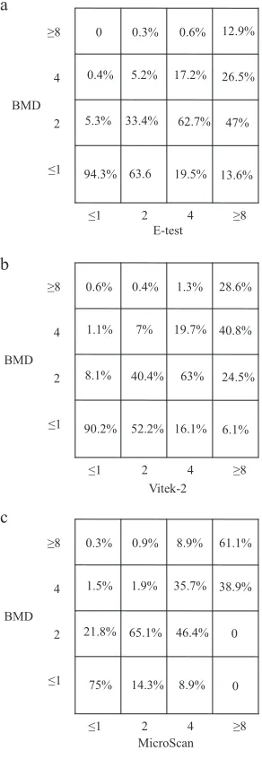
FIG 1Variations in the MICs of tigecycline amongAcinetobacter baumannii
isolates as determined by three methodologies, Etest versus BMD (a), Vitek-2 versus BMD (b), and MicroScan versus BMD (c).
on May 16, 2020 by guest
http://jcm.asm.org/
[image:3.585.349.495.274.697.2]among the various testing methodologies, but none reached
sta-tistical significance.
Figure 1a
displays comparative MICs determined by Etest and
BMD for
A. baumannii
isolates.
Figure 1b
displays the variations
between the MICs determined by Vitek-2 and BMD. For both
comparisons, the discordances between the methods were
partic-ularly evident in the higher-MIC range, as determined by Etest or
Vitek-2.
Figure 1c
displays the variations between the MICs
deter-mined by MicroScan and BMD. Variations in the MICs were
in-significantly discordant for MicroScan and BMD.
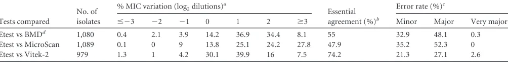
Table 3
shows the various correlations among
A. baumannii
MICs determined by Etest versus those determined by the other
methodologies. An essential agreement of only 55% was noted
between Etest and BMD; compared to MicroScan, the essential
agreement between the tests (Etest versus MicroScan) was
⬍50%.
Major errors were noted, again, between MICs as determined by
Etest compared to the MICs determined by broth microdilution
methodologies. There were only a few very major errors when the
MICs for
A. baumannii
were determined by Etest. The rate of very
major errors was 2.6% when Etest was compared to Vitek-2.
DISCUSSION
This cohort consisting of nearly 4,500 Gram-negative isolates
from unique patients, collected from 25 hospitals during a
20-month period, clearly demonstrates that discordances and
ele-vated MICs of tigecycline determined by Etests are issues that are
unique to the testing of
A. baumannii
isolates among the bacteria
that were trailed. MICs also varied among
Enterobacteriaceae
iso-lates as a result of the susceptibility-testing methodology, but the
Etest MIC results were not exceptionally higher. The reason for
this unique phenomenon in
A. baumannii
has been reported
be-fore in small single-center studies (
20
,
21
), but this report,
con-ducted on a large multicenter cohort, confirms this finding and
provides important new information. Previous reports have
asso-ciated the variation in MIC results with different compositions of
the Mueller-Hinton medium used for Etesting (particularly the
manganese concentration) (
22
,
24
). This study demonstrates that
similar results are observed even when using testing plates with a
low manganese concentration.
One plausible explanation for the high MICs reported via the
Etest methodology might be that tigecycline heteroresistance is
particularly common among
A. baumannii
isolates and that
het-eroresistant colonies are identified by Etest, which elevates the
absolute MIC values (
25
). This theory, however, should be
sub-jected to future advanced population analysis investigations.
Interestingly, there were also significant variations among the
tigecycline MICs for
A. baumannii
isolates when the MICs were
determined by Vitek-2 compared to those determined by BMD.
The reason for this discordance is unclear, and it suggests that
Vitek-2 might not be appropriate for determining tigecycline
MICs for
A. baumannii
isolates. Clinicians who utilize Vitek-2 test
results should be aware of this issue related to tigecycline
suscep-tibility testing for
A. baumannii
isolates.
The discordances between MICs determined by Etest and the
other test methods are cause for concern, as many clinical
micro-biology laboratories utilize Etest for tigecycline testing of
A.
bau-mannii
isolates. Future studies should analyze the impact of MICs
of tigecycline determined by Etest on the outcomes of patients
with infection due to
A. baumannii
who are treated with
tigecy-cline. As tigecycline is often one of the only available agents
avail-able to treat some strains of
A. baumannii
, it is critically important
for health care providers to be aware of the limitations and
discor-dances associated with
in vitro
susceptibility testing performed
using the Etest methodology.
ACKNOWLEDGMENTS
Keith S. Kaye is supported by the National Institute of Allergy and Infec-tious Diseases (NIAID), DMID protocol 10-0065. Marcus J. Zervos has received grant support from Merck, Cubist, and Forrest.
This study was partially funded by Pfizer Inc. The company had no access to or involvement in laboratory testing, data collection, or data interpretation and was not involved in the process of drafting the manu-script.
Keith S. Kaye and Jason M. Pogue serve as consultants and on the speakers’ bureau of Pfizer, and Keith S. Kaye received grant support from Pfizer. Marcus J. Zervos has received grant support from Pfizer. Anurag N. Malani is a shareholder of Pfizer. The other authors have no potential conflicts of interest to declare in relation to the content of this article.
REFERENCES
1.Gilad J, Carmeli Y. 2008. Treatment options for multidrug-resistant
Acinetobacter species. Drugs 68:165–189. http://dx.doi.org/10.2165 /00003495-200868020-00003.
2.Peleg AY, Hooper DC.2010. Hospital-acquired infections due to
Gram-negative bacteria. N. Engl. J. Med.362:1804 –1813.http://dx.doi.org/10 .1056/NEJMra0904124.
3.Ku K, Pogue JM, Moshos J, Bheemreddy S, Wang Y, Bhargava A,
Campbell M, Khandker N, Lephart PR, Chopra T, Hayakawa K, Martin
ET, Abreu-Lanfranco O, Dhar S, Kaye KS, Marchaim D.2012.
Retro-TABLE 3Correlation between Etest and other susceptibility testing methods forAcinetobacter baumannii, southeast Michigan, January 2010 to
August 2011
Tests compared
No. of isolates
% MIC variation (log2dilutions)a Essential
agreement (%)b
Error rate (%)c
ⱕ⫺3 ⫺2 ⫺1 0 1 2 ⱖ3 Minor Major Very major
Etest vs BMDd 1,080 0.4 2.1 3.9 14.2 36.9 34.4 8.1 55 32.9 48.1 0.3
Etest vs MicroScan 1,089 0.1 0 9 13.8 25.1 24.2 27.8 47.9 35.2 52.3 0
Etest vs Vitek-2 979 1.3 1 4.2 30.1 39.9 16 7.5 74.2 21.3 27.1 2.6
aA negative number indicates that the Etest MIC was lower than the MIC of the test to which it was being compared. A positive number indicates that the Etest MIC was higher
than the MIC of the test to which it was being compared. Zero indicates that the Etest MIC and the MIC of the test to which it was being compared were equal.
bEssential agreement is indicated by Etest MICs that are either identical or 1 doubling dilution from the MIC of the test to which Etest was being compared. c
Minor error, isolate interpreted by Etest as intermediate (MIC⫽4 mg/L), susceptible (MICⱕ2 mg/L), or resistant (MICⱖ8 mg/L) by the test to which Etest was being compared; major error, isolate interpreted as false resistant/nonsusceptible by Etest (MICⱖ4 mg/L); very major error, isolate interpreted as false susceptible (MICⱕ2 mg/L) by Etest. NoAcinetobacterspecies MIC breakpoints for tigecycline exist. The common practice at the participating centers at the time of the study was to use the FDA breakpoints set
forEnterobacteriaceaeandA. baumannii.
d
BMD, broth microdilution.
on May 16, 2020 by guest
http://jcm.asm.org/
[image:4.585.42.544.88.150.2]spective evaluation of colistin versus tigecycline for the treatment of Acin-etobacter baumanniiand/or carbapenem-resistantEnterobacteriaceae in-fections. Am. J. Infect. Control40:983–987.http://dx.doi.org/10.1016/j .ajic.2011.12.014.
4.Magiorakos AP, Srinivasan A, Carey RB, Carmeli Y, Falagas ME, Giske
CG, Harbarth S, Hindler JF, Kahlmeter G, Olsson-Liljequist B, Pater-son DL, Rice LB, Stelling J, Struelens MJ, Vatopoulos A, Weber JT,
Monnet DL. 2012. Multidrug-resistant, extensively drug-resistant and
pandrug-resistant bacteria: an international expert proposal for interim standard definitions for acquired resistance. Clin. Microbiol. Infect.18:
268 –281.http://dx.doi.org/10.1111/j.1469-0691.2011.03570.x.
5.Fishbain J, Peleg AY.2010. Treatment ofAcinetobacterinfections. Clin.
Infect. Dis.51:79 – 84.http://dx.doi.org/10.1086/653120.
6.Schwaber MJ, Lev B, Israeli A, Solter E, Smollan G, Rubinovitch B,
Shalit I, Carmeli Y.2011. Containment of a country-wide outbreak of
carbapenem-resistantKlebsiella pneumoniaein Israeli hospitals via a na-tionally implemented intervention. Clin. Infect. Dis.52:848 – 855.http: //dx.doi.org/10.1093/cid/cir025.
7. Marchaim D, Chopra T, Perez F, Hayakawa K, Lephart PR,
Bheemreddy S, Blunden C, Hujer AM, Rudin S, Shango M, Campbell M, Varkey J, Slim J, Ahmad F, Patel D, Chen TY, Pogue JM, Salimnia
H, Dhar S, Bonomo RA, Kaye KS.2011. Outcomes and genetic
related-ness of carbapenem-resistantEnterobacteriaceaeat Detroit medical center. Infect. Control Hosp. Epidemiol.32:861– 871.http://dx.doi.org/10.1086 /661597.
8.Marchaim D, Gottesman T, Schwartz O, Korem M, Maor Y, Rahav G,
Karplus R, Lazarovitch T, Braun E, Sprecher H, Lachish T, Wiener-Well Y, Alon D, Chowers M, Ciobotaro P, Bardenstein R, Paz A, Potasman I, Giladi M, Schechner V, Schwaber MJ, Klarfeld-Lidji S,
Carmeli Y.2010. National multicenter study of predictors and outcomes
of bacteremia upon hospital admission caused byEnterobacteriaceae pro-ducing extended-spectrum beta-lactamases. Antimicrob. Agents Che-mother.54:5099 –5104.http://dx.doi.org/10.1128/AAC.00565-10.
9.Ramirez J, Dartois N, Gandjini H, Yan JL, Korth-Bradley J, McGovern
PC.2013. Randomized phase 2 trial to evaluate the clinical efficacy of two high-dosage tigecycline regimens versus imipenem-cilastatin for treat-ment of hospital-acquired pneumonia. Antimicrob. Agents Chemother.
57:1756 –1762.http://dx.doi.org/10.1128/AAC.01232-12.
10. Cai Y, Wang R, Liang B, Bai N, Liu Y. 2011. Systematic review and
meta-analysis of the effectiveness and safety of tigecycline for treatment of infectious disease. Antimicrob. Agents Chemother.55:1162–1172.http: //dx.doi.org/10.1128/AAC.01402-10.
11. Giamarellou H, Poulakou G.2009. Multidrug-resistant Gram-negative
infections: what are the treatment options? Drugs69:1879 –1901.http: //dx.doi.org/10.2165/11315690-000000000-00000.
12. Nicasio AM, Kuti JL, Nicolau DP.2008. The current state of
multidrug-resistant Gram-negative bacilli in North America. Pharmacotherapy28:
235–249.http://dx.doi.org/10.1592/phco.28.2.235.
13. Tasina E, Haidich AB, Kokkali S, Arvanitidou M.2011. Efficacy and
safety of tigecycline for the treatment of infectious diseases: a meta-analysis. Lancet Infect. Dis.11:834 – 844.http://dx.doi.org/10.1016/S1473 -3099(11)70177-3.
14. Yahav D, Lador A, Paul M, Leibovici L.2011. Efficacy and safety of
tigecycline: a systematic review and meta-analysis. J. Antimicrob. Che-mother.66:1963–1971.http://dx.doi.org/10.1093/jac/dkr242.
15. Rello J.2005. Pharmacokinetics, pharmacodynamics, safety and
tolera-bility of tigecycline. J. Chemother.17(Suppl 1):12–22.http://dx.doi.org /10.1179/joc.2005.17.Supplement-1.12.
16. FDA.2010. Drug safety communication—increased risk of death with Tygacil (tigecycline) compared to other antibiotics used to treat similar infections. FDA, Washington, DC.
17. Navon-Venezia S, Leavitt A, Carmeli Y.2007. High tigecycline resistance
in multidrug-resistantAcinetobacter baumannii. J. Antimicrob. Che-mother.59:772–774.http://dx.doi.org/10.1093/jac/dkm018.
18. Lu CT, Chuang YC, Sun W, Liu YC, Cheng YJ, Lu PL, Chen CM, Hsu
GJ, Jang TN, Lee CM, Chiang PC, Shi ZY, Wang LS, Kung HC, Lin HC,
Liao CH, Liu JW, Huang CH, Tsao SM, Hsueh PR.2008. Nationwide
surveillance in Taiwan of the in-vitro activity of tigecycline against clinical isolates of extended-spectrum beta-lactamase-producing Enterobacteria-ceae. Int. J. Antimicrob. Agents32(Suppl 3):S179 –S183.http://dx.doi.org /10.1016/S0924-8579(08)70024-4.
19. Spanu T, De Angelis G, Cipriani M, Pedruzzi B, D’Inzeo T, Cataldo
MA, Sganga G, Tacconelli E.2012. In vivoemergence of tigecycline
resistance in multidrug-resistantKlebsiella pneumoniaeandEscherichia coli. Antimicrob. Agents Chemother.56:4516 – 4518.http://dx.doi.org/10 .1128/AAC.00234-12.
20. Pillar CM, Draghi DC, Dowzicky MJ, Sahm DF.2008.In vitroactivity of
tigecycline against Gram-positive and Gram-negative pathogens as evalu-ated by broth microdilution and Etest. J. Clin. Microbiol.46:2862–2867. http://dx.doi.org/10.1128/JCM.00637-08.
21. Casal M, Rodriguez F, Johnson B, Garduno E, Tubau F, de Lejarazu
RO, Tenorio A, Gimenez MJ, Bartolome R, Garcia-Rey C, Aguilar L,
Garcia-Escribano N.2009. Influence of testing methodology on the
tige-cycline activity profile against presumably tigetige-cycline-non-susceptible Acinetobacterspp. J. Antimicrob. Chemother.64:69 –72.http://dx.doi.org /10.1093/jac/dkp169.
22. Fernandez-Mazarrasa C, Mazarrasa O, Calvo J, del Arco A,
Martinez-Martinez L.2009. High concentrations of manganese in Mueller-Hinton
agar increase MICs of tigecycline determined by Etest. J. Clin. Microbiol.
47:827– 829.http://dx.doi.org/10.1128/JCM.02464-08.
23. Clinical and Laboratory Standards Institute.2009. Performance
stan-dards for antimicrobial susceptibility testing. 19th informational supple-ment. Approved standard M100-S19. Clinical and Laboratory Standards Institute, Wayne, PA.
24. Veenemans J, Mouton JW, Kluytmans JA, Donnely R, Verhulst C, van
Keulen PH.2012. Effect of manganese in test media onin vitro
suscepti-bility ofEnterobacteriaceaeandAcinetobacter baumanniito tigecycline. J. Clin. Microbiol.50:3077–3079.http://dx.doi.org/10.1128/JCM.01485-12.
25. Lo-Ten-Foe JR, de Smet AM, Diederen BM, Kluytmans JA, van Keulen
PH.2007. Comparative evaluation of the Vitek 2, disk diffusion, Etest, broth microdilution, and agar dilution susceptibility testing methods for colistin in clinical isolates, including heteroresistantEnterobacter cloacae andAcinetobacter baumanniistrains. Antimicrob. Agents Chemother.51:
3726 –3730.http://dx.doi.org/10.1128/AAC.01406-06.