Evaluation of the cobas Cdiff Test for
Detection of Toxigenic
Clostridium
difficile
in Stool Samples
Lance R. Peterson,
aStephen A. Young,
b,cThomas E. Davis, Jr.,
dZi-Xuam Wang,
eJohn Duncan,
fChristopher Noutsios,
fOliver Liesenfeld,
fJohn C. Osiecki,
fMichael A. Lewinski
fDepartment of Laboratory Medicine and Pathology, NorthShore University HealthSystem, Evanston, Illinois, USAa; Department of Pathology, University of New Mexico HSC, Albuquerque, New Mexico, USAb; TriCore Reference Laboratories, Albuquerque, New Mexico, USAc; Department of Pathology and Laboratory Medicine, Indiana University School of Medicine, Indianapolis, Indiana, USAd; Thomas Jefferson University, Philadelphia, Pennsylvania, USAe; Medical and Scientific Affairs, Roche Molecular Diagnostics, Pleasanton, California, USAf
ABSTRACT
Nucleic acid amplification tests (NAATs) are reliable tools for the
detec-tion of toxigenic
Clostridium difficile
from unformed (liquid or soft) stool samples.
The objective of this study was to evaluate performance of the cobas Cdiff test on
the cobas 4800 system using prospectively collected stool specimens from patients
suspected of having
C. difficile
infection (CDI). The performance of the cobas Cdiff
test was compared to the results of combined direct and broth-enriched toxigenic
culture methods in a large, multicenter clinical trial. Additional discrepancy analysis
was performed by using the Xpert
C. difficile
Epi test. Sample storage was evaluated
by using contrived and fresh samples before and after storage at
⫺
20°C. Testing
was performed on samples from 683 subjects (306 males and 377 females); 113
(16.5%) of 683 subjects were positive for toxigenic
C. difficile
by direct toxigenic
cul-ture, and 141 of 682 subjects were positive by using the combined direct and
en-riched toxigenic culture method (reference method), for a prevalence rate of 20.7%.
The sensitivity and specificity of the cobas Cdiff test compared to the combined
di-rect and enriched culture method were 92.9% (131/141; 95% confidence interval [CI],
87.4% to 96.1%) and 98.7% (534/541; 95% CI, 97.4% to 99.4%), respectively.
Discrep-ancy analysis using results for retested samples from a second NAAT (Xpert C.
diffi-cile/Epi test; Cepheid, Sunnyvale, CA) found no false-negative and 4 false-positive
cobas Cdiff test results. There was no difference in positive and negative results in
comparisons of fresh and stored samples. These results support the use of the cobas
Cdiff test as a robust aid in the diagnosis of CDI.
KEYWORDS
Clostridium difficile
infection (CDI), health care-associated infection,
nucleic acid amplification test, active surveillance testing (AST), toxin B (
tcdB
) gene
C
lostridium difficile
is a Gram-positive, anaerobic, spore-forming bacillus identified as
an etiological agent of antibiotic-associated diarrhea and pseudomembranous
colitis in the late 1970s (1, 2). Historically, the incidence of
C. difficile
infection (CDI)
ranged from 30 to 40 cases per 100,000 population in acute-care hospitals in the United
States (3). The incidence rose to more than 140 cases per 100,000 population by 2013,
with 60.7% clearly being health care associated (4). Currently, CDI is the most
com-monly diagnosed infectious cause of health care-associated diarrhea and is a significant
cause of morbidity and mortality (5). Toxigenic strains of
C. difficile
typically produce
two toxins, toxin A, which is an enterotoxin, and toxin B, which is a cytotoxin (6). A small
percentage of strains produce only toxin B (7). There are no known naturally occurring
toxin A-positive, toxin B-negative strains associated with clinical disease (8, 9).
Diagno-Received18 July 2017Returned for modification8 August 2017 Accepted22 September 2017
Accepted manuscript posted online27 September 2017
CitationPeterson LR, Young SA, Davis TE, Jr, Wang Z-X, Duncan J, Noutsios C, Liesenfeld O, Osiecki JC, Lewinski MA. 2017. Evaluation of the cobas Cdiff test for detection of toxigenic
Clostridium difficilein stool samples. J Clin
Microbiol 55:3426 –3436.https://doi.org/10
.1128/JCM.01135-17.
EditorBetty A. Forbes, Virginia Commonwealth University Medical Center
Copyright© 2017 American Society for
Microbiology.All Rights Reserved.
Address correspondence to Lance R. Peterson, [email protected].
crossm
on May 16, 2020 by guest
http://jcm.asm.org/
sis of CDI may be established by the presence of toxin B or toxins A and B in stool
samples from patients with appropriate clinical symptoms, driving the need for
accu-rate testing.
Early detection of CDI with the rapid implementation of contact precautions is
considered critical for infection control to prevent transmission in the health care
setting (10). There are various diagnostic approaches for CDI (11), and real-time PCR
(quantitative PCR [qPCR]) is considered a reliable and rapid single test for the detection
of
C. difficile
toxin (12–15). The purpose of this study was to compare the performance
of the cobas Cdiff test to that of the direct and enriched toxigenic culture method for
the detection of toxigenic
C. difficile
in stool samples.
(The data were presented in part at annual meetings of the American Society for
Microbiology [16] and the Association for Molecular Pathology [17].)
RESULTS
Study population.
A clinical utility study was performed by using 683 specimens
collected from 4 collection sites, and all specimens had valid test results with the cobas
Cdiff test as well as direct culture from initial testing. One sample lacked a sufficient
volume for repeat direct and enrichment culture methods and was not included in the
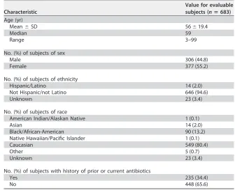
final statistical analysis. Demographics and baseline characteristics of evaluable
sub-jects are shown in Table 1. The age range of the patients was between 3 and 99 years,
with a median age of 59 years. The gender distribution was 306 males (44.8%) and 377
females (55.2%).
Comparison of the cobas Cdiff test with direct culture methods.
The
perfor-mance of the cobas Cdiff test compared to that of the direct culture method is shown
in Table 2. The overall percent agreement (OPA) was 95.3%. The three specimens with
false-negative cobas Cdiff test results relative to the direct culture method were all
negative by the Xpert Cdiff test. Of the 29 specimens with false-positive cobas Cdiff test
results relative to direct culture, 15 were positive by the Xpert Cdiff test; 1 sample was
not tested because of an insufficient specimen volume.
[image:2.585.36.372.83.356.2]The clinical performance of the cobas Cdiff test compared with the combined results
of initial and repeat direct and enriched toxigenic culture methods is shown in Table 3.
TABLE 1Demographics and baseline characteristics of evaluable subjects
Characteristic
Value for evaluable subjects (nⴝ683)
Age (yr)
Mean⫾SD 56⫾19.4
Median 59
Range 3–99
No. (%) of subjects of sex
Male 306 (44.8)
Female 377 (55.2)
No. (%) of subjects of ethnicity
Hispanic/Latino 14 (2.0) Not Hispanic/not Latino 646 (94.6)
Unknown 23 (3.4)
No. (%) of subjects of race
American Indian/Alaskan Native 1 (0.1)
Asian 14 (2.0)
Black/African-American 90 (13.2) Native Hawaiian/Pacific Islander 1 (0.1)
Caucasian 549 (80.4)
Other 5 (0.7)
Unknown 23 (3.4)
No. (%) of subjects with history of prior or current antibiotics
Yes 235 (34.4)
No 448 (65.6)
on May 16, 2020 by guest
http://jcm.asm.org/
These data demonstrate the benefit of the use of enrichment culture methods to
enhance the recovery of
C. difficile
when cultures are used as a reference method. The
sensitivity and specificity of the cobas Cdiff test were 92.9 and 98.7%, respectively; the
positive predictive value (PPV) and negative predictive value (NPV) were 94.9 and
98.2%, respectively. In the discrepancy analysis, of the 7 specimens with false-positive
cobas Cdiff test results relative to combined direct and enrichment culture methods, 3
were positive by the Xpert Cdiff test. The 10 specimens with false-negative cobas Cdiff
test results were all negative by the Xpert
C. difficile
Epi test.
Impact of sample storage.
The results for 124 random samples returned to the
reference laboratory for retesting had positive percent agreement, negative percent
agreement, and overall percent agreement values between fresh and frozen stool
specimens of 97.1% (33/34 samples), 97.8% (88/90), and 97.6% (121/124), respectively
(chi-square statistic, 0.02;
P
⫽
0.89). For the 100 contrived specimens, the positive
percent agreement between the cobas Cdiff test results before and those after one
freeze-thaw cycle was 100.0% (100/100; 95% confidence interval [CI], 96.4 to 100.0%).
There was no difference in the distributions of positive and negative cobas Cdiff test
results when fresh and frozen specimens from the multicenter trial of 580 samples were
compared (
P
⫽
1 by Fisher’s exact test;
P
⫽
0.93 by the chi-square test likelihood ratio).
Finally, there was 100% agreement between direct and repeat toxigenic culture results
for 87 samples cultured fresh and following storage for at least 60 days at
⫺
20°C or
colder (Kappa statistic, 1.0).
Analytical performance.
Analysis of the precision study data showed that at
⬍
1
⫻
the limit of detection (LOD), the positivity rate was 29%, and at 1
⫻
or 3
⫻
the LOD, it
was 100%. The mean cycle threshold (
C
T) values were 38.5 at 1
⫻
the LOD and 37.5 at
3
⫻
the LOD, with standard deviations of 1.5% and 1.1%, respectively (18).
[image:3.585.39.370.83.161.2]In the analysis of clinical reproducibility, the positive percent agreement values for
results below the LOD, 1
⫻
the LOD, and 3
⫻
the LOD were 66.1% (95% CI, 58.7% to
73.0%), 100.0% (95% CI, 98.0% to 100.0%), and 100.0% (95% CI, 97.9% to 100.0%),
respectively. The negative percent agreement was 100.0% (95% CI, 97.9% to 100.0%).
TABLE 2Comparison of the cobas Cdiff test with direct culturea
cobas Cdiff test result
No. of specimens with result
Total no. of specimens Positive by
direct culture
Negative by direct culture
Positive 110 29 139
Negative 3 541 544
Total 113 570 683
aThe positive percent agreement between the results of the cobas Cdiff test and direct culture was 97.3%
(95% CI, 92.5% to 99.1%). The negative percent agreement between the results of the cobas Cdiff test and direct culture was 94.9% (95% CI, 92.8% to 96.4%). The overall percent agreement between the results of the cobas Cdiff test and direct culture was 95.3% (95% CI, 93.5% to 96.7%).
TABLE 3Comparison of the cobas Cdiff test with combined direct and enrichment cultureb
cobas Cdiff test result
No. of speicmens with result
Total no. of specimens Positive by combined direct
and enrichment culture
Negative by combined direct and enrichment culture
Positive 131 7 138
Negative 10 534 544
Total 141 541 682a
aOne sample had an insufficient sample volume for repeat testing and is not included.
bThe sensitivity and specificity of the results of the cobas Cdiff test were 92.9% (131/141 samples; 95% CI,
87.4% to 96.1%) and 98.7% (534/541 samples; 95% CI, 97.4% to 99.4%), respectively. The positive and negative predictive values of the cobas Cdiff test were 94.9% (95% CI, 89.9% to 97.5%) and 98.2% (95% CI, 96.6% to 99.0%), respectively.
on May 16, 2020 by guest
http://jcm.asm.org/
[image:3.585.41.372.618.694.2]The LOD for
C. difficile
strains ranged from 54 to 225 CFU per swab. The inclusivity study
found that all 28
C. difficile
strains were determined to be positive in
ⱖ
95% of replicates
at densities ranging from 77.9 to 460 CFU/swab; 15 were determined to be positive
100% of the time (18).
Analytical specificity testing found that that none of the organisms tested interfered
with the detection of the intended
C. difficile
targets, and there were no false-positive
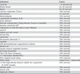
results (19). The compounds listed in Table 4 were tested for interference. There was no
interference found for any material other than mucin, which interfered with the
detection of toxigenic
C. difficile
isolates at a 50% (vol/vol) concentration. For the
cross-contamination analysis, in the nine checkerboard runs, 1 out of 423 negative
samples produced a positive result, for a cross-contamination rate of
ⱕ
0.24%. None of
the 282 negative samples from the carryover contamination runs produced a positive
result (19).
Rate of invalid results.
Among 683 specimens tested for toxigenic
C. difficile
, the
initial failure rate was 0% (0/683 specimens; 95% CI, 0% to 0.7%), and no retesting was
needed, providing a final failure (invalid) rate of 0%. Thus, there were no invalid runs
during this study.
Statistical opinion.
The performance of the cobas Cdiff test was comparable to that
of toxigenic culture using unformed stool samples from subjects suspected of having
CDI and met the criteria specified in the study protocol, assay product requirements,
and clinical validation plan. The results support the intended use of the cobas Cdiff test
as an aid in the diagnosis of CDI in humans in conjunction with clinical and
epidemi-ological risk factors.
DISCUSSION
[image:4.585.43.368.83.386.2]In our large, prospective, multisite clinical investigation, we demonstrated that the
cobas Cdiff test is a robust test for the detection of toxigenic
C. difficile
. Our results
extend those reported previously by Moure and colleagues, who found that the
TABLE 4Compounds tested for possible interference
Substance Concn
Fecal fat ⬃4–28 (wt/vol)
Whole blood 25, 50 (vol/vol)
Mucin 25, 50 (wt/vol)
Calcium carbonate (Tums) 10% (wt/vol)
Vancomycin 1% (wt/vol)
Metronidazole 10% (wt/vol) Loperamide (Imodium A-D) 10% (wt/vol) Stool softener 10% (wt/vol) Bismuth subsalicylate (Pepto-Bismol; Procter & Gamble) 10% (vol/vol) Nystatin ointment USP 10% (wt/vol) Preparation H with Bio-Dyne cream (Wyeth) 10% (wt/vol)
Gynol II 10% (wt/vol)
Vagisil anti-itch cream 10% (wt/vol)
Anusol Plus 10% (wt/vol)
Sunscreen 1% (wt/vol)
Miconazole (Monistat 7) 10% (wt/vol)
Vaseline 10% (wt/vol)
SAB-dimenhydrinate suppositories (Sabex) 10% (wt/vol) Mineral oil 10% (vol/vol) Equate natural vegetable laxative 10% (wt/vol) Bisacodyl (Dulcolax) 10% (wt/vol) Fleet (CB Fleet Company) 10% (wt/vol) K-Y Jelly/Gelée (McNeil-PPC) 1% (wt/vol) Afrin original nasal spray 10% (vol/vol)
Witch hazel Liquid from 1 wipe/swab E-Z-HD high-density barium sulfate for suspension
(E-Z-EM Canada)
20% (wt/vol) Palmitic acid 10% (wt/vol) Stearic acid 10% (wt/vol) Naproxen (Aleve) 10% (wt/vol)
on May 16, 2020 by guest
http://jcm.asm.org/
concordance between cobas Cdiff test results and glutamate dehydrogenase (GDH)/
toxin gene screening was 97.6% (20). Also, we demonstrated the stability of sample
storage, which will be useful for future test development of next-generation molecular
and culture-based CDI diagnostics.
The genomics of
C. difficile
and the molecular structure of its toxin genes are well
known (6, 9, 21, 22). Thus, the design of primers and probes for a robust real-time PCR
assay is relatively straightforward. We demonstrated that the cobas Cdiff test had
excellent analytical sensitivity, inclusivity, and reproducibility prior to beginning the
clinical trial. We also demonstrated that there was minimal interference from other
substances possibly found in the stool specimens of patients. Therefore, it was not
surprising that the clinical performance of this test was robust. From the laboratory use
perspective, we also demonstrated that the new assay is reliable in that the initial
failure rate of the test was 0% (0 of 683 tests), which facilitates deployment in a
diagnostic microbiology laboratory for clinical use.
For our literature review, we used PubMed.gov (MEDLINE) with the search terms
Clostridium difficile
diagnosis,
Clostridium difficile
genetics, and real-time PCR and
the names of U.S. Food and Drug Administration (FDA)-cleared molecular tests that
detect
C. difficile
to search the medical literature from 2005 through June 2017. We
also searched Google using these terms as well as the FDA website (
https://www
.fda.gov/MedicalDevices/ProductsandMedicalProcedures/InVitroDiagnostics/
ucm330711.htm
) to determine which commercial tests were available as CDI assays.
From this review, it is clear that there are potentially three main issues to consider when
selecting a testing program for facilitating the diagnosis of CDI. One issue is how well
the various molecular tests perform. The second issue is what the best testing approach
is. The third issue, as an emerging approach to CDI control, is how the chosen test
might perform in an admission active surveillance testing (AST) program to prevent the
spread of
C. difficile
in acute-care hospitals.
The reported performances of the available molecular tests for the detection of
toxigenic
C. difficile
were recently reviewed (11). Of the 11 tests reviewed, the assays
that consistently showed
⬎
90% sensitivity and specificity were limited to the Cepheid
Xpert
C. difficile
, Portrait Toxigenic
C. difficile
, and Illumigene
C. difficile
assays (22). In our
large, multicenter trial, the cobas Cdiff test performed at least as well as the other
assays when our results were compared to those results.
The question of which test or series of tests (e.g., algorithm) should a clinical
laboratory implement is critical for this disease and very complex for the laboratory. A
key argument has been that molecular qPCR tests are too sensitive and detect not only
disease but also colonization. However, there is no evidence that disease is related to
the level of toxin detected in stool (23). One of the frequently referenced clinical reports
on this topic is that by Polage and colleagues, who concluded that PCR tests were too
sensitive and detected an excess of colonized patients as well as those with disease
(24). Unfortunately, those researchers did not use any clinical criteria or laboratory
assessment of specimen quality to determine who should actually be evaluated (e.g.,
included in their data assessment were patients with
⬍
3 diarrheal stools per day who
were not excluded from potential CDI cases) in the data that were critically analyzed
(24). Thus, it is not surprising that those researchers found a high number of positive
specimens from patients with no significant diarrhea, given that it is not unusual for
hospitalized patients to be colonized with this organism (25). Our own data suggest
that PCR-positive, direct stool toxin-negative patients have a significant risk for future
complications if not treated (26). We found a significant positive effect of receiving
treatment on reducing 90-day readmission for any reason, decreasing the inpatient
length of stay (LOS) and total LOS (inpatient plus readmission LOS), and reduced
charges in the enzyme-immunoassay-negative (EIA
⫺)/PCR
⫹group versus the EIA
⫹/
PCR
⫹group, suggesting that at least some of these patients actually had clinically
significant CDI. Importantly, a recent clinical effectiveness review (CER) from the Agency
for Healthcare Research and Quality (AHRQ) found that nucleic acid amplification tests
(NAATs) had high sensitivity and specificity for the laboratory detection of CDI with the
on May 16, 2020 by guest
http://jcm.asm.org/
highest-quality evidence (27). The lowest-quality evidence was the use of algorithms
for this endeavor, and while the algorithms were specific, this approach lacked
sensitivity (27). Clinical diagnosis is essential for the diagnosis of CDI (25), as is well
stated in another recent review: “The diagnosis of CDI is primarily based on the
clinical signs and symptoms and is only confirmed by laboratory testing” (11). Thus,
in the appropriate clinical setting, the use of the most sensitive assay for the
detection of toxigenic
C. difficile
in a patient’s stool sample should remain the goal
of the laboratory.
A novel question being asked of these tests is whether NAATs can be useful for the
control of CDI in the acute-care setting by using them as screening tests (AST) at the
time of admission for the detection of
C. difficile
carriers, who can then be subjected to
contact precautions to prevent spread to other patients (10). A recent report
illumi-nating the better understanding of CDI epidemiology indicates that asymptomatic
carriers (e.g., colonized persons) are important sources of ongoing hospital
transmis-sion (28). Another investigation comparing the likely transmistransmis-sion of
C. difficile
from
patients whose stool samples were positive by direct toxin testing as well as positive
(by qPCR) for a toxin gene versus those that were positive only for the toxin gene found
that symptomatic patients with qPCR-positive toxigenic
C. difficile
infection (without
fecal toxin being detected) accounted for at least one-quarter of potential hospital
transmission events (29). A third important study demonstrated that at the time of
admission, patients acquire new organisms from their surrounding environment but
that after the first night, they contribute bacteria (e.g., contaminate the room
environ-ment) until the microbiological representation of the patient and the room becomes
quite homogeneous (30). This implies that if one is contemplating AST for pathogens
that can spread within the hospital, a rapid (
⬍
24-h), sensitive test such as qPCR is
needed. There has been one report that showed success in the use of the AST
approach, where the investigators found an overall reduction in the number of patients
who developed hospital-associated CDI from 416 patients (6.9 per 10,000 patient-days)
during the control period to 38 patients (3.0 per 10,000 patient-days; 56.5% reduction)
during the admission testing period (
P
⬍
0.001), suggesting that AST for toxigenic
C.
difficile
is useful (31).
Our research had limitations in that there is no “perfect gold standard” for
compar-ison with any new test developed to diagnose
Clostridium difficile
infection, but rather,
they target toxigenic
C. difficile
. We used toxigenic culture as an accepted reference
method for detecting toxigenic
C. difficile
(32), combined with broth enrichment to
enhance sensitivity (14). We also demonstrated a potential cross-contamination rate of
0.24% (1 of 423 negative samples) when using high-signal-positive samples,
demon-strating that cross-contamination is possible but that the risk is low. Thus, we believe
that the clinical trial data are valid as a result of all the comparisons performed and that
this study demonstrates the reliability of the cobas Cdiff test as a new automated test
for the laboratory confirmation of CDI.
Future research will be useful in developing laboratory diagnostics that are able to
separate colonization from disease attributed to
C. difficile
. This will be a challenge,
since at this time, there is no acceptable, clear standard for definitively classifying who
does and who does not have CDI. Also, defining the role of rapid molecular diagnostics
in the prevention and control of health-care-onset CDI should be a priority, as this
disease remains an increasing threat in our current environment. The best practice(s) to
minimize this threat remains uncertain (10). Third, validation of assay performance
using perirectal admission swabs for active surveillance testing to detect colonization
is needed for this and other tests targeting toxigenic
C. difficile
.
In conclusion, we have demonstrated that the cobas Cdiff test is a reliable and
robust automated assay for the detection of toxigenic
C. difficile
in stool samples from
patients with diarrhea. It has entered the commercial testing market with other
laboratory tests for confirming the diagnosis of CDI as a useful clinical diagnostic tool.
on May 16, 2020 by guest
http://jcm.asm.org/
MATERIALS AND METHODS
Study population.Stool samples were collected from 683 eligible patients over a 5-month period at 4 sites in the United States (NorthShore University HealthSystem, Evanston, IL; hospitals using TriCore Reference Laboratories located in Albuquerque, NM; Indiana University School of Medicine, Indianapolis, IN; and Thomas Jefferson University, Philadelphia, PA). Patients⬎12 months of age who were being tested for the diagnosis of CDI were identified and enrolled in the study. Each patient entering the study had an unformed stool sample of⬎5 ml submitted for testing. Patients were excluded from the study if they (i) submitted a formed stool sample, (ii) had been administered antibiotic therapy againstC. difficile(e.g., oral and/or parenteral metronidazole, oral vancomycin, or fidaxomicin) during their present hospitalization, (iii) were previously enrolled in this study, (iv) were enrolled in any other study focused on the prevention or treatment of CDI within the previous 12 months, or (v) had a contraindication for the collection of stool samples according to institution policies and procedures.
Clinical trial design.The clinical performance of the cobas Cdiff test was evaluated in a prospective, multisite investigation comparing the results of this test with those of toxigenic culture methods using remnant, deidentified, unformed stool samples from eligible subjects. Toxigenic culture was performed at a single site designated the reference laboratory (NorthShore University HealthSystem). The cobas Cdiff test was performed at the other three sites, according to the manufacturer’s instructions.
This study was conducted in compliance with International Conference on Harmonization (ICH) guidelines, good clinical practice (GCP) guidelines, and regulations of the FDA. Approval by the institutional review boards at all four sites was obtained. This study was performed to obtain clearance of the cobas Cdiff test by the FDA.
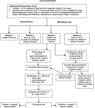
Stool sample processing.On the day of sample collection, fresh stool specimens were transported to the collection site laboratory at temperatures of between 2°C and 30°C. Upon receipt, fresh stool samples were processed within 48 h of collection. The accepted liquid and unformed samples of fresh stool were stirred to homogenize them, and aliquots were then prepared by transferring ⬃1.0-ml portions into 4 separate, sterile, 5-ml cryogenic vials, as depicted in Fig. 1. Aliquots 1 and 2 were shipped on cold packs to one of the designated test sites forC. difficilePCR testing within 5 days of sample collection. Aliquots 3 and 4 were shipped on ice to the reference laboratory to arrive within the same time frame.
Toxigenic culture.The toxigenic culture included initial and repeat direct and enrichment cultures of stool specimens followed by cytotoxicity testing. Direct culture was done by inoculating a portion of the sample into prereduced anaerobically sterilized (PRAS) selective medium and cycloserine-cefoxitin-fructose agar with horse blood and taurocholate (CCFA-HT; Anaerobe Systems, Morgan Hill, CA), followed by cytotoxicity testing on recoveredC. difficile bacteria. Briefly, after incubation at 35°C, suspected colonies were identified as beingC. difficileby Gram staining, aerotolerance testing, and testing using the Pro Disk test (Hardy Diagnostics, Santa Maria, CA) and then inoculated into anaerobic chopped-meat broth (Anaerobe Systems), which was incubated for 5 to 7 days at 35°C for cytotoxicity testing. The supernatant from the anaerobic chopped-meat broth was processed for the detection ofC. difficiletoxin B by cell culture cytotoxicity testing according to the manufacturer’s instructions (C. Difficile Tox-B test; Techlab, Blacksburg, VA). An enriched toxigenic culture method was performed simultaneously using anaerobic cycloserine-cefoxitin-mannitol broth with taurocholate, lysozyme, and cysteine (CCMB-TAL; Anaerobe Systems), followed by subculturing on anaerobicBrucellaagar plates (Anaerobe Systems), with identification and cytotoxicity testing of all recoveredC. difficileisolates.
A specimen was considered positive for toxigenicC. difficile if a toxin-producing organism was recovered from a stool specimen in a direct or enriched toxigenic culture (any positive rule). IfC. difficile
was isolated from the direct culture and the isolate tested positive by a cell cytotoxicity assay, the enrichment culture was not further analyzed. By using aliquot 4 specimens that were frozen at⫺70°C for up to 5 months, 580 study samples were retested by direct and enriched culture methods. If a stool specimen was positive by the cobas Cdiff test but negative by the initial culture, a repeat culture was done (n⫽29). Also, all other samples with a negative initial direct culture result (n⫽541) had a repeat direct and enriched culture performed. Additionally, 10 specimens with a positive initial direct culture result were tested as positive controls. Specimens were classified as being negative for toxigenicC. difficileonly if they tested negative by both initial as well repeat direct and enrichment culture methods.
Testing of patient samples on the cobas 4800 system.After receipt of patient specimens in the laboratory, test orders were logged into the cobas 4800 system either manually, with a work list, or via a Laboratory Information System (LIS) interface and then tested. The cobas 4800 system is a walk-away platform to perform nucleic acid purification and real-time PCR (qPCR) set up in a 96-well plate, followed by manual sealing and transferring of the plate to the amplification and detection system (33). The system currently is available for methicillin-resistantStaphylococcus aureus (MRSA)-S. aureus(34),C. difficile(this study), herpes simplex virus 1 (HSV-1) and HSV-2 (35, 36),Chlamydia trachomatis-Neisseria gonorrhoeae(37–42), and high-risk human papillomavirus (43–45) testing and BRAF (46), epidermal growth factor receptor (EGFR) (47), and KRAS (48) mutation detection. The cobas MRSA/SA test, the cobas Cdiff test, and the cobas HSV 1 and 2 test can be performed simultaneously in a single testing run. This system uses the cobas x 480 instrument, which includes an automated pipetter to extract, purify, and prepare target nucleic acid for qPCR (e.g., automated specimen preparation). Bacterial lysis of the stool specimen is achieved with proteinase K, detergent, and a chaotropic agent. The purified DNA is bound to magnetic glass particles (MGPs), washed, and eluted. The cobas x 480 instrument next transfers the working master mix reagent and moves the specimen to the qPCR microwell plate. The plate is then transferred to the cobas z 480 instrument where the amplification and detection of target DNA and controls occur by using specific primers and probes. Data analysis and report generation are performed
on May 16, 2020 by guest
http://jcm.asm.org/
with cobas 4800 software. The cobas Cdiff test uses positive and negative controls (cobas 4800 Cdiff Controls and Cofactor kit) and an internal control (cobas 4800 system internal control kit). One set of positive and negative controls is included in each run. Valid results must be obtained for both the positive and negative controls for the cobas 4800 software to display a reportable result.
Discrepancy analysis.Discrepancy analysis using the Xpert C. difficile/Epi test (Cepheid, Sunnyvale, CA) was performed on all samples with discordant results between the cobas Cdiff test and the combined direct and enrichment culture method. Eight concordant positive and eight concordant negative samples were also tested as controls. The results of the Xpert C. difficile/Epi test determined the final reference result for the discrepancy analysis.
Testing of fresh and archived (frozen) stool specimens.To evaluate the effect of specimen storage on the results of toxigenic culture and the cobas Cdiff test, aliquots from contrived specimens and unformed stool samples prospectively collected from patients suspected of having CDI were tested fresh and following storage at⫺20°C or colder. Briefly, four studies were performed; specifically, (i) toxigenic culture was performed on fresh samples at the reference laboratory before the samples were shipped frozen on dry ice to Roche Molecular Systems, Inc. (Pleasanton, CA), for PCR, and following PCR, a subset was returned for a repeat culture; (ii) a panel of 100 contrived specimens was prepared by using fresh negative stool specimens spiked with toxigenic C. difficile at various densities (2.5-, 5.0-, 10-, and 20-fold-higher densities than the assay limit of detection) and tested fresh plus following storage for 32 days at⫺20°C⫾5°C; (iii) the assay also was run on the 580 prospectively collected samples, both fresh and following storage at⫺70°C for up to 5 months; and (iv) a repeat toxigenic culture was performed on a subset of clinical samples (15 culture-positive and 72 culture-negative samples) stored at⫺20°C or colder forⱖ60 days.
FIG 1Processing of unformed stool specimens from subjects with suspectedClostridium difficileinfection.
on May 16, 2020 by guest
http://jcm.asm.org/
[image:8.585.41.406.74.491.2]Analytical performance.The technical performance verification (TPV) study was conducted by using a panel composed ofC. difficilecultures diluted into a negative stool matrix in cobas PCR medium to a density below the LOD, near the LOD, and above the LOD as well as a negative level composed of only the stool suspension in cobas PCR medium. We used three unique lots of cobas Cdiff test reagents and three instruments for a total of 36 runs over 12 days.
The reproducibility of the results of the cobas 4800 system was established by a multisite investi-gation using simulated clinical samples evaluated across lot, site/instrument, operator, and day and within a run. Reproducibility test panels consisted of four specimens, with three replicates each, using various densities ofC. difficilestrain ATCC 43255 (negative, below the LOD, 1⫻the LOD, and 3⫻the LOD) in a pooled,C. difficile-negative, unformed stool matrix in cobas PCR medium. These specimens were tested at three sites by two operators/day (six operators total) for 5 days per lot over two lots for an overall total of 712 valid tests. We modeled the targetCTvalue as the dependent variable using the
random effects lot, site, operator, and day.
The LOD was determined by analyzing quantified cultures of seven toxigenicC. difficilestrains diluted into a pooled negative stool specimen matrix in cobas PCR medium. All densities were analyzed by using three unique reagent lots. At least 21 replicates per reagent lot were tested at each density. To assess inclusivity, the cobas Cdiff test was evaluated with 28 additional strains of toxigenicC. difficiletested at a minimum of three densities and with 40 replicates per density level. The lowest density that had a ⱖ95% hit (positive-detection) rate was considered the LOD for each strain.
Analytical specificity was evaluated with a panel that consisted of 103 bacteria, fungi, and viruses as well as one human tissue cell line (18). In addition, 28Clostridiumgenus organisms, including nontoxi-genicC. difficile, were tested (18). Bacteria were quantified as CFU per milliliter, human cells were quantified as cells per milliliter, and viruses were quantified as PFU per milliliter. The only exceptions were for the following microorganisms:Chlamydia trachomatiswas quantified as elementary bodies (EB) per milliliter, and cytomegalovirus, human echovirus, and human enterovirus were quantified as IU per milliliter. Bacteria and human cells were spiked to 1⫻106CFU/ml and 1⫻106cells/ml, respectively, and all viruses were spiked to 1⫻105PFU/ml, except for adenovirus type 40, cytomegalovirus (human herpesvirus 5 [HHV5]), and human rotavirus, which were spiked to lower densities due to stock density limitations. Testing was performed with the organisms alone or with twoC. difficileisolates present individually at 3⫻the LOD.
Interference was evaluated for 26 medications, fecal fat, whole blood, and mucin. The effects of the potentially interfering substances were evaluated in the presence and absence of two toxigenicC. difficile
isolates spiked to⬃3⫻the LOD of the cobas Cdiff test (54 CFU/ml and 113 CFU/ml for the two strains). Potential cross-contamination was assessed by testing a series of high-positive toxigenicC. difficile
samples and negative samples in a checkerboard configuration on the cobas 4800 system. High-positive samples were prepared by spiking a pooled negative stool matrix sufficient to generate aCTvalue (i.e.,
the number of PCR cycles until the PCR growth curve was classified as positive) of the 95th percentile of the clinical specimen population. A total of 94 samples were included per run, with nine runs being performed, using three cobas 4800 systems with alternating checkerboard configurations. A total of three runs of negative samples following these checkerboard runs were performed to assess carryover contamination.
Statistical analysis.SAS/STAT software (SAS, Cary, NC) was used to perform all analyses. Statistical analyses of clinical samples were chosen based on statistical guidance from the FDA (49, 50) and in accordance with the guidelines of Clinical and Laboratory Standards Institute method EP12-A2 for evaluating qualitative test performance (19). For the clinical reproducibility study, 95% two-sided exact binomial confidence intervals were used. In the studies designed to examine the difference between results from fresh and those from frozen samples, statistical analysis was done by using two-sided Fisher’s exact test and the likelihood ratio of the chi-square test.
ACKNOWLEDGMENTS
We sincerely appreciate the contributions of Yosh Ohhashi and all the laboratory
personnel from the study testing sites to this project.
This work was supported by Roche Molecular Diagnostics, Pleasanton, CA.
For potential conflicts of interest, L.R.P. has received speaking honoraria from Becton
Dickinson, Cepheid, Roche, and CareFusion. L.R.P. has received research funding from
Becton Dickinson, Cepheid, Nanosphere, 3M, GeneWEAVE, and Roche. S.A.Y. has
re-ceived research funding from Roche. He has also consulted for Roche and Quidel, Inc.
T.E.D. has received research funding from BioFire, Becton Dickinson, Cepheid, GenMark,
Hologic, Luminex, Nanosphere, and Roche. Z.-X.W. has received research funding from
Roche and has consulted for Roche. J.D., C.N., O.L., J.C.O., and M.A.L. are employees of
Roche Molecular Diagnostics.
L.R.P. had full access to all of the data in the study and takes responsibility for the
integrity of the data and the accuracy of the data analysis. L.R.P., O.L., and M.A.L. were
responsible for the study concept and design. L.R.P., S.A.Y., T.E.D., Z.-X.W., and M.A.L.
performed acquisition, analysis, or interpretation of data. L.R.P., O.L., and M.A.L. drafted
the manuscript. L.R.P., S.A.Y., T.E.D., Z.-X.Y., J.D., C.N., O.L., J.C.O., and M.A.L. performed
on May 16, 2020 by guest
http://jcm.asm.org/
critical revision of the manuscript for important intellectual content. J.D. performed
statistical analysis. C.N., O.L., J.C.O., and M.A.L. provided administrative, technical, or
material support. L.R.P., O.L., and M.A.L. supervised the study.
REFERENCES
1. Bartlett JG, Chang TW, Moon N, Onderdonk AB. 1978. Antibiotic-induced lethal enterocolitis in hamsters: studies with eleven agents and evidence to support the pathogenic role of toxin-producing clostridia. Am J Vet Res 39:1525–1530.
2. Larson HE, Price AB, Honour P, Borriello SP. 1978.Clostridium difficileand the aetiology of pseudomembranous colitis. Lancet i:1063–1066.https:// doi.org/10.1016/S0140-6736(78)90912-1.
3. McDonald LC, Owings M, Jernigan DB. 2006.Clostridium difficileinfection in patients discharged from US short-stay hospitals, 1996-2003. Emerg Infect Dis 12:409 – 415.https://doi.org/10.3201/eid1205.051064. 4. Centers for Disease Control and Prevention. 2013. Annual report for the
Emerging Infections Program forClostridium difficileinfection.https:// www.cdc.gov/hai/eip/pdf/cdiff/2013-annual-report.pdf. Accessed 8 May 2017.
5. Lessa FC, Mu Y, Bamberg WM, Beldavs ZG, Dumyati GK, Dunn JR, Farley MM, Holzbauer SM, Meek JI, Phipps EC, Wilson LE, Winston LG, Cohen JA, Limbago BM, Fridkin SK, Gerding DN, McDonald LC. 2015. Burden of
Clostridium difficileinfection in the United States. N Engl J Med 372: 825– 834.https://doi.org/10.1056/NEJMoa1408913.
6. Voth DE, Ballard JD. 2005. Clostridium difficile toxins: mechanism of action and role in disease. Clin Microbiol Rev 18:247–263.https://doi .org/10.1128/CMR.18.2.247-263.2005.
7. Johnson S, Sambol SP, Brazier JS, Delmee M, Avesani V, Merrigan MM, Gerding DN. 2003. International typing study of toxin A-negative, toxin B-positiveClostridium difficile variants. J Clin Microbiol 41:1543–1547.
https://doi.org/10.1128/JCM.41.4.1543-1547.2003.
8. Kawa D. 1 June 2011. Molecular detection of toxigenicC. difficile: toxin A or B gene? Med Lab Observer https://www.mlo-online.com/molecular -detection-of-toxigenic-c-difficile-toxin-a-or-b-gene.php#respond. Accessed 5 May 2017.
9. Lyras D, O’Connor JR, Howarth PM, Sambol SP, Carter GP, Phumoonna T, Poon R, Adams V, Vedantam G, Johnson S, Gerding DN, Rood JI. 2009. Toxin B is essential for virulence ofClostridium difficile. Nature 458: 1176 –1179.https://doi.org/10.1038/nature07822.
10. McDonald LC, Kutty PK. 25 July 2017, posting date.Clostridium difficile
infection: prevention and control. Topic 2687, p 1–12. UpToDate, version 26.0. Wolters Kluwer, Philadelphia, PA. https://www.uptodate.com/ contents/2687.
11. Martínez-Meléndez A, Camacho-Ortiz A, Morfin-Otero R, Maldonado-Garza HJ, Villarreal-Treviño L, Maldonado-Garza-González E. 2017. Current knowl-edge on the laboratory diagnosis ofClostridium difficileinfection. World J Gastroenterol 23:1552–1567.https://doi.org/10.3748/wjg.v23.i9.1552. 12. Deshpande A, Pasupuleti V, Rolston DD, Jain A, Deshpande N, Pant C,
Hernandez AV. 2011. Diagnostic accuracy of real-time polymerase chain reaction in detection ofClostridium difficile in the stool samples of patients with suspectedClostridium difficileinfection: a meta-analysis. Clin Infect Dis 53:e81– e90.https://doi.org/10.1093/cid/cir505. 13. Kufelnicka AM, Kirn TJ. 2011. Effective utilization of evolving methods for
the laboratory diagnosis ofClostridium difficileinfection. Clin Infect Dis 52:1451–1457.https://doi.org/10.1093/cid/cir201.
14. Peterson LR, Mehta MS, Patel PA, Hacek DM, Harazin M, Nagwekar PP, Thomson RB, Jr, Robicsek A. 2011. Laboratory testing forClostridium difficile infection: light at the end of the tunnel. Am J Clin Pathol 136:372–380.https://doi.org/10.1309/AJCPTP5XKRSNXVIL.
15. Tenover FC, Baron EJ, Peterson LR, Persing DH. 2011. Laboratory diag-nosis ofClostridium difficileinfection: can molecular amplification meth-ods move us out of uncertainty? J Mol Diagn 13:573–582.https://doi .org/10.1016/j.jmoldx.2011.06.001.
16. Peterson L, Young S, Davis T, Wang Z-X, Duncan J, Noutsios C, Ohhashi Y, Liesenfeld O, Osiecki J, Lewinski M. 2015. Evaluation of stool speci-mens with the cobasC. difficiletest performed on the cobas 4800 system for the detection ofClostridium difficiletoxin B compared with toxigenic culture, abstr 2420. Abstr 115th Gen Meet Am Soc Microbiol, New Orleans, LA, 30 May to 2 June 2015.
17. Peterson LR, Young S, Davis T, Jr, Wang ZX, Duncan J, Noutsios C, Ohhashi Y, Liesenfeld O, Osiecki J, Lewinski MA. 2015. Detection of
toxigenicClostridium difficile from fresh and archived (frozen) stool specimens by toxigenic culture and the cobas Cdiff test performed on the cobas 4800 system, abstr ID69. Abstr 2015 Annu Meet Assoc Mol Pathol, Austin, TX, 5 to 7 November 2015.
18. US Food and Drug Administration. 2014. 510(k) substantial equivalence determination decision summary. US Food and Drug Administration, Washington, DC. https://www.accessdata.fda.gov/cdrh_docs/reviews/ K142422.pdf. Accessed 25 August 2017.
19. CLSI. 2008. EP12-A2: user protocol for evaluation of qualitative test performance; approved guideline. CLSI, Wayne PA.
20. Moure R, Cañizares Á, Muíño M, Lobato M, Fernández A, Rodríguez M, Gude MJ, Tomás M, Bou G. 2016. Use of the cobas 4800 system for the rapid detection of toxigenicClostridium difficileand methicillin-resistant
Staphylococcus aureus. J Microbiol Methods 120:50 –52.https://doi.org/ 10.1016/j.mimet.2015.11.014.
21. Knight DR, Elliott B, Chang BJ, Perkins TT, Riley TV. 2015. Diversity and evolution in the genome of Clostridium difficile. Clin Microbiol Rev 28:721–741.https://doi.org/10.1128/CMR.00127-14.
22. Elliott B, Androga GO, Knight DR, Riley TV. 2017. Clostridium difficile
infection: evolution, phylogeny and molecular epidemiology. Infect Genet Evol 49:1–11.https://doi.org/10.1016/j.meegid.2016.12.018. 23. Anikst VE, Gaur RL, Schroeder LF, Banaei N. 2016. Organism burden,
toxin concentration, and lactoferrin concentration do not distinguish between clinically significant and nonsignificant diarrhea in patients withClostridium difficile. Diagn Microbiol Infect Dis 84:343–346.https:// doi.org/10.1016/j.diagmicrobio.2015.11.022.
24. Polage CR, Gyorke CE, Kennedy MA, Leslie JL, Chin DL, Wang S, Nguyen HH, Huang B, Tang YW, Lee LW, Kim K, Taylor S, Romano PS, Panacek EA, Goodell PB, Solnick JV, Cohen SH. 2015. Overdiagnosis ofClostridium difficile infection in the molecular test era. JAMA Intern Med 175: 1792–1801.https://doi.org/10.1001/jamainternmed.2015.4114. 25. Curry SR. 2017.Clostridium difficile. Clin Lab Med 37:341–369.https://doi
.org/10.1016/j.cll.2017.01.007.
26. Smith B, Schaefer E, Liu E, Schoonmaker M, Ridgway J, Robicsek A, Peterson L. 2015. The significance and utility of a highly sensitive PCR test for the diagnosis ofClostridium difficileinfection (CDI). Open Forum Infect Dis 2(Suppl 1):949.https://doi.org/10.1093/ofid/ofv133.665. 27. Butler M, Olson A, Drekonja D, Shaukat A, Schwehr N, Shippee N, Wilt TJ.
2016. Early diagnosis, prevention, and treatment ofClostridium difficile: update. Comparative effectiveness review no. 172. (Prepared by the Min-nesota Evidence-Based Practice Center under contract no. 290-2012-00016-I.) AHRQ publication no. 16-EHC012-EF. Agency for Healthcare Research and Quality, Rockville, MD.https://www.ncbi.nlm.nih.gov/books/ NBK361173/pdf/Bookshelf_NBK361173.pdf. Accessed 6 June 2017. 28. Caroff DA, Yokoe DS, Klompas M. 17 May 2017. Evolving insights into the
epidemiology and control ofClostridium difficilein hospitals. Clin Infect Dishttps://doi.org/10.1093/cid/cix456.
29. Mawer DPC, Eyre DW, Griffiths D, Fawley WN, Martin JSH, Quan TP, Peto TEA, Crook DW, Walker AS, Wilcox MH. 2017. Contribution toClostridium difficiletransmission of symptomatic patients with toxigenic strains who are fecal toxin negative. Clin Infect Dis 64:1163–1170.https://doi.org/10 .1093/cid/cix079.
30. Lax S, Sangwan N, Smith D, Larsen P, Handley KM, Richardson M, Guyton K, Krezalek M, Shogan BD, Defazio J, Flemming I, Shakhsheer B, Weber S, Landon E, Garcia-Houchins S, Siegel J, Alverdy J, Knight R, Stephens B, Gilbert JA. 2017. Bacterial colonization and succession in a newly opened hospital. Sci Transl Med 9:eaah6500.https://doi.org/10.1126/ scitranslmed.aah6500.
31. Longtin Y, Paquet-Bolduc B, Gilca R, Garenc C, Fortin E, Longtin J, Trottier S, Gervais P, Roussy JF, Lévesque S, Ben-David D, Cloutier I, Loo VG. 2016. Effect of detecting and isolatingClostridium difficile
carriers at hospital admission on the incidence of C. difficile
infections: a quasi-experimental controlled study. JAMA Intern Med 176:796 – 804.https://doi.org/10.1001/jamainternmed.2016.0177. 32. Burnham C-AD, Carroll KC. 2013. Diagnosis of Clostridium difficile
infection: an ongoing conundrum for clinicians and for clinical
on May 16, 2020 by guest
http://jcm.asm.org/
tories. Clin Microbiol Rev 26:604 – 630. https://doi.org/10.1128/CMR .00016-13.
33. Van Der Pol B. 2013. Cobas 4800: a fully automated system for the detection ofChlamydia trachomatisandNeisseria gonorrhoeae. Expert Rev Mol Diagn 13:131–140.https://doi.org/10.1586/erm.12.141. 34. Peterson LR, Woods CW, Davis TE, Jr, Wang Z-X, Young SA, Osiecki JC,
Lewinski MA, Liesenfeld O. 13 July 2017. Performance of the cobas MRSA/SA test for simultaneous detection of methicillin-susceptible and methicillin-resistantStaphylococcus aureusfrom nasal swabs. Am J Clin Patholhttps://doi.org/10.1093/ajcp/aqx040.
35. Binnicker MJ, Espy MJ, Duresko B, Irish C, Mandrekar J. 2017. Automated processing, extraction and detection of herpes simplex virus types 1 and 2: a comparative evaluation of three commercial platforms using clinical specimens. J Clin Virol 89:30 –33.https://doi.org/10.1016/j.jcv.2017.02.006. 36. Van Der Pol B. 2016. Type-specific detection of herpes simplex virus type
1 and type 2 using the cobas HSV 1 and 2 test on the cobas 4800 platform. Expert Rev Mol Diagn 16:1145–1154.https://doi.org/10.1080/ 14737159.2016.1243473.
37. Chernesky MA, Jang D, Gilchrist J, Smieja M, Arias M, Hatchette T, Poirier A, Mayne D, Ratnam S. 2017. Comparison of cobas 4800, m2000, Viper XTR, and Infinity 80 automated instruments when processing urine specimens for the diagnosis ofChlamydia trachomatisand Neisseria gonorrhoeae. Sex Transm Dis 44(3):161–165.
38. Peuchant O, de Diego S, Le Roy C, Frantz-Blancpain S, Hocké C, Bébéar C, de Barbeyrac B. 2015. Comparison of three real-time PCR assays for the detection ofChlamydia trachomatisandNeisseria gonorrhoeaein young pregnant women. Diagn Microbiol Infect Dis 83:335–337.https:// doi.org/10.1016/j.diagmicrobio.2015.09.002.
39. Bromhead C, Liyanarachchy N, Mayes J, Upton A, Balm M. 2015. Sup-plementary testing is not required in the cobas 4800 CT/NG test for
Neisseria gonorrhoeaeweak-positive urogenital samples. J Clin Microbiol 53:327–328.https://doi.org/10.1128/JCM.01976-14.
40. Ratnam S, Jang D, Gilchrist J, Smieja M, Poirier A, Hatchette T, Flandin JF, Chernesky M. 2014. Workflow and maintenance characteristics of five automated laboratory instruments for the diagnosis of sexually transmitted infections. J Clin Microbiol 52:2299 –2304. https://doi .org/10.1128/JCM.03549-13.
41. Chernesky M, Jang D, Gilchrist J, Hatchette T, Poirier A, Flandin JF, Smieja M, Ratnam S. 2014. Head-to-head comparison of second-generation nucleic acid amplification tests for detection ofChlamydia trachomatis
andNeisseria gonorrhoeaeon urine samples from female subjects and self-collected vaginal swabs. J Clin Microbiol 52:2305–2310.https://doi .org/10.1128/JCM.03552-13.
42. Geelen TH, Rossen JW, Beerens AM, Poort L, Morré SA, Ritmeester WS,
van Kruchten HE, van de Pas MM, Savelkoul PH. 2013. Performance of cobas 4800 and m2000 real-time assays for detection ofChlamydia trachomatisandNeisseria gonorrhoeaein rectal and self-collected vagi-nal specimen. Diagn Microbiol Infect Dis 77:101–105.https://doi.org/10 .1016/j.diagmicrobio.2013.06.020.
43. Pettus JR, Wilson TL, Steinmetz HB, Lefferts JA, Tafe LJ. 2016. Utility of the Roche Cobas 4800 for detection of high-risk human papillomavirus in formalin-fixed paraffin-embedded oropharyngeal squamous cell carcinoma. Exp Mol Pathol 102:47– 49.https://doi.org/10.1016/j.yexmp.2016.12.004. 44. Lee DH, Hwang NR, Lim MC, Yoo CW, Joo J, Kim JY, Park SY, Hwang SH.
2016. Comparison of the performance of Anyplex II HPV HR, the Cobas 4800 human papillomavirus test and Hybrid Capture 2. Ann Clin Biochem 53:561–567.https://doi.org/10.1177/0004563215614036. 45. Álvarez-Argüelles ME, de Oña-Navarro M, Rojo-Alba S, Torrens-Muns M,
Junquera-Llaneza ML, Antonio-Boga J, Pérez-Castro S, Melón-García S. 2015. Quantification of human papilloma virus (HPV) DNA using the Cobas 4800 system in women with and without pathological alterations attributable to the virus. J Virol Methods 222:95–102.https://doi.org/10 .1016/j.jviromet.2015.05.016.
46. Mourah S, Denis MG, Narducci FE, Solassol J, Merlin JL, Sabourin JC, Scoazec JY, Ouafik L, Emile JF, Heller R, Souvignet C, Bergougnoux L, Merlio JP. 2015. Detection of BRAF V600 mutations in melanoma: eval-uation of concordance between the Cobas 4800 BRAF V600 mutation test and the methods used in French National Cancer Institute (INCa) platforms in a real-life setting. PLoS One 10:e0120232.https://doi.org/ 10.1371/journal.pone.0120232.
47. Ardakani NM, Giardina T, Grieu-Iacopetta F, Tesfai Y, Carrello A, Taylor J, Robinson C, Spagnolo D, Amanuel B. 2016. Detection of epidermal growth factor receptor mutations in lung adenocarcinoma: comparing Cobas 4800 EGFR assay with Sanger bidirectional sequencing. Clin Lung Cancer 17:e113– e119.https://doi.org/10.1016/j.cllc.2016.02.002. 48. Harlé A, Busser B, Rouyer M, Harter V, Genin P, Leroux A, Merlin JL. 2013.
Comparison of COBAS 4800 KRAS, TaqMan PCR and high resolution melting PCR assays for the detection of KRAS somatic mutations in formalin-fixed paraffin embedded colorectal carcinomas. Virchows Arch 462:329 –335.https://doi.org/10.1007/s00428-013-1380-x.
49. US Food and Drug Administration. 2007. Statistical guidance on reporting results from studies evaluating diagnostic tests. US Food and Drug Admin-istration, Washington, DC. https://www.fda.gov/RegulatoryInformation/ Guidances/ucm071148.htm. Accessed 6 June 2017.
50. US Food and Drug Administration. 2015. Guideline for industry and Food and Drug Administration staff. Class II special controls guideline document: toxin gene amplification assays for the detection of Clostrid-ium difficile. US Food and Drug Administration, Washington, DC.