S20
DO2= oxygen delivery; EGDT = early goal-directed therapy; MAP = mean arterial pressure; MMDS = microcirculatory and mitochondrial distress syndrome; OPS imaging = orthogonal polarization spectral imaging; pCO2 = partial pressure of CO2; PGI2 = prostacyclin; pHi = gastric intra-mucosal pH; PslCO2= sublingual pCO2; ROSC = return of spontaneous circulation; SvO2= mixed venous oxygen saturation; VO2= oxygen consumption.
Abstract
Microcirculatory dysfunction plays a pivotal role in the development of the clinical manifestations of severe sepsis. Prior to the advent of new imaging technologies, clinicians had been limited in their ability to assess the microcirculation at the bedside. Clinical evidence of microcirculatory perfusion has historically been limited to physical examination findings or surrogates that could be derived from global parameters of oxygen transport. This review explores: (1) the clinical manifestations of severe sepsis that can be linked to microcirculatory dysfunction; (2) the relationship between conventional hemodynamic parameters and micro-circulatory blood flow indices; (3) the incorporation of microcirculatory function into the definition of ‘shock’ in the sepsis syndrome; (4) the role of the microcirculation in oxygen transport; and (5) the potential impact of novel sepsis therapies on microcirculatory flow. Although the study of the microcirculation has long been the domain of basic science, newly developed imaging technologies, such as orthogonal polarization spectral imaging, have now given us the ability to directly visualize and analyze microcirculatory blood flow at the bedside, and see the microcirculatory response to therapeutic interventions. Disordered microcirculatory flow can now be associated with systemic inflammation, acute organ dysfunction, and increased mortality. Using new technologies to directly image microcirculatory blood flow will help define the role of microcirculatory dysfunction in oxygen transport and circulatory support in severe sepsis.
Introduction
Microcirculatory blood flow is markedly impaired in sepsis [1-3], and microcirculatory dysfunction plays a pivotal role in the development of the clinical manifestations of severe sepsis and septic shock. In the past, direct visualization of microcirculatory networks was only possible in experimental models of sepsis using intravital videomicroscopy, which is not possible in human subjects. Newly developed imaging technologies, such as orthogonal polarization spectral (OPS)
imaging, have brought the ability to directly visualize the microcirculation to the bedside. Microcirculatory imaging, however, is still investigational in human sepsis and has not yet been incorporated into routine clinical practice. The purpose of this review, therefore, is to bridge the experimental and clinical aspects of microcirculatory science, and discuss the concepts that can be incorporated into bedside assessments of patients with severe sepsis and septic shock.
“What lies beneath?” The clinical
manifestations of microcirculatory
dysfunction in severe sepsis
Although the hemodynamic profile and clinical management of septic shock are typically characterized solely in terms of global hemodynamic (i.e. macrovascular) parameters, it is the microcirculation that is responsible for delivering blood flow from the cardiovascular system to the tissues. After conventional cardiovascular support measures achieve restoration of an acceptable arterial blood pressure in a septic shock patient, a clinician may be falsely reassured that the patient is clinically ‘stable’. However, there are a myriad of possible pathogenic mechanisms occurring in the micro-circulation that are difficult to detect with conventional clinical means and are, in effect, hidden from the clinician. In fact, much of the pathophysiology of severe sepsis can be explained by ‘what lies beneath’ in the microcirculation [4]. In severe sepsis this can include: (1) global tissue hypoxia [5]; (2) pan-endothelial cell injury [6]; (3) activation of the coagulation cascade [7]; and (4) “microcirculatory and mitochondrial distress syndrome” (MMDS) [8]. These factors, either individually or in some combination, likely represent primary determinants of acute organ dysfunction in severe sepsis.
Review
Clinical manifestations of disordered microcirculatory perfusion
in severe sepsis
Stephen Trzeciak
1and Emanuel P Rivers
21Section of Critical Care Medicine and the Department of Emergency Medicine, UMDNJ-Robert Wood Johnson Medical School at Camden, Cooper University Hospital, Camden, New Jersey, USA
2Departments of Emergency Medicine and Surgery, Henry Ford Hospital, Detroit, Michigan, USA
Corresponding author: Stephen Trzeciak, [email protected]
Published online: 25 August 2005 Critical Care2005, 9(suppl 4):S20-S26 (DOI 10.1186/cc3744) This article is online at http://ccforum.com/supplements/9/S4/S20
S21 Many of the pathogenic mechanisms of severe sepsis take
place in the microcirculatory unit [4], which is comprised of the arteriole, the capillary bed, and the postcapillary venule. The role of microcirculatory dysfunction in the pathogenesis of acute organ system dysfunction has been discussed else-where in this supplement [9]. However, there are micro-circulatory derangements pertaining to acute cardiovascular system dysfunction that merit special discussion here. It is the microcirculatory unit, and specifically the arterioles, where vasoactive mediators of sepsis exert their vasodilatory effects. This manifests clinically as a low systemic vascular resistance and profound arterial hypotension requiring the administration of vasoconstrictor agents. It is the capillary network where endothelial injury and ongoing capillary leakage occur in severe sepsis, which can manifest clinically as profound hypovolemia and persistently low cardiac filling pressures despite efforts to expand intravascular volume [10]. Therefore, basic elements of the hemodynamic profile, such as hypotension and hypo-volemia thought typically to be macrovascular derangements in nature, are actually rooted in the microcirculation.
The relationship of conventional hemodynamic
parameters and tissue perfusion in severe sepsis
Shock is typically defined as a failure of the cardiovascular system to maintain effective tissue perfusion, resulting in cellular dysfunction and acute organ system failure. If effective tissue perfusion is persistently impaired and not corrected, the cellular dysfunction and organ failure may become irreversible. Some shock etiologies will impair tissue perfusion because of a markedly low cardiac output. However, septic shock can cause an impairment of effective tissue perfusion because of: (1) a maldistribution of blood flow (despite a normal or elevated cardiac output) secondary to microcirculatory dysfunction; or (2) impaired use of substrate due to defects in cellular oxygen utilization. In shock profiles other than sepsis where the microcirculation is not affected to such an extent, correction of global hemodynamic parameters would likely ensure adequate oxygenation of tissues. This is not true in severe sepsis, where regional hypoperfusion abnormalities can persist even after global optimization of conventional hemodynamic and oxygen-derived parameters.
Historically, the conventional clinical signs of hypoperfusion that can be easily assessed at the bedside have included hypotension, tachycardia, oliguria, encephalopathy, cool extremities, slow cutaneous capillary refill, and metabolic (lactic) acidosis. Although the presence of arterial hypo-tension is clearly an ominous sign indicating a high severity of illness, arterial blood pressure alone is an insensitive indicator of tissue hypoperfusion in sepsis. It has been well docu-mented in experimental and clinical studies alike that tissue hypoperfusion abnormalities, either with or without an impairment in cardiac output, can occur long before the manifestation of hypotension. Therefore, blood pressure parameters alone are insufficient to identify the need for aggressive resuscitation [5,11,12].
In the absence of profound hypotension, there appears to be a complex relationship between macrocirculatory hemo-dynamic parameters and microcirculatory blood flow. In a study by LeDoux and colleagues [13], 10 patients with septic shock received increasing doses of norepinephrine to drive up the mean arterial pressure (MAP) from 65 to 85 mmHg, and no changes were reported in indices of regional perfusion (as measured by skin capillary blood flow, red blood cell velocity, urine output, and gastric mucosal partial pressure of CO2 [pCO2]). The authors concluded that the use of vasopressors to elevate the arterial blood pressure to above 65 mmHg in septic shock did not affect regional blood flow [13]. This was recently supported by data from Bourgoin and colleagues [14]. Two recent studies of OPS imaging in septic shock by De Backer and colleagues [1] and Sakr and colleagues [3] reported that microcirculatory perfused vessel density was independent of arterial blood pressure. The data suggest that microcirculatory blood flow in severe sepsis is not simply a function of local perfusion pressures. There appears to be a degree of disconnection between the macro-circulation and the micromacro-circulation, indicating that blood pressure-targeted management strategies in severe sepsis will not necessarily ensure adequate regional perfusion.
Recognition of shock in sepsis syndrome
S22
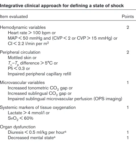
Incorporating additional indicators of tissue perfusion into our bedside assessments may help to improve the identification of the hypoperfused patient. An optimal strategy for recog-nizing shock in sepsis would include sensitive means of detecting regional and microcirculatory perfusion abnor-malities. A perfusion-based scoring system for shock recognition has been proposed by Spronk and colleagues [22] (Table 1). Although exact calculation of a perfusion-based score is not likely to be necessary at the bedside, this type of conceptual framework can be useful to the clinician in bedside assessments by extending the recognition of shock severity to markers of small vessel perfusion (in addition to global hemodynamic and oxygen-derived parameters).
Oxygen transport and the role of the
microcirculation
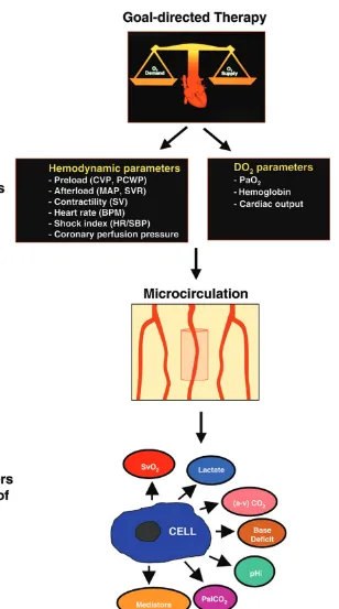
The microcirculation plays a critical role in oxygen transport to tissues. Although the provision of goal-directed circulatory support is typically focused on achieving target values for global hemodynamic and oxygen-derived parameters (here-after referred to as ‘upstream’ endpoints of resuscitation), as well as tracking the response of variables directly related to tissue perfusion such as acid–base parameters and gastric intramucosal pH (pHi)/sublingual pCO2 (PslCO2) (hereafter referred to as ‘downstream’ markers of effective resuscita-tion), an intact microcirculatory network is a critical inter-mediary between the cardiovascular system and effective tissue oxygenation, effectively serving as the bridge between the upstream and downstream parameters (Fig. 1). The Krogh model of microcirculatory oxygen diffusion [23] is a conceptual framework that underscores the importance of the microcirculation in oxygen transport. Oxygen is transported to tissues by diffusion from capillary (and arteriolar [24]) blood. The diffusion distance for oxygen increases as the density of perfused capillaries decreases (Fig. 2). If the diffusion distance for oxygen exceeds a critical point in tissues, then anaerobic metabolism will likely occur. In severe sepsis, this may occur regionally either with or without marked derangements of global hemodynamic or oxygen-derived parameters.
Although global parameters of oxygen transport are typically thought to be insensitive indicators of change in regional blood flow, there are some bedside observations that can be made from derangements of global oxygen-derived para-meters that are central to the concept of microcirculatory oxygen transport. For example, profoundly low mixed venous oxygen saturation (SvO2), a global parameter that represents the oxygen saturation of pooled blood from all the postcapillary venules in the body, may identify when anaerobic metabolism is likely to be occurring in the tissues (i.e. global tissue hypoxia) [5]. Regardless of the oxygen saturation on the arteriolar side of capillaries, a markedly low SvO2 identifies that the venular ends of capillaries likely contain deoxygenated blood. Conceptually speaking, the cylindrical area of tissue in the Krogh model (Fig. 3) that is supplied by this deoxygenated
capillary blood (the so-called “lethal corner”) is at risk for anaerobic metabolism. This has clinical importance, as global tissue hypoxia has been associated with endothelial cell activation and injury, triggering of systemic inflammation, activation of the coagulation cascade, and multiorgan dysfunction [6,25,26]. This cascade of events caused by global tissue hypoxia bears a striking resemblance to key pathogenic elements in the microcirculation that are directly attributable to sepsis. Avoidance of global tissue hypoxia, therefore, is an important goal of cardiovascular support in clinical practice, as failure to correct this physiologic derangement may compound the pathophysiologic effects of severe sepsis in the microcirculation.
Finding a normal or high SvO2value (i.e. venous hyperoxia) at
[image:3.612.315.554.101.352.2]the bedside does not preclude that tissue dysoxia is occurring, because derangements of cellular oxygen utilization (i.e. cytopathic hypoxia [27]) may be playing a significant role (especially in established or late-phase sepsis). Although cytopathic hypoxia is a well recognized mechanism that can cause venous hyperoxia, an alternative hypothesis to explain this finding is that microcirculatory shunting may also be contributing, such that some degree of venous hyperoxia measured at the bedside may actually be caused to some extent by microcirculatory dysfunction. In this model, blood is shunted to open areas of the microcirculation when weak microcirculatory units are effectively shut down Table 1
Integrative clinical approach for defining a state of shock
Item evaluated Points
Hemodynamic variables 2
Heart rate > 100 bpm or
MAP < 50 mmHg and (CVP < 2 or CVP > 15 mmHg) or CI < 2.2 l/min per m2
Peripheral circulation 2
Mottled skin or
Tc–Tpdifference > 5°C or Pfi < 0.3 or
Impaired peripheral capillary refill
Microvascular variables 1
Increased tonometric CO2gap or Increased sublingual CO2gap or
Impaired sublingual microvascular perfusion (OPS imaging) Systemic markers of tissue oxygenation 1
Lactate > 4 mmol/l or SvO2< 60% Organ dysfunction
Diuresis < 0.5 ml/kg per houra 1
Decreased mental statea 1
A state of shock is present if the score exceeds 2 points. CI, cardiac index; CVP, central venous pressure; MAP, mean arterial pressure; OPS, orthogonal polarization spectral imaging; Pfi, peripheral perfusion index; SvO2, mixed venous oxygen saturation; Tc, core temperature;
S23 Figure 1
S24
because of severe flow impairment [28]. Shunting could potentially contribute to decreased systemic oxygen utilization, venous hyperoxia observed at the bedside, and an elevation of serum lactate concentration (Fig. 4). Venous hyperoxia has been observed in patients with cardiac arrest after return of spontaneous circulation (ROSC) [29], and appears to be significantly associated with the amount of epinephrine administered during resuscitative efforts [30]. However, it is not clear to what extent (if any) microcirculatory dysfunction plays a role in the post-ROSC patient, as derangements of cellular oxygen utilization have been hypothesized to be the driving force of this observation. In severe sepsis, where microcirculatory derangements are a key component of the pathophysiology, microcirculatory shunting and cytopathic hypoxia are likely both occurring, and are probably not mutually exclusive processes. The relative contribution of each is currently unclear.
Novel sepsis therapies and their potential
impact on microcirculatory blood flow
Effectively serving as the bridge between the upstream and downstream endpoints of resuscitation, the microcirculation could be an attractive target for novel sepsis therapies. This may be particularly true if downstream markers of the effectiveness of resuscitation (i.e. lactate, base deficit, pHi/PslCO2) fail to normalize despite successful achievement of predefined target values for the upstream endpoints of resuscitation in goal-directed therapy (Fig. 1). A persistent derangement of downstream markers may be attributable to MMDS [8], and may identify the ideal individual for demon-strating the specific effects of novel agents to recruit the Figure 3
A conceptual model of capillary flow and oxygen diffusion. A cylinder represents the area of tissue surrounding an individual capillary. A markedly low value for the (global) mixed venous oxygen saturation (SvO2) reflects markedly deoxygenated blood in the pooled post-capillary venules. Regardless of how well oxygenated the blood may be on the arteriolar side of the capillary (A), a very low SvO2indicates that tissues near the venous end of the capillary (V) are supplied by deoxygenated blood (‘lethal corner’). This conceptual model explains how a low SvO2is associated with tissue dysoxia.
Figure 4
[image:5.612.62.290.90.450.2]The microcirculatory shunting model of sepsis. Severe flow impairment (denoted by X) in weak microcirculatory units causes a shunting of blood to open microcirculatory units. Elevation of serum lactate concentration (from the microcirculatory units with impaired flow) may be simultaneously observed along with venous hyperoxia from the shunted units. Adapted with permission [22].
Figure 2
S25 microcirculation. In theory, novel agents that could specifically
augment microcirculatory blood flow would probably be either: (1) agents that improve flow by modulating actions on the endothelial cell surfaces; or (2) vasodilators. The potential effects of activated protein C on microcirculatory flow are discussed elsewhere in this supplement [31]. Vasodilators that have been studied in human septic shock include prostacyclin, dobutamine, and nitric oxide donor agents, and these agents merit special discussion here because of their potential effects on microcirculatory flow.
Clinical studies of vasodilator agents in
septic shock
The clinical use of vasodilator agents in shock states has typically been limited to macrocirculatory effects, such as modulation of afterload on the heart in cardiogenic shock. There is limited clinical data on the use of vasodilators in sepsis. In a recent study using OPS imaging, De Backer and colleagues demonstrated that a marked impairment of sublingual microcirculatory blood flow in patients with septic shock was reversible with local administration of acetylcholine [1]. This indicates that the endothelium in septic shock is still responsive to mediators of vascular tone and suggests that vasodilatory response in small vessels can be manipulated with therapeutic agents. In theory, the systemic administration of vasodilator agents in septic shock could recruit micro-circulatory units, decrease micromicro-circulatory shunting, and improve regional tissue oxygenation [32]. Because systemic oxygen consumption (VO2) only occurs in the capillaries and terminal arterioles, a true increase in VO2(as opposed to one that is only apparent because of mathematical coupling of the variables) associated with the use of a vasodilator agent in septic shock could indicate that oxygen needs are being met with increasing microcirculatory blood flow, and this finding could support the theory that microcirculatory shunting in severe sepsis plays a significant role in oxygen transport.
Clinical studies of prostacyclin (PGI2) in severe sepsis have demonstrated a change in global parameters of oxygen transport, including a rise in VO2[33-35]. Bihari and colleagues [35] demonstrated that PGI2 administration was associated
with a marked increase in both oxygen delivery (DO2) and VO2, and the authors concluded that PGI2could identify a change in VO2 that represented an occult oxygen debt. In a study by
Radermacher and colleagues [36], septic shock patients were treated with intravenous volume expansion and inotropic support to maximize DO2, and then PGI2 was administered. Prostacyclin improved gastric pHi and the authors concluded that this represented a recruitment of splanchnic perfusion (despite the fact that global oxygen uptake directly measured from respiratory gases was unchanged). Prostacyclin has been poorly tolerated in clinical studies because of inducing or exacerbating arterial hypotension.
Nitroglycerin and dobutamine administration have been associated with increases in microcirculatory blood flow in
sepsis [2,37,38]. In a recent study of resuscitated septic shock patients, Spronk and colleagues [2] demonstrated a severe impairment in sublingual microcirculatory blood flow that was sharply improved with an infusion of nitroglycerin. De Backer and colleagues administered both PGI2 and dobutamine in a clinical study of severe sepsis [33], and dobutamine significantly increased both DO2 and VO2. Shoemaker and colleagues [39] also demonstrated that giving dobutamine to critically ill surgical patients resulted in an improvement in global parameters of oxygen transport, including a rise in both DO2and VO2. The authors concluded that this rise in VO2 indicated an improvement in tissue
perfusion. Note that the results of these studies of vaso-dilators and the potential relationship (if any) between the change in VO2 and presumed microcirculatory recruitment should be interpreted with caution. Because a minority of studies of vasodilators directly measured VO2via respiratory
gases, observed changes in VO2could have more to do with the calculation (mathematical coupling of the oxygen transport variables) than an actual improvement in small-vessel perfusion.
The clinical impact of using nitric oxide donor agents (such as nitroglycerin) or dobutamine to open the microcirculation [32] and improve tissue perfusion is unclear at present. However, it is notable that in the randomized controlled trial of EGDT, the goal-directed protocol included: (1) nitroglycerin for a MAP > 90 mmHg; and/or (2) dobutamine for a central venous oxygen saturation < 70% (after correction of central venous pressure, MAP, and hematocrit) [5]. For individuals in the EGDT group, the impact that these agents had on microcirculatory blood flow is unknown.
New technologies and future directions
New technologies such as OPS imaging have recently brought to the bedside the ability to image the micro-circulation, and have allowed the direct visualization of marked microcirculatory flow impairments that characterize severe sepsis [1]. Although it has been debated that microcirculatory dysfunction and the clinical manifestations of severe sepsis may just be epiphenomena, De Backer and colleagues used OPS imaging and logistic regression analysis to identify that an impairment of capillary perfusion was an independent predictor of mortality in a series of severe sepsis patients [40]. Sakr and colleagues [3] reported that nonsurvivors who died with persistent multiorgan failure due to sepsis had markedly impaired small-vessel perfusion over time compared with survivors. The degree of improvement in small-vessel perfusion over the first 24 hours of therapy was a good predictor of mortality, suggesting that the capacity to impact clinical outcome via restoration of microcirculatory perfusion may be time sensitive [3]. This supports the concept that optimal circulatory support in sepsis should be achieved early.
S26
microcirculatory dysfunction in oxygen transport during conventional goal-directed circulatory support; (2) the prog-nostic value of microcirculatory dysfunction for subsequent acute organ dysfunction and mortality; and (3) the effects of novel antisepsis therapies on microcirculatory blood flow. Although the optimal agents for achieving microcirculatory recruitment, as well as the optimal place in our current goal-directed therapy algorithms for use of such agents, have yet to be determined, these new bedside imaging modalities will provide a window of the microcirculation to help answer these questions.
Competing interests
EPR is a speaker for Edwards Lifesciences.
References
1. De Backer D, Creteur J, Preiser JC, Dubois MJ, Vincent JL: Microvascular blood flow is altered in patients with sepsis. Am J Respir Crit Care Med2002, 166:98-104.
2. Spronk PE, Ince C, Gardien MJ, Mathura KR, Oudemans-van Straaten HM, Zandstra DF: Nitroglycerin in septic shock after intravascular volume resuscitation.Lancet 2002, 360:1395-1396. 3. Sakr Y, Dubois MJ, De Backer D, Creteur J, Vincent JL: Persis-tent microcirculatory alterations are associated with organ failure and death in patients with septic shock.Crit Care Med
2004, 32:1825-1831.
4. Bateman RM, Sharpe MD, Ellis CG: Bench-to-bedside review: microvascular dysfunction in sepsis – hemodynamics, oxygen transport, and nitric oxide.Crit Care2003, 7:359-373. 5. Rivers E, Nguyen B, Havstad S, Ressler J, Muzzin A, Knoblich B,
Peterson E, Tomlanovich M, Early Goal-Directed Therapy Collabora-tive Group: Early goal-directed therapy in the treatment of severe sepsis and septic shock. N Engl J Med2001, 345:1368-1377. 6. Aird WC: The role of the endothelium in severe sepsis and
mul-tiple organ dysfunction syndrome. Blood2003, 101:3765-3777. 7. Yan SB, Helterbrand JD, Hartman DL, Wright TJ, Bernard GR:
Low levels of protein C are associated with poor outcome in severe sepsis.Chest2001, 120:915-922.
8. Ince C: Microcirculation in distress: a new resuscitation end point?Crit Care Med2004, 32:1963-1964.
9. Vincent J-L, De Backer D: Microvascular dysfunction as a cause of organ dysfunction in severe sepsis.Critical Care 2005, 9 (suppl 4):S9-S12.
10. Dellinger RP: Cardiovascular management of septic shock.
Crit Care Med2003, 31:946-955.
11. Nguyen HB, Rivers EP, Knoblich BP, Jacobsen G, Muzzin A, Ressler JA, Tomlanovich MC: Early lactate clearance is associ-ated with improved outcome in severe sepsis and septic shock.Crit Care Med 2004, 32:1637-1642.
12. Wo CC, Shoemaker WC, Appel PL, Bishop MH, Kram HB, Hardin E: Unreliability of blood pressure and heart rate to evaluate cardiac output in emergency resuscitation and criti-cal illness.Crit Care Med1993, 21:218-223.
13. LeDoux D, Astiz ME, Carpati CM, Rackow EC: Effects of perfu-sion pressure on tissue perfuperfu-sion in septic shock.Crit Care Med2000, 28:2729-2732.
14. Bourgoin A, Leone M, Delmas A, Garnier F, Albanese J, Martin C: Increasing mean arterial pressure in patients with septic shock: effects on oxygen variables and renal function.Crit Care Med2005, 33:780-786.
15. Donnino MW, Nguyen HB, Jacobsen G, Tomlanovich M, Rivers EP: Cryptic septic shock: a sub-analysis of early goal-directed therapy [abstract].Chest 2003, 124:90S.
16. Dellinger RP, Carlet JM, Masur H, Gerlach H, Calandra T, Cohen J, Gea-Banacloche J, Keh D, Marshall JC, Parker MM, et al., Surviving Sepsis Campaign Management Guidelines Committee: Surviving sepsis campaign guidelines for management of severe sepsis and septic shock.Crit Care Med 2004, 32:858-873.
17. Vincent JL, Abraham E, Annane D, Bernard G, Rivers E, Van den Berghe G: Reducing mortality in sepsis: new directions.Crit Care2002, 6(suppl 3):S1-18.
18. Trzeciak S: Lac-time?Crit Care Med2004, 32:1785-1786. 19. Aduen J, Bernstein WK, Khastgir T, Miller J, Kerzner R, Bhatiani A,
Lustgarten J, Bassin AS, Davison L, Chernow B: The use and clinical importance of a substrate-specific electrode for rapid determination of blood lactate concentrations. JAMA 1994, 272:1678-1685.
20. Bakker J, Coffernils M, Leon M, Gris P, Vincent JL: Blood lactate levels are superior to oxygen-derived variables in predicting outcome in human septic shock.Chest 1991, 99:956-962. 21. Bakker J, Gris P, Coffernils M, Kahn RJ, Vincent JL: Serial blood
lactate levels can predict the development of multiple organ failure following septic shock.Am J Surg1996, 171:221-226. 22. Spronk PE, Zandstra DF, Ince C: Bench-to-bedside review: sepsis
is a disease of the microcirculation. Crit Care2004, 8:462-468. 23. Krogh A: The number and the distribution of capillaries in
muscle with the calculation of the oxygen pressure necessary for supplying tissue.J Physiol1919, 52:409-515.
24. Ellsworth ML, Pittman RN: Arterioles supply oxygen to capillar-ies by diffusion as well as by convection. Am J Physiol1990, 258:H1240-H1243.
25. Beal AL, Cerra FB: Multiple organ failure syndrome in the 1990s. Systemic inflammatory response and organ dysfunc-tion.JAMA1994, 271:226-233.
26. Karimova A, Pinsky DJ: The endothelial response to oxygen deprivation: biology and clinical implications. Intensive Care Med2001, 27:19-31.
27. Fink MP: Bench-to-bedside review: Cytopathic hypoxia. Crit Care2002, 6:491-499.
28. Ince C, Sinaasappel M: Microcirculatory oxygenation and shunt-ing in sepsis and shock. Crit Care Med1999, 27:1369-1377. 29. Rivers EP, Rady MY, Martin GB, Fenn NM, Smithline HA,
Alexan-der ME, Nowak RM: Venous hyperoxia after cardiac arrest. Characterization of a defect in systemic oxygen utilization.
Chest 1992, 102:1787-1793.
30. Rivers EP, Wortsman J, Rady MY, Blake HC, McGeorge FT, Buderer NM: The effect of the total cumulative epinephrine dose administered during human CPR on hemodynamic, oxygen transport, and utilization variables in the postresusci-tation period.Chest 1994, 106:1499-1507.
31. Macias WL, Yan SB, Williams ND, Um SL, Sandusky GE, Ballard DW, Planquois J-MS: New insights into the protein C pathway: potential implications for the biological activities of drotreco-gin alfa (activated).Critical Care2005, 9(suppl 4):S38-S45. 32. Buwalda M, Ince C: Opening the microcirculation: can
vaso-dilators be useful in sepsis? Intensive Care Med 2002, 28: 1208-1217.
33. De Backer D, Berre J, Zhang H, Kahn RJ, Vincent JL: Relation-ship between oxygen uptake and oxygen delivery in septic patients: effects of prostacyclin versus dobutamine.Crit Care Med1993, 21:1658-1664.
34. Pittet JF, Lacroix JS, Gunning K, Laverriere MC, Morel DR, Suter PM: Prostacyclin but not phentolamine increases oxygen con-sumption and skin microvascular blood flow in patients with sepsis and respiratory failure.Chest 1990, 98:1467-1472. 35. Bihari D, Smithies M, Gimson A, Tinker J: The effects of
vasodi-lation with prostacyclin on oxygen delivery and uptake in criti-cally ill patients.N Engl J Med1987, 317:397-403.
36. Radermacher P, Buhl R, Santak B, Klein M, Kniemeyer HW, Becker H, Tarnow J: The effects of prostacyclin on gastric intramucosal pH in patients with septic shock.Intensive Care Med1995, 21:414-421.
37. Secchi A, Wellmann R, Martin E, Schmidt H: Dobutamine main-tains intestinal villus blood flow during normotensive endo-toxemia: an intravital microscopic study in the rat.J Crit Care
1997, 12:137-141.
38. Duranteau J, Sitbon P, Teboul JL, Vicaut E, Anguel N, Richard C, Samii K: Effects of epinephrine, norepinephrine, or the combi-nation of norepinephrine and dobutamine on gastric mucosa in septic shock. Crit Care Med1999, 27:893-900.
39. Shoemaker WC, Appel PL, Kram HB: Hemodynamic and oxygen transport effects of dobutamine in critically ill general surgical patients.Crit Care Med1986, 14:1032-1037. 40. De Backer D, Dubois MJ, Creteur J, Sakr Y, Vincent JL: