Serotype and Genotype (Multilocus Sequence Type) of
Streptococcus suis
Isolates from the United States Serve as
Predictors of Pathotype
April A. Estrada,aMarcelo Gottschalk,bStephanie Rossow,cAaron Rendahl,aConnie Gebhart,a,cDouglas G. Marthalerc,d
aDepartment of Veterinary and Biomedical Sciences, College of Veterinary Medicine, University of Minnesota, St. Paul, Minnesota, USA bFaculty of Veterinary Medicine, University of Montreal, Saint-Hyacinthe, Quebec, Canada
cVeterinary Diagnostic Laboratory, College of Veterinary Medicine, University of Minnesota, St. Paul, Minnesota, USA dVeterinary Diagnostic Laboratory, College of Veterinary Medicine, Kansas State University, Manhattan, Kansas, USA
ABSTRACT Streptococcus suisis a significant cause of mortality in piglets and
grow-ing pigs worldwide. The species contains pathogenic and commensal strains, with pathogenic strains causing meningitis, arthritis, endocarditis, polyserositis, and septi-cemia. Serotyping and multilocus sequence typing (MLST) are primary methods to differentiate strains, but the information is limited for strains found in the United States. The objective of this study was to characterize the diversity of 208S. suis iso-lates collected between 2014 and 2017 across North America (mainly the United States) by serotyping and MLST and to investigate associations between subtype and pathotype classifications (pathogenic, possibly opportunistic, and commensal), based on clinical information and site of isolation. Twenty serotypes were identified, and the predominant serotypes were 1/2 and 7. Fifty-eight sequence types (STs) were identi-fied, and the predominant ST was ST28. Associations among serotypes, STs, and pathotypes were investigated using odds ratio and clustering analyses. Evaluation of serotype and ST with pathotype identified a majority of isolates of serotypes 1, 1/2, 2, 7, 14, and 23 and ST1, ST13, ST25, ST28, ST29, ST94, ST108, ST117, ST225, ST373, ST961, and ST977 as associated with the pathogenic pathotype. Serotypes 21 and 31, ST750, and ST821 were associated with the commensal pathotype, which is com-posed of isolates from farms with no known history ofS. suis-associated disease. Our study demonstrates the use of serotyping and MLST to differentiate pathogenic from commensal isolates and establish links between pathotype and subtype, thus increasing the knowledge aboutS. suisstrains circulating in the United States.
KEYWORDS MLST,Streptococcus suis, multilocus sequence typing, pathogenic,
pathotype, porcine, serotyping
D
isease caused byStreptococcus suisis a significant economic and welfare concern in the swine industry.S. suisis a Gram-positive bacterium, and the species contains pathogenic and commensal strains. Pathogenic S. suis strains are associated with meningitis, arthritis, endocarditis, polyserositis, and septicemia in piglets and growing pigs (1, 2), and S. suis strains isolated from neurological or systemic tissues (brain/ meninges, joints, and heart) are commonly considered the primary pathogens (2–4). Commensal strains normally reside in the upper respiratory tract of pigs, with pigs commonly serving as carriers (1, 5, 6).S. suiscan be an opportunistic pathogen associated with coinfections with other bacterial and viral pathogens (2, 3). In addition, someS. suis strains have zoonotic potential, causing meningitis in humans (7).Serotyping and multilocus sequence typing (MLST) are commonly used to differen-tiateS. suisstrains, and numerous subtypes exist within theS. suisspecies. Traditionally,
CitationEstrada AA, Gottschalk M, Rossow S, Rendahl A, Gebhart C, Marthaler DG. 2019. Serotype and genotype (multilocus sequence
type) ofStreptococcus suisisolates from the
United States serve as predictors of pathotype.
J Clin Microbiol 57:e00377-19.https://doi.org/
10.1128/JCM.00377-19.
EditorBrad Fenwick, University of Tennessee at Knoxville
Copyright© 2019 Estrada et al. This is an open-access article distributed under the terms
of theCreative Commons Attribution 4.0
International license.
Address correspondence to Douglas G. Marthaler, [email protected].
Received12 March 2019
Returned for modification5 April 2019
Accepted18 June 2019
Accepted manuscript posted online26 June 2019
Published
CLINICAL VETERINARY MICROBIOLOGY
crossm
26 August 2019
on May 17, 2020 by guest
http://jcm.asm.org/
serotyping uses the coagglutination test with reference antisera to subtype S. suis strains, and 35 knownS. suisserotypes (1 to 34 and 1/2) exist. Six of those serotypes have been reclassified as Streptococcus orisratti(serotypes 32 and 34),Streptococcus parasuis(serotypes 20, 22, and 26), orStreptococcus ruminantium(serotype 33) (8–10). The development of PCR-based and whole-genome sequencing (WGS) techniques can also be used to serotype and identify strains that were previously nontypeable through traditional serological methods (11, 12).
A 1992 United States study investigated the serotype distribution of S. suis in porcine samples from Minnesota and reported the prevalence of serotypes 2 to 9 and 11, of which serotype 2 was the predominant serotype associated with neurological disease (3). A 1993 U.S. study identified serotypes 1 to 8 and 1/2 in naturally infected pigs primarily from a single state, with serotype 2 being the predominant serotype, followed by serotypes 3, 4, 7, 8, 1, 5, 1/2, and 6 (13). A large U.S. study in 2009 investigated the serotype distribution ofS. suisstrains collected from 2003 to 2005 from 17 states, illustrating that the distribution of strains was similar to Canada (14). In both countries, serotypes 1/2, 2, 3, 7, and 8 were most prevalent in diseased pigs (14, 15) which is dissimilar to the distribution in Europe, in which serotype 2 occurs at a considerably higher percentage of isolates than in North America (16).
MLST is a nucleotide sequence-based technique for subtyping bacteria, and a standard MLST scheme has been developed forS. suis, with 1,161 registered sequence type (ST) profiles as of 28 February 2019 (17) (pubmlst.org). Global MLST studies ofS. suisidentified ST1, ST25, and ST28 as the most prevalent STs in swine (18–21). In North America, ST25 and ST28 are more common among strains recovered from diseased animals, while ST1 strains are more prevalent in Europe and Asia (18, 20, 22). However, these studies address MLST for serotype 2 strains and may not apply to the remaining serotypes.
Previously, studies have classified isolates into pathotypes based on clinical infor-mation and site of isolation (3, 4). Our objective was to combine inforinfor-mation on pathotype with serotype and ST to address the limited information on currentS. suis strains circulating within the United States. In total, 208 porcineS. suis isolates from North America were characterized by serotyping and MLST to determine the popula-tion and distribupopula-tion ofS. suis in the United States. Furthermore, the serotype and MLST data were used to investigate associations with the pathogenic and commensal pathotypes with the goal to identify pathogenic- and commensal-specific serotype and MLST patterns. Identifying the major disease-causing strains can promote the devel-opment of treatment and control plans. Our research seeks to identify pathogenic strains to track isolates in an outbreak, select strains for a vaccine, and develop effective treatment and control plans.
MATERIALS AND METHODS
Selection ofS. suisisolates.A total of 208S. suisisolates were selected for the project. Most of the
S. suisisolates were obtained from routine diagnostic cases submitted between April 2014 and July 2017 to the University of Minnesota Veterinary Diagnostic Laboratory (UMNVDL) or the Kansas State Veterinary Diagnostic Lab (KSVDL). Further commensal isolates were collected from 9 different farms with a lack of systemicS. suisclinical disease. Isolates that met our pathotype criteria (defined below) were selected from as many states as possible (n⫽20) to minimize sample bias and increase geographic diversity to represent the major regions of the U.S. swine industry.S. suisisolates were verified to the species level by matrix-assisted laser desorption ionization–time of flight mass spectrometry (MALDI-TOF MS) (Micro-flex device, Bruker Daltonics GmbH, Germany) (23).
Multiple isolates may be recovered from healthy pigs due to the native microflora of the upper respiratory tract, while a single isolate is generally responsible for systemic infections (24). To limit the bias in isolating and selecting strains associated with clinical signs, a pathotype category system was developed for theS. suisisolates similar to previously published methods (4, 25). “Pathogenic” isolates were obtained from the brain/meninges, joint, heart, or liver and reported as the primary cause of meningitis, arthritis, epicarditis, or septicemia in diagnostic reports by pathologists. “Possibly opportu-nistic” isolates were from lung samples submitted to the diagnostic lab from pigs without signs of neurological or systemic disease and included two isolates from nasal samples from farms with a clinical outbreak ofS. suisdisease. “Commensal” isolates were from laryngeal, tonsil, or nasal samples retrieved from farms with no known history or current control methods forS. suisdisease.
Estrada et al. Journal of Clinical Microbiology
on May 17, 2020 by guest
http://jcm.asm.org/
Serotyping, MLST via whole-genome sequencing.Isolates were recultured for 24 to 48 h at 37°C on blood agar plates (tryptic soy agar [TSA] with 5% sheep blood) (Thermo Fisher Scientific, Waltham, MA, USA) and sent for serotyping to the bacterial serology laboratory at the Diagnostic Service of the Faculty of Veterinary Medicine of the Université de Montréal, Canada. The serotyping was done through the coagglutination test with reference antisera (26–29). Nontypeable samples (samples which failed to react with the serum panel, autoagglutinated, or reacted to several sera) were further serotyped by PCR (30), a technique that cannot differentiate serotype 2 from 1/2 and serotype 1 from 14.
TheS. suisDNA was extracted using the protocol for cultured cells from the QIAamp DNA kit (Qiagen Inc., Germantown, MD, USA) and submitted to the University of Minnesota Genomic Center (UMGC, St. Paul, MN, USA) for library preparation using Nexture TX (Illumina, San Diego, CA), and next-generation sequencing was performed on a HiSeq 2500 instrument (Illumina) with 250-bp paired-end reads. Illumina sequencing reads for each isolate were processed using Trimmomatic (31) with an average quality cutoff of 20 (2.3 million average reads per sample). Strains were again confirmed asS. suisby having a 96.6% to 100% nucleotide identity to the 1,662-bpS. suis-specific recombination/repair protein (recN) sequence (Streptococcus suis05HAS68, GenBank accession numberCP002007) using theS. suisserotyping pipeline (32).
The isolates with serotypes 2 or 1/2, 1 or 14, or lacking a subtype using traditional serotyping or PCR methods were serotyped using the WGS data and theS. suisserotyping pipeline (https://github.com/ streplab/SsuisSerotyping_pipeline) (12). The pipeline uses thecpsKgene (Streptococcus suisNSUI002, GenBank accession numberCP011419) missense mutation at position 161 to differentiate serotypes 2 and 1/2 and serotypes 1 and 14. Isolates undifferentiable by WGS were categorized as serotype “1or14” or NT (nontypeable) in the downstream analysis.
In silicoMLST analysis was performed using the Short Read Sequence Typing for Bacterial Pathogens (SRST2) program (http://katholt.github.io/srst2), which maps reads to MLST references (33). The ST allele sequences and profiles were obtained from theS. suisMLST database (https://pubmlst.org/ssuis/) (34). Novel ST allele sequences were confirmed by PCR amplification and Sanger sequencing of thearoA,
cpn60,dpr,gki,mutS,recA, orthrAgenes (17). The primers used for the amplification and sequencing of the mutS gene were mutS forward (5=-AAGCAGGCAGTCGGCGTGGT-3=) and mutS reverse (5= -AGTACAAACTACCATGCTTC-3=) as described (35). STs were grouped into major clonal complexes (CCs) using the entire MLST database and the eBURST software (36). Groups were defined with the strict parameters for determining single-locus variants (match of 6 or more loci). The entireS. suisMLST database was displayed as a single eBURST diagram by setting the group definition to zero of seven shared alleles.
MLST clustering analysis.Alignments, sequence identity calculations, and construction of the MLST sequence identity heatmap for basic clustering analysis were performed with R software (v.3.4.3) (37) and R packages (38–41). The concatenated sequences of the seven MLST alleles were aligned with MUSCLE (v.3.8.31) (42), and sequence identities were calculated. The sequence identity scores were used to generate a heatmap based on Euclidian distances and neighbor joining clustering.
Statistical analysis.Basic data transformation and plotting for statistical analyses were performed using R software and R packages (43–45). Ternary plots of subtypes and pathotypes were gener-ated using the R package Ternary (v.1.0.2) (46). The pathotype boundaries were assigned and color-coded using 50% as a cutoff. Odds ratio (OR) analysis was used to test all pathotype-subtype combina-tions containing more than a single isolate, and 95% confidence intervals (CIs) were generated using Fisher’s exact test. For each combination, the 2 by 2 table was created comparing that pathotype and subtype against all others. Similar 2 by 2 tables were generated for testing pathotype and serotype-ST-combinations by chi-square and Fisher’s exact tests. ORs greater than 1 with a 0.3 minimum lower limit were considered biologically significant. The minimum lower limit of 0.3 was calculated as the average lower limit among the combinations, is specific to our data set, and was selected for the identification of biologically meaningful relationships. An infinite (Inf) OR for a pathotype-subtype combination refers to a subtype that occurred in only one pathotype. The associations within and between types were investigated using multiple correspondence analysis (MCA), with the FactoMineR (v.1.41) and factoextra (v.1.0.5) packages (47, 48), by setting the serotype, ST, and pathotype as the three variables.
Data availability.The reads associated with the samples were deposited in the NCBI Sequence Read Archive under accession numbersSRR9123061toSRR9123268(see Table S1).
RESULTS
Serotype and ST distributions ofS. suisin the United States. Characterization of S. suis isolates by serotyping and MLST. A total of 208 S. suis isolates were characterized, of which 203 were from the United States, 4 from Canada, and 1 from Mexico (Fig. 1). The clinical history and tissue of origin of the isolates were used to determine the pathotype, and the 208 isolates were classified as pathogenic (n⫽139), possibly opportunistic (n⫽47), and commensal (n⫽22) (Table 1). TherecNsegment fromS. suiswas identified in the whole-genome sequences of all the 208 strains (⬎99% coverage of the gene and 40⫻to 314⫻depth), indicating that the isolates wereS. suis. Serotyping identified 20 different serotypes representing 94.2% of the strains, while 5.3% were nontypeable (indicated as NT) and 0.5% could not be differentiated between
Serotype, MLST, and Pathotype ofStreptococcus suis Journal of Clinical Microbiology
on May 17, 2020 by guest
http://jcm.asm.org/
serotype 1 and 14 (n⫽1) by coagglutination, PCR, or WGS (see Table S2 in the supple-mental material). The predominant serotypes were 1/2 (n⫽54) and 7 (n⫽23).
In silicoMLST analyses were performed on the WGS data, and the samples had an average depth of 155⫻across the seven loci. STs could not be determined for four isolates because one housekeeping gene necessary for MLST classification was not identified in these isolates (referred to as NF, see Table S1 in the supplemental material). Fifty-eight different STs were identified for the remaining 204 isolates, indicating high diversity among the isolates (see Table S3 in the supplemental material). Twenty of these STs were previously defined, while 38 were newly identified (961 to 969, 971 to 998, and 1001; n⫽56). The predominant ST was ST28 (n⫽52), followed by ST94 (n⫽18), ST1 and ST108 (n⫽17 each).
[image:4.585.50.539.70.404.2]FIG 1The number and percentage ofS. suisisolates characterized from each state. States are colored according to the number of isolates characterized from each state. States without isolates are gray. Isolates from Canada (n⫽4) and Mexico (n⫽1) are not shown.
TABLE 1Distribution ofS. suisisolates by pathotype classification and tissue of origin
Pathotype Tissue of origin n
Pathogenic (n⫽139, 66.8%) Brain/meninges/spinal cord 56
Heart 25
Joint/synovial fluid 23
Liver 35
Possibly opportunistic (n⫽47, 22.6%) Lung 45
Nasal 2
Commensal (n⫽22, 10.6%) Laryngeal 1
Nasal 17
Tonsil 4
Estrada et al. Journal of Clinical Microbiology
on May 17, 2020 by guest
http://jcm.asm.org/
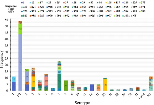
[image:4.585.40.371.619.739.2]Relationship between serotypes and STs. The distribution of STs by serotype illustrated the diversity of theS. suisstrains (Fig. 2). Fifteen of the 20 serotypes identified contained multiple STs, with the number of different STs within a single serotype ranging from 2 to 8. The predominant serotype 1/2 contained three STs (ST28 [n⫽44], ST961 [n⫽8], and ST982 [n⫽1]). Serotypes 8, 14, 24, 28, and 29 contained a single ST each, namely, ST87, ST1, ST94, ST968, and ST972, respectively. However, serotypes 24, 28, 29, and 1or14 contained only a single isolate.
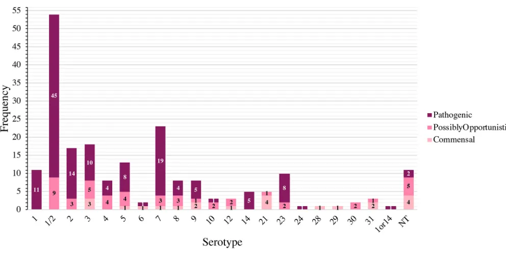
Distribution of pathotypes by serotype and ST.The distribution ofS. suis patho-types by serotype is shown in Fig. 3. Serotype 1/2 contained the most pathogenic isolates (n⫽45/54), followed by serotypes 7 (n⫽19/23), 2 (n⫽14/17), and 1 (n⫽11/ 11). Serotypes 1 and serotype 14 (n⫽5) were composed entirely of pathogenic isolates. The distribution of pathotypes by ST is shown in Fig. 4. ST28 contained the most pathogenic isolates (n⫽42/52), followed by ST1 (n⫽17/17), ST94 (n⫽14/18), and ST108 (n⫽14/17), of which only ST1 was composed entirely of pathogenic isolates. Additional pathogenic STs included ST961 (n⫽9/10), ST977 (n⫽7/9), and ST13 (n⫽5/ 5). Twelve STs contained isolates only classified as commensal, with ST750 and ST821 containing more than a single commensal isolate.
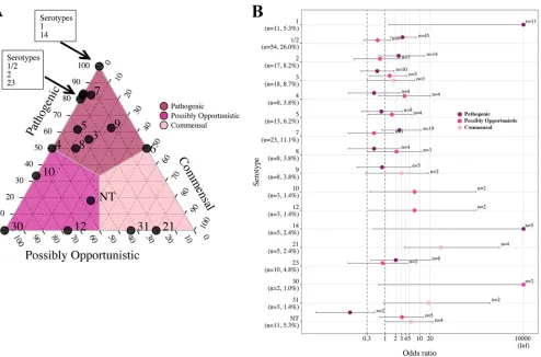
Associations among pathotypes, serotypes, and STs by analysis of proportions and OR. Associations between pathotype and serotype.Proportions and OR anal-yses were used to investigate pathotype associations with serotype for serotypes (proportions) or serotype-pathotype combinations (OR analysis) that contained more than one isolate. Between 80% and 100% of serotypes 1, 1/2, 2, 7, 14, and 23 were
FIG 2Distribution ofS. suisSTs by serotype. The stacked histogram illustrates the serotypes identified in this study, which were subdivided by STs. Thexaxis represents each serotype while theyaxis represents the frequency of each serotype. Bar sections are labeled with their respective STs. The category 1or14 and (nontypeable (NT) represents isolates with serotypes that could not be differentiated by coagglutination, PCR, or WGS.
Serotype, MLST, and Pathotype ofStreptococcus suis Journal of Clinical Microbiology
on May 17, 2020 by guest
http://jcm.asm.org/
[image:5.585.43.542.69.411.2]classified as the pathogenic pathotype (Fig. 5A), and these associations were supported by OR analysis (Fig. 5B). In the ternary plot, serotypes 3, 5, and 9 demonstrated a moderate association with the pathogenic pathotype, with 56% to 63% of isolates classified as pathogenic. However, the association between pathotype and serotype was not supported by OR analysis. OR analysis supported associations of serotypes 10 and 12 with the possibly opportunistic pathotype, with 67% of isolates classified as possibly opportunistic in the ternary plot. Serotypes 21 and 31, with 67% to 80% of
FIG 3Distribution ofS. suispathotypes by serotype. The stacked histogram illustrates the serotypes identified in this study, which were subdivided by pathotype (pathogenic, possibly opportunistic, and commensal). Thexaxis represents each serotype while theyaxis represents the frequency of each pathotype. Bar sections are labeled with their respective pathotypes. The category 1or14 and NT (nontypeable) represents isolates with serotypes that could not be differentiated by coagglutination, PCR, or WGS.
FIG 4Distribution ofS. suispathotypes by ST. The stacked histogram illustrates the STs identified in this study, which were subdivided by pathotype (pathogenic, possibly opportunistic, and commensal). Thexaxis represents each ST while theyaxis represents the frequency of each pathotype. Bar sections are labeled with their respective pathotypes. Not found (NF) indicates ST could not be determined because one housekeeping gene could not be identified for MLST classification.
Estrada et al. Journal of Clinical Microbiology
on May 17, 2020 by guest
http://jcm.asm.org/
[image:6.585.42.538.67.315.2] [image:6.585.41.538.502.697.2]isolates classified as commensal in the ternary plot, were supported as commensal pathotypes by OR analysis.
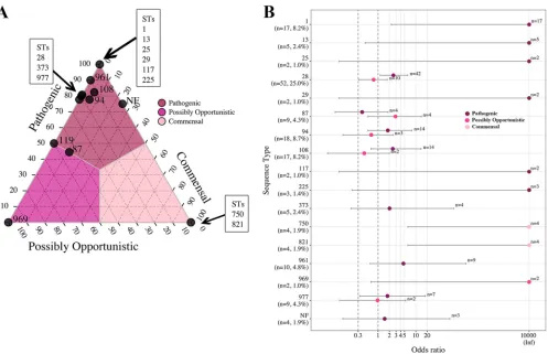
Associations between pathotype and ST.Proportions and OR analysis were used to investigate pathotype associations with ST for STs (proportions) or ST-pathotype combinations (OR analysis) that contained more than one isolate. The ternary plot of the 58 STs (and the NF category) illustrated a clear differentiation by pathotype for all STs except ST87 and ST119 (approximately 50% pathogenic) (Fig. 6A). Twelve STs and the NF category contained over 75% of isolates classified as pathogenic, including ST1, ST13, ST25, ST28, ST29, ST94, ST108, ST117, ST225, ST373, ST961, and ST977, which demonstrated the same associations by OR (Fig. 6B). ST969 had an association with the possibly opportunistic pathotype, which was supported by OR. The commensal patho-type demonstrated a strong association with ST750 and ST821, which was supported by OR analysis.
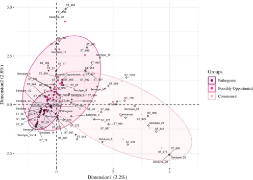
Odds ratio and MCA of pathotypes, serotypes, and STs.Initially, OR was used to investigate the relationships between pathotype and serotype-ST-combinations, but significance relationships were lacking for the combinations (OR data not shown). Then, MCA was performed to analyze the possible relationships among all serotypes, STs, and pathotypes (Fig. 7). The first and second dimensions of the analysis only represent 6% of the data. The ellipses represent 95% of isolates in each pathotype. All the subtypes demonstrating a strong association with the pathogenic pathotype by OR analysis (Fig. 5 and 6) fell within the overlapping 95% ellipses for multiple pathotypes by MCA
FIG 5Ternary and OR plots summarizing the associations betweenS. suispathotype and serotype. Only serotypes containing more than a single isolate are illustrated in the ternary plot, and only pathotype-serotype combinations containing more than a single isolate are illustrated in the OR plot. (A) The proportions of isolates classified as the pathogenic, possibly opportunistic, and commensal pathotype in each of the 16 serotypes (and the NT category) were plotted. The gray lines and color shading denote pathotype boundaries. (B) OR plot for 16 serotypes (and the NT category) versus pathotype. The dotted lines illustrate the minimum lower limit (OR, 0.3) and typical threshold (OR, 1) for identifying significant ORs. Error bars represent the 95% confidence intervals. Inf, Infinite. Nontypeable (NT) represents isolates which could not be serotyped using coagglutination, PCR, or WGS.
Serotype, MLST, and Pathotype ofStreptococcus suis Journal of Clinical Microbiology
on May 17, 2020 by guest
http://jcm.asm.org/
[image:7.585.51.545.70.396.2](Fig. 7). Five serotypes and 13 STs in the commensal pathotype lacked overlapping ellipses. Serotypes 21 and 31 lacked any isolates with the pathogenic pathotype (Fig. 3), while ST750 and ST821 contained only isolates with the commensal pathotype (Fig. 4). The limited representations of the MCA data (6% variance) and the overlapping ellipses indicate a lack of relationship between serotype, ST, and pathotype, highlighting potential confounding factors for predicting pathogenic isolates based on both sero-typing and MLST together. Thus, the relationship between pathotype, serotype, and ST is lacking for the pathogenic and possibly opportunistic pathotypes.
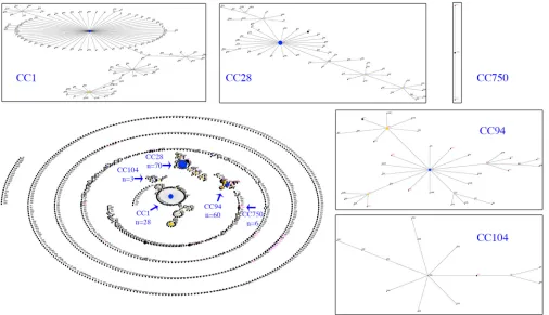
Associations between pathotype and MLST CC by analysis of proportions and OR. Identification ofS. suisCCs.To investigate the population structure of ourS. suis isolates by MLST, the STs were assigned into CCs defined by eBURST, using the entire S. suisMLST database and our 58 STs (Fig. 8 and Table S3). Using the stringent definition (six of seven shared alleles) for defining a CC, five CCs (CC1, CC28, CC94, CC104, and CC750) with a primary founder were identified from our set of STs. However, multiple STs (n⫽30) did not form a CC or formed a CC without a primary founder (Table 2). The most diverse CC (CC94) contained isolates from 13 of the 28 STs assigned into CC, compared with CC1, CC28, CC104, and CC750, which contained isolates from 4, 7, 1, and 3 STs, respectively.
Associations between pathotype and CC.Patterns between CC and pathotype were investigated by proportions and OR analysis. CC1, CC28, CC94, and CC104 were associated with the pathogenic pathotype, and the association was supported by OR
FIG 6Ternary and OR plots summarizing the associations betweenS. suispathotype and ST. Only STs containing more than a single isolate are illustrated in the ternary plot, and only pathotype-ST combinations containing more than a single isolate are illustrated in the OR plot. (A) The proportions of isolates classified as the pathogenic, possibly opportunistic, and commensal pathotype in each of the 17 STs (and the NF category) were plotted. The gray lines and color shading denote pathotype boundaries. (B) OR plot for 16 STs (and the NF category) versus pathotype. ST119 contained a single isolate of each pathogenetic and possibly opportunistic pathotype failing to meet the criteria for the plot. The dotted lines illustrate the minimum lower limit (OR, 0.3) and typical threshold (OR, 1) for identifying significant ORs. Error bars represent the 95% confidence intervals. Infinite (Inf) is represented by the value 10,000 for visualization purposes. Not found (NF) indicates ST could not be determined because one housekeeping gene could not be identified for MLST classification.
Estrada et al. Journal of Clinical Microbiology
on May 17, 2020 by guest
http://jcm.asm.org/
[image:8.585.47.544.69.390.2]analysis (Fig. 9). CC750 was associated with the commensal pathotype and was supported by OR analysis, with 83% of isolates classified as the commensal pathotype. The STs among the group of isolates lacking a CC did not associate with any pathotype. Associations among pathotypes, serotypes, and MLST by clustering analysis. To investigate genetic relationships among the samples and possible associations among serotype, genotype, and pathotype classifications, the MLST allelic sequences were clustered, illustrating 75% to 100% nucleotide sequence identity (Fig. 10). The five main CCs were identifiable in the MLST sequence identity heatmap as clusters of genetically similar strains. CC28 contained the most isolates with the pathogenic pathotype (n⫽58/70) (Table 2) and was mostly composed of ST28 (predominant ST, n⫽52) belonging to serotype 1/2 (predominant serotype,n⫽44) and serotype 2 (n⫽8) (Fig. 10). CC28 also contained ST961 (n⫽10) belonging to serotype 1/2 (n⫽8) and serotype 2 (n⫽2). MLST clustering analysis demonstrated clustering of CC28 and CC104, and the latter consisted of only three isolates with the pathogenic pathotype (serotype 7, ST225).
CC1 was divided into two groups and clustered with CC750 and isolates without a CC. The first cluster of CC1 contained a concentration of isolates in the pathogenic pathotype (n⫽17/28), while the second cluster contained 4 pathogenic isolates, 6 possibly opportunistic isolates, and a single isolate with the commensal pathotype (Table 2). The isolates within the first cluster of CC1 were predominantly characterized as ST1 (serotype 1 [n⫽7/17], serotype 2 [n⫽5/17], and serotype 14 [n⫽5/17]).
FIG 7Three-way MCA analyzing the associations among pathotypes, serotypes, and STs. The ellipses represent 95% of isolates in each pathotype. Dots represent isolates colored by their respective pathotype, and hollow diamonds represent the three active variables (pathotype, serotype, and ST). Nontypeable (NT) represents isolates which could not be serotyped using coagglutination, PCR, or WGS. Not found (NF) indicates ST could not be determined because one housekeeping gene could not be identified for MLST classification.
Serotype, MLST, and Pathotype ofStreptococcus suis Journal of Clinical Microbiology
on May 17, 2020 by guest
http://jcm.asm.org/
[image:9.585.43.539.72.425.2]Lacking a CC, the ST13 isolates (n⫽5; serotype 1 [n⫽4] and serotype 1or14 [n⫽1]) clustered with CC1 isolates, demonstrating a possible genetic relatedness to isolates of CC1 and the pathogenic pathotype. Serotypes 1, 2, and 14 and ST1 and ST13 were also associated with isolates of the pathogenic pathotype by proportions and OR. Inversely, CC750 (n⫽6) consisted of isolates with the commensal (n⫽5) and possibly opportu-nistic (n⫽1) pathotypes and was predominantly composed of isolates characterized as nontypeable (n⫽5/6) and ST750 (n⫽4/6). Interestingly, CC750 was closely related to the group of isolates lacking a CC (n⫽31), which consisted of isolates with the commensal pathotype (n⫽12/31, multiple serotypes and novel STs), providing further evidence for the association between CC750 and the commensal pathotype.
CC94 was predominantly composed of isolates with the pathogenic pathotype (n⫽43/60) but contained isolates with all three pathotypes (Table 2). The isolates within CC94 with the pathogenic pathotype were predominantly characterized as ST94
[image:10.585.42.548.72.363.2]FIG 8eBURST illustration of the globalS. suispopulation. Primary founders (blue) are positioned at the center of the cluster and subgroup founders are shown in yellow. Clusters of linked STs correspond to CCs. Magenta dots mark the STs identified in our study and arrows mark the CCs relevant to this study. Individual CCs have been expanded to illustrate relationships among STs.
TABLE 2PrimaryS. suisCCs identified in this study
No. of CCs
Total no. of isolatesa
No. of pathogenic isolates
No. of possibly opportunistic isolates
No. of commensal
isolates ST(s)
1 28 21 6 1 1, 17, 23, 87
28 70 58 12 0 25, 27–29, 117, 961b, 973b
94 60 43 14 3 94, 108, 119, 373, 839, 962b, 964b, 966b, 969b, 977b, 980–982b
104 3 3 0 0 225
750 6 0 1 5 750, 972b, 992b
noCCc 37 11 14 12 13, 821, 948, 949, 963b, 965b, 967b, 968b, 971b, 974–976b,
978b, 979b, 983–991b, 993–998b, 1001b
aSTs could not be determined for 4 of the 208 isolates because 1 housekeeping gene could not be identified for MLST classification. bNovel ST(s).
cCCs were lacking for STs that occurred as singletons or had no determined founder.
Estrada et al. Journal of Clinical Microbiology
on May 17, 2020 by guest
http://jcm.asm.org/
[image:10.585.41.561.609.711.2](n⫽14/43; serotype 7 [n⫽10/14]), ST108 (n⫽14/43; serotype 23 [n⫽8/14]), and ST977 (n⫽7/43; serotype 5 [n⫽6/7]). Serotype 7 was the second-most predominant serotype and was associated with the pathogenic pathotype by proportions and OR. Clustering analysis identified CC1, CC28, CC104, and a subset of CC94 as corresponding to the pathogenic pathotype. Isolates within these CCs were predominantly character-ized as serotypes 1, 1/2, 2, 7, 14, and 23 and ST1, ST13, ST28, ST94, ST108, ST961, and ST977, providing further evidence of these subtypes corresponding to the pathogenic pathotype.
DISCUSSION
S. suis is an important swine pathogen, often resulting in neurological and sys-temic disease caused by pathogenic strains. However, much is still unknown about the population structure ofS. suisin the United States. In this study, we utilized serological and molecular typing techniques to investigate the serotype and ST distributions of U.S. isolates. Fourteen of the 20 S. suisserotypes identified in this study were recovered from pigs with clinical disease (n⫽139). The predominant pathogenic serotypes identified in this study were 1/2 (n⫽45), 7 (n⫽19), and 2 (n⫽14), which have been previously identified as the predominant serotypes from diseased pigs in North Amer-ica (14, 15, 49, 50). While serotypes 2 and 3 are considered predominant pathogenic serotypes in North America, only 10.6% of the strains in our study were recovered from diseased pigs. Furthermore, the serotype distribution from our study differed from European studies, in which serotypes 2 and 9 are predominant (50, 51). The higher prevalence of serotype 1/2 in North America could be due to a common
evolu-FIG 9Ternary and OR plots summarizing the associations between pathotype and CC. (A) The proportions of isolates classified as the pathogenic, possibly opportunistic, and commensal pathotype in each of the five CCs (and the noCC category) were plotted. The gray lines and color shading denote pathotype boundaries. (B) OR plot for all five CCs (and the noCC category) versus pathotype. The dotted lines illustrate the minimum lower limit (OR, 0.3) and typical threshold (OR, 1) for identifying significant ORs. Error bars represent the 95% confidence intervals. Infinite (Inf) is represented by the value 10,000 for visualization purposes. noCC represents the group of isolates lacking a CC.
Serotype, MLST, and Pathotype ofStreptococcus suis Journal of Clinical Microbiology
on May 17, 2020 by guest
http://jcm.asm.org/
[image:11.585.44.543.71.388.2]tionary lineage with serotype 2. Genetic analysis by PCR-based serotyping of thecps loci demonstrated serotypes 1/2 and 2 share the same genetic profile and cannot be differentiated by serotype-specificcpsloci (11, 12). Sequencing of thecpsKgene reveals a missense mutation permitting the differentiation of serotypes 2 and 1/2 (12), but a PCR protocol has not been implemented yet to differentiate these serotypes.
In our study, the geographic distribution ofS. suiswas from 20 different states (Table S1), which represent the major swine-producing states in the United States. Variability in the serotype distribution ofS. suishas been reported within the same country, which is likely due to natural differences in geographic distribution (13). Geographic distri-bution of theS. suisserotypes in our study identified serotype 1/2 in 13 of the 20 states, with a concentration in 5 of the 20 states, possibly displaying a geographic distribution pattern of serotype 1/2 in the United States. Serotype 1/2 is also a frequent serotype
FIG 10MLST sequence identity heatmap ofS. suis. Isolates are annotated (colored rectangles) by pathotype (1), serotype (2), and ST (3). The five CCs are indicated by black brackets, with the number of isolates in the CC. Blue brackets represent clusters of isolates without a CC. Nontypeable (NT) represents isolates which could not be serotyped using coagglutination, PCR, or WGS. #, group of isolates lacking a CC;⫹, ST13 not within a CC but closest to CC1;⬃, ST979 not within a CC but closest to CC94.
Estrada et al. Journal of Clinical Microbiology
on May 17, 2020 by guest
http://jcm.asm.org/
[image:12.585.43.535.71.523.2]found in Canada, although at lower levels than serotypes 2 and 3 (52). This prevalence of serotype 1/2 in Canada may contribute to the U.S. serotype distribution through the transport of pigs between the two countries (50). Transport of livestock has been associated with geographic invasion or the emergence of a pathogen in a novel geographic area (53–55). While most transport of pigs to the United States head to harvest facilities, new breeding stock of pigs could be colonialized with new S. suis strains, which could result in the spread of new strains to downstream swine farms. Whole-genome analysis of the U.S. and Canadian serotype 1/2 strains would further clarify the relationship between U.S. and Canadian 1/2 strains.
We anticipated identifying a large number of novel ST profiles due to the inclusion of commensal and possibly opportunistic samples, which are not generally subjected to subtyping by MLST. As a result of this study, 38 novel ST profiles were submitted to the S. suisMLST database. Of the 58 STs identified here, 24 STs were isolated from pigs with clinical disease, and the predominant STs were ST28 (n⫽42), followed by ST1 (n⫽17), ST94 (n⫽14), and ST108 (n⫽14). In a previous Canadian study in 2011, ST25 was the predominant ST found in Canada, while ST28 was the predominant ST found in the United States (22). Our results confirm ST28 as a predominant pathogenic pathotype, while ST25 represents only 1% of the strains recovered from diseased pigs (n⫽2). The reason for this low percentage of ST25 isolates in the United States is unclear, and updated ST analysis ofS. suis strains from Canada is needed to confirm ST25 as the predominant ST in that country. Our ST distribution also differs from that of European and Asian countries in which ST1 strains, largely characterized as serotype 2, are predominant in diseased pigs (50, 56).
Proportions, OR, and clustering analysis illustrated potential relationships among pathotypes, serotypes, and STs. While multiple pathogenic serotypes and STs were identified in our study, this discussion focuses on serotype and STs with more than four isolates in the pathogenic pathotype. Serotypes 1, 1/2, 2, 7, 14, and 23 as well as ST1, ST13, ST28, ST94, ST108, ST961, and ST977 were frequently identified as pathogenic strains. Based on our pathotype classifications, isolates characterized as pathogenic were linked to neurological or systemic disease, and our analyses provide evidence that these subtypes are potential indicators of virulence. As discussed previously, serotypes 2 and 1/2 are predominant serotypes identified from diseased pigs in North America, supporting our observations of these serotypes as pathogenic strains by proportions, OR, and clustering analysis (14, 15, 49, 50, 52).
Serotypes 1 and 7 are more prevalent in diseased pigs in some European countries than in North America, and pathogenic serotype 1 strains have been linked to the production of muramidase-released protein (MRP), extracellular-factor protein (EF), and suilysin (SLY). Pathogenic serotype 1 strains have been characterized as producing both MRP and EF, with variable production of SLY (16, 18). In one study (18), four of the six serotype 1 strains were MRP⫹EF⫹SLY⫹and five of the six were either ST1 or ST13,
indicating a correlation between serotype 1, ST1, ST13, and virulence. Interestingly, the serotype 1 isolates in the current study were either ST1 (n⫽7/11) or ST13 (n⫽4/11) and were associated with the pathogenic pathotype, supporting the previous study. Serotype 7 was the second-most common serotype identified in this study, and 19/23 isolates were characterized as the pathogenic pathotype. Virulence studies on serotype 7 strains demonstrating clinical disease in pigs are limited, but a previousin vivostudy associated serotype 7 with septicemia and arthritis, with rare cases of meningitis (57). These findings support the classification of serotype 7 as pathogenic.
This study demonstrates that ST appears to be a stronger predictor of pathotype than serotype. While experimental mouse models have demonstrated the virulence of serotype 2 ST1, ST25, and ST28 (22, 56), our analyses also illustrated ST1, ST13, ST28, ST94, ST108, ST961, and ST977 (of various serotypes) as pathogenic. As mentioned previously, we hypothesize that Canadian and U.S. serotype 2 and serotype 1/2 strains share a evolutionary lineage. If so, the observed virulence of serotype 2 ST28 in previous studies may support the virulence of serotype 1/2 ST28, as predicted in our study. Whole-genome single nucleotide polymorphism (SNP)-based phylogenetic analysis of
Serotype, MLST, and Pathotype ofStreptococcus suis Journal of Clinical Microbiology
on May 17, 2020 by guest
http://jcm.asm.org/
S. suisserotype 2 ST28 strains revealed a unique clade composed of virulent strains capable of inducing severe disease in a murine infection model (58). These strains demonstrated differences in virulence to reference serotype 2 ST28 strains of low virulence. Recently, a study characterized pathogenic Australian serotype 1/2 ST1 strains by core genome single nucleotide polymorphisms and linked the genetic similarity to pathogenic serotype 1/2 ST1 strains from the United Kingdom and Vietnam (59). Our clustering analysis indicates that ST1, ST13, ST94, ST108, ST961, and ST977 may also be pathogenic. It would be of interest to further investigate the virulence properties of serotype 1/2 ST28, as well as ST1, ST13, ST94, ST108, ST961, and ST977 strains isolated in the United States.
In addition to strains in CC1, CC28, and CC104, serotype 9 strains belonging to CC16 (previously CC87) have been isolated from pigs with invasive disease (20). However, the low percentage of serotype 9 strains in our study is reasonable because serotype 9 is predominant in diseased pigs from the Netherlands (16). The serotype 9 strains in this study belong to multiple CCs or occur as singletons and did not demonstrate associ-ations with pathotype. Serotype 9 isolates from diseased and healthy pigs in China were characterized into multiple STs and demonstrated high diversity among the isolates (60). The majority of these serotype 9 isolate STs occurred as singletons and did not form major clonal complexes.
Inversely, commensalS. suisserotypes 21 and 31 and ST750 and ST821 were identified by proportions, OR, and cluster analysis. Studies on S. suis from North America have observed a prevalence of serotype 21 from healthy pigs (26, 27). However, previous studies have identified a limited number of serotype 31 strains from pigs with typical clinical signs of S. suisdisease (49, 52, 61, 62). The association between serotype 31 and pathotype remains unclear and requires further investigation.
Associations among serotypes, STs, and pathotypes, although identified by individ-ual analyses, were not evident in the MCA, indicating both serotype and ST together could not indicate pathotype. We investigated additional approaches, such as chi-square and Fisher’s exact tests, but these tests failed to generate significant relation-ships between both serotype and ST. In addition, we investigated associations between serotype-ST combinations and pathotype by chi-square and Fisher’s exact tests and did not identify any significant associations. One possible explanation for this is the lack of discrimination due to the limitations of sample size within each subtype. Traditional chi-square and Fisher’s exact tests work best on nonsparse data (few zero values) (63, 64). These tests have been used to identify associations betweenS. suissubtypes and characteristics of pathogenicity. However, most studies involved a limited number of subtypes of interest, while our study focused on all serotypes and STs identified in our sample set. Due to the diversity of theS. suisstrains in this study and the large number of subtypes evaluated, the division of our data by pathotype resulted in sparse data. Thus, sparse data limits our ability to conduct certain analyses using common ap-proaches for S. suis. An OR formula was used to evaluate statistical significance of subtype with pathotype, as well as the size of the possible effect, to limit the misiden-tification of associations due to sample size. For this reason, proportions were used for basic identification of relationships and OR analysis was used for further discrimination of strains.
In summary, our study increases the knowledge onS. suisstrains circulating in the United States between 2014 and 2017 by investigating serotype and ST distributions. We identified a diverse set of strains, predominantly serotypes 1/2, 3, and 7, and as ST1, ST28, and ST94. Further investigation by pathotype classification (defined in this study) identified STs that could be differentiated as pathogenic or commensal pathotypes. The predominance of serotype 1/2 strains from clinically affected pigs in our study stresses the importance of expanding studies of virulence traits to other serotypes and STs of S. suis. These findings can be applied to improve the prevention and control ofS. suis by selecting strains for diagnostics and vaccine development.
Estrada et al. Journal of Clinical Microbiology
on May 17, 2020 by guest
http://jcm.asm.org/
SUPPLEMENTAL MATERIAL
Supplemental material for this article may be found athttps://doi.org/10.1128/JCM .00377-19.
SUPPLEMENTAL FILE 1, XLSX file, 0.03 MB. SUPPLEMENTAL FILE 2, XLSX file, 0.01 MB. SUPPLEMENTAL FILE 3, XLSX file, 0.01 MB.
ACKNOWLEDGMENTS
We thank the bacteriology sections at UMNVDL and KSVDL for their technical assistance. In addition, we thank Lacey Marshall-Lund (University of Minnesota, St. Paul, MN) and Marta Pérez-Sancho (Universidad Complutense de Madrid, Madrid, Spain) for technical assistance and Richard Gebhart (University of Minnesota, St. Paul, MN) and Laura Bruner (Swine Vet Center) for critical discussion of the manuscript.
The research project was funded by the Rapid Agricultural Response Fund, estab-lished by the Minnesota legislature and administered by the University of Minnesota Agricultural Experiment Station. April A. Estrada’s stipend was funded by the USDA National Needs Fellowship Award, 2016-38420-25288.
REFERENCES
1. Clifton-Hadley FA. 1984. Studies ofStreptococcus suistype 2 infection in pigs. Vet Res Commun 8:217–227.https://doi.org/10.1007/BF02214715. 2. Reams RY, Glickman LT, Harrington DD, Thacker HL, Bowersock TL. 1994.
Streptococcus suisinfection in swine: a retrospective study of 256 cases. Part II. Clinical signs, gross and microscopic lesions, and coexisting microorganisms. J Vet Diagn Invest 6:326 –334.https://doi.org/10.1177/ 104063879400600308.
3. Galina L, Collins JE, Pijoan C. 1992. Porcine Streptococcus suis in Minnesota. J Vet Diagn Invest 4:195–196. https://doi.org/10.1177/ 104063879200400216.
4. Weinert LA, Chaudhuri RR, Wang J, Peters SE, Corander J, Jombart T, Baig A, Howell KJ, Vehkala M, Välimäki N, Harris D, Chieu TTB, Van Vinh Chau N, Campbell J, Schultsz C, Parkhill J, Bentley SD, Langford PR, Rycroft AN, Wren BW, Farrar J, Baker S, Hoa NT, Holden MTG, Tucker AW, Maskell DJ. 2015. Genomic signatures of human and animal disease in the zoonotic pathogenStreptococcus suis. Nat Commun 6:6740 – 6748.https://doi.org/ 10.1038/ncomms8272.
5. Clifton-Hadley FA, Alexander TJ. 1980. The carrier site and carrier rate of
Streptococcus suistype II in pigs. Vet Rec 107:40 – 41.https://doi.org/10 .1136/vr.107.2.40.
6. Arends JP, Hartwig N, Rudolphy M, Zanen HC. 1984. Carrier rate of
Streptococcus suiscapsular type 2 in palatine tonsils of slaughtered pigs. J Clin Microbiol 20:945–947.
7. Gottschalk M, Segura M, Xu J. 2007.Streptococcus suisinfections in humans: the Chinese experience and the situation in North America. Anim Health Res Rev 8:29 – 45.https://doi.org/10.1017/S1466252307001247.
8. Hill JE, Gottschalk M, Brousseau R, Harel J, Hemmingsen SM, Goh SH. 2005. Biochemical analysis, cpn60 and 16S rDNA sequence data indicate thatStreptococcus suis serotypes 32 and 34, isolated from pigs, are
Streptococcus orisratti. Vet Microbiol 107:63– 69.https://doi.org/10.1016/ j.vetmic.2005.01.003.
9. Tien LHT, Nishibori T, Nishitani Y, Nomoto R, Osawa R. 2013. Reappraisal of the taxonomy ofStreptococcus suisserotypes 20, 22, 26, and 33 based on DNA–DNA homology and sodA and recN phylogenies. Vet Microbiol 162:842– 849.https://doi.org/10.1016/j.vetmic.2012.11.001.
10. Tohya M, Arai S, Tomida J, Watanabe T, Kawamura Y, Katsumi M, Ushimizu M, Ishida-Kuroki K, Yoshizumi M, Uzawa Y, Iguchi S, Yoshida A, Kikuchi K, Sekizaki T. 2017. Defining the taxonomic status ofStreptococcus suis sero-type 33: the proposal forStreptococcus ruminantiumsp. nov. Int J Syst Evol Microbiol 67:3660 –3665.https://doi.org/10.1099/ijsem.0.002204. 11. Kerdsin A, Akeda Y, Hatrongjit R, Detchawna U, Sekizaki T, Hamada S,
Gottschalk M, Oishi K. 2014.Streptococcus suisserotyping by a new multiplex PCR. J Med Microbiol 63:824 – 830.https://doi.org/10.1099/ jmm.0.069757-0.
12. Athey TBT, Teatero S, Lacouture S, Takamatsu D, Gottschalk M, Fittipaldi N. 2016. DeterminingStreptococcus suisserotype from short-read whole-genome sequencing data. BMC Microbiol 16:162–169.https://doi.org/10 .1186/s12866-016-0782-8.
13. Reams RY, Glickman LT, Harrington DD, Bowersock TL, Thacker HL. 1993.
Streptococcus suisinfection in swine: a retrospective study of 256 cases. Part I. Epidemiologic factors and antibiotic susceptibility patterns. J Vet Diagn Invest 5:363–367.https://doi.org/10.1177/104063879300500310. 14. Fittipaldi N, Fuller TE, Teel JF, Wilson TL, Wolfram TJ, Lowery DE,
Gottschalk M. 2009. Serotype distribution and production of muramidase-released protein, extracellular factor and suilysin by field strains ofStreptococcus suisisolated in the United States. Vet Microbiol 139:310 –317.https://doi.org/10.1016/j.vetmic.2009.06.024.
15. Messier S, Lacouture S, Gottschalk M. 2008. Distribution ofStreptococcus suiscapsular types from 2001 to 2007. Can Vet J 49:461– 462. 16. Wisselink HJ, Smith HE, Stockhofe-Zurwieden N, Peperkamp K, Vecht U.
2000. Distribution of capsular types and production of muramidase-released protein (MRP) and extracellular factor (EF) ofStreptococcus suis
strains isolated from diseased pigs in seven European countries. Vet Microbiol 74:237–248.https://doi.org/10.1016/S0378-1135(00)00188-7. 17. King SJ, Leigh JA, Heath PJ, Luque I, Tarradas C, Dowson CG, Whatmore
AM. 2002. Development of a multilocus sequence typing scheme for the pig pathogenStreptococcus suis: identification of virulent clones and potential capsular serotype exchange. J Clin Microbiol 40:3671–3680. https://doi.org/10.1128/jcm.40.10.3671-3680.2002.
18. de Greeff A, Wisselink HJ, de Bree FM, Schultsz C, Baums CG, Thi HN, Stockhofe-Zurwieden N, Smith HE. 2011. Genetic diversity of Streptococ-cus suisisolates as determined by comparative genome hybridization. BMC Microbiol 11:161–175.https://doi.org/10.1186/1471-2180-11-161. 19. Onishi H, Sugawara M, Okura M, Osaki M, Takamatsu D. 2012. Prevalence of
Streptococcus Suisgenotypes in isolates from porcine endocarditis in East Japan. J Vet Med Sci 74:1681–1684.https://doi.org/10.1292/jvms.12-0301. 20. Schultsz C, Jansen E, Keijzers W, Rothkamp A, Duim B, Wagenaar JA, van
der Ende A. 2012. Differences in the population structure of invasive
Streptococcus suisstrains isolated from pigs and from humans in the Netherlands. PLoS One 7:e33854.https://doi.org/10.1371/journal.pone .0033854.
21. Zhu W, Wu C, Sun X, Zhang A, Zhu J, Hua Y, Chen H, Jin M. 2013. Characterization ofStreptococcus suisserotype 2 isolates from China. Vet Microbiol 166:527–534.https://doi.org/10.1016/j.vetmic.2013.06.009. 22. Fittipaldi N, Xu J, Lacouture S, Tharavichitkul P, Osaki M, Sekizaki T,
Takamatsu D, Gottschalk M. 2011. Lineage and virulence ofStreptococcus suis serotype 2 isolates from North America. Emerg Infect Dis 17: 2239 –2244.https://doi.org/10.3201/eid1712.110609.
23. Pérez-Sancho M, Vela AI, García-Seco T, González S, Domínguez L, Fernández-Garayzábal JF. 2017. Usefulness of MALDI-TOF MS as a diagnos-tic tool for the identification ofStreptococcusspecies recovered from clinical specimens of pigs. PLoS One 12:e0170784.https://doi.org/10.1371/journal .pone.0170784.
24. Staats JJ, Feder I, Okwumabua O, Chengappa MM. 1997.Streptococcus suis: past and present. Vet Res Commun 21:381– 407.https://doi.org/10 .1023/A:1005870317757.
Serotype, MLST, and Pathotype ofStreptococcus suis Journal of Clinical Microbiology
on May 17, 2020 by guest
http://jcm.asm.org/
25. Higgins R, Gottschalk M, Beaudoin M, Rawluk SA. 1992. Distribution of
Streptococcus suiscapsular types in Quebec and western Canada. Can Vet J 33:27–30.
26. Gottschalk M, Higgins R, Jacques M, Mittal KR, Henrichsen J. 1989. Description of 14 new capsular types ofStreptococcus suis. J Clin Micro-biol 27:2633–2636.
27. Gottschalk M, Higgins R, Jacques M, Beaudoin M, Henrichsen J. 1991. Isolation and characterization ofStreptococcus suiscapsular types 9 –22. J Vet Diagn Invest 3:60 – 65.https://doi.org/10.1177/104063879100300113. 28. Higgins R, Gottschalk M, Boudreau M, Lebrun A, Henrichsen J. 1995.
Description of six new capsular types (29 –34) ofStreptococcus suis. J Vet Diagn Invest 7:405– 406.https://doi.org/10.1177/104063879500700322. 29. Gottschalk M, Higgins R, Jacques M, Beaudoin M, Henrichsen J. 1991. Characterization of six new capsular types (23 through 28) of Strepto-coccus suis. J Clin Microbiol 29:2590 –2594.
30. Okura M, Lachance C, Osaki M, Sekizaki T, Maruyama F, Nozawa T, Nakagawa I, Hamada S, Rossignol C, Gottschalk M, Takamatsu D. 2014. Development of a two-step multiplex PCR assay for typing of capsular polysaccharide synthesis gene clusters ofStreptococcus suis. J Clin Mi-crobiol 52:1714 –1719.https://doi.org/10.1128/JCM.03411-13. 31. Bolger AM, Lohse M, Usadel B. 2014. Trimmomatic: a flexible trimmer for
Illumina sequence data. Bioinformatics 30:2114 –2120.https://doi.org/10 .1093/bioinformatics/btu170.
32. Ishida S, Tien LHT, Osawa R, Tohya M, Nomoto R, Kawamura Y, Takahashi T, Kikuchi N, Kikuchi K, Sekizaki T. 2014. Development of an appropriate PCR system for the reclassification of Streptococcus suis. J Microbiol Methods 107:66 –70.https://doi.org/10.1016/j.mimet.2014.09.003. 33. Inouye M, Dashnow H, Raven L-A, Schultz MB, Pope BJ, Tomita T, Zobel
J, Holt KE. 2014. SRST2: rapid genomic surveillance for public health and hospital microbiology labs. Genome Med 6:90 –105.https://doi.org/10 .1186/s13073-014-0090-6.
34. Jolley KA, Maiden MC. 2010. BIGSdb: scalable analysis of bacterial ge-nome variation at the population level. BMC Bioinformatics 11:595– 605. https://doi.org/10.1186/1471-2105-11-595.
35. Rehm T, Baums CG, Strommenger B, Beyerbach M, Valentin-Weigand P, Goethe R. 2007. Amplified fragment length polymorphism of Strepto-coccus suisstrains correlates with their profile of virulence-associated genes and clinical background. J Med Microbiol 56:102–109.https://doi .org/10.1099/jmm.0.46616-0.
36. Feil EJ, Li BC, Aanensen DM, Hanage WP, Spratt BG. 2004. eBURST: inferring patterns of evolutionary descent among clusters of related bacterial genotypes from multilocus sequence typing data. J Bacteriol 186:1518 –1530.https://doi.org/10.1128/JB.186.5.1518-1530.2004. 37. R Core Team. 2017. R: a language and environment for statistical
com-puting. R Foundation for Statistical Computing, Vienna, Austria. 38. Charif D, Lobry JR. 2007. SeqinR 1.0-2: a contributed package to the R
project for statistical computing devoted to biological sequences re-trieval and analysis, p. 207–232. InBastolla U, Porto M, Roman HE, Vendruscolo M (ed). Structural approaches to sequence evolution: mol-ecules, networks, populations. Springer Verlag, New York.
39. Day A. 2012 heatmap.plus.package: heatmap with more sensible behav-ior. The Comprehensive R Archive Network (CRAN), R Foundation for Statistical Computing, Vienna, Austria.
40. Bodenhofer U, Bonatesta E, Horejs-Kainrath C, Hochreiter S. 2015. msa: an R package for multiple sequence alignment. Bioinformatics 31: 3997–3999.https://doi.org/10.1093/bioinformatics/btv494.
41. Warnes GR, Bolker B, Bonebakker L, Gentleman R, Liaw WHA, Lumley T, Maechler M, Magnusson A, Moeller S, Schwartz M, Venables B. 2016. gplots: various R programming tools for plotting data.https://rdrr.io/ cran/gplots/.
42. Edgar RC. 2004. MUSCLE: multiple sequence alignment with high accu-racy and high throughput. Nucleic Acids Res 32:1792–1797.https://doi .org/10.1093/nar/gkh340.
43. Wickham H. 2009. ggplot2: elegant graphics for data analysis. Springer-Verlag, New York.
44. Wickham H. 2017 tidyverse: easily install and load the “tidyverse.” https://tidyverse.tidyverse.org/.
45. Chen W, Simpson J, Levesque CA. 2018. RAM: R for amplicon-sequencing-based microbial-ecology. Agriculture and Agri-Food Canada.
46. Smith MR. 2017. Ternary: an R package for creating ternary plots. Zenodo.
47. Lê S, Josse J, Husson F. 2008. FactoMineR: a package for multivariate analysis. J Stat Softw 25:1–18.https://doi.org/10.18637/jss.v025.i01. 48. Kassambara A, Mundt F. 2017 factoextra: extract and visualize the results
of multivariate data analyses.https://rpkgs.datanovia.com/factoextra/. 49. Gottschalk M, Lacouture S, Bonifait L, Roy D, Fittipaldi N, Grenier D. 2013.
Characterization ofStreptococcus suisisolates recovered between 2008 and 2011 from diseased pigs in Québec, Canada. Vet Microbiol 162: 819 – 825.https://doi.org/10.1016/j.vetmic.2012.10.028.
50. Goyette-Desjardins G, Auger J-P, Xu J, Segura M, Gottschalk M. 2014.
Streptococcus suis, an important pig pathogen and emerging zoonotic agent-an update on the worldwide distribution based on serotyping and sequence typing. Emerg Microbes Infect 3:e45.https://doi.org/10.1038/ emi.2014.45.
51. Vela AI, Goyache J, Tarradas C, Luque I, Mateos A, Moreno MA, Borge C, Perea JA, Domínguez L, Fernández-Garayzábal JF. 2003. Analysis of genetic diversity ofStreptococcus suisclinical isolates from pigs in Spain by pulsed-field gel electrophoresis. J Clin Microbiol 41:2498 –2502. https://doi.org/10.1128/jcm.41.6.2498-2502.2003.
52. Gottschalk M, Lacouture S. 2015. Canada: distribution ofStreptococcus suis
(from 2012 to 2014) and Actinobacillus pleuropneumoniae (from 2011 to 2014) serotypes isolated from diseased pigs. Can Vet J Rev Veterinaire Can 56:1093–1094.
53. Pasma T, Davidson S, Shaw SL. 2008. Idiopathic vesicular disease in swine in Manitoba. Can Vet J 49:84 – 85.
54. Nelson MI, Lemey P, Tan Y, Vincent A, Lam TT-Y, Detmer S, Viboud C, Suchard MA, Rambaut A, Holmes EC, Gramer M. 2011. Spatial dynamics of human-origin H1 influenza A virus in North American swine. PLoS Pathog 7:e1002077.https://doi.org/10.1371/journal.ppat.1002077. 55. Lowe J, Gauger P, Harmon K, Zhang J, Connor J, Yeske P, Loula T, Levis
I, Dufresne L, Main R. 2014. Role of transportation in spread of porcine epidemic diarrhea virus infection, United States. Emerg Infect Dis 20: 872– 874.https://doi.org/10.3201/eid2005.131628.
56. Auger J-P, Fittipaldi N, Benoit-Biancamano M-O, Segura M, Gottschalk M. 2016. Virulence studies of different sequence types and geographical origins ofStreptococcus suisserotype 2 in a mouse model of infection. Pathog Basel Switz 5:E48.https://doi.org/10.3390/pathogens5030048. 57. Boetner AG, Binder M, Bille-Hansen V. 1987.Streptococcus suisinfections
in Danish pigs and experimental infection with Streptococcus suis se-rotype 7. Acta Pathol Microbiol Scand Ser B Microbiol 95B:233–239. https://doi.org/10.1111/j.1699-0463.1987.tb03118.x.
58. Athey TBT, Auger J-P, Teatero S, Dumesnil A, Takamatsu D, Wasserscheid J, Dewar K, Gottschalk M, Fittipaldi N. 2015. Complex population struc-ture and virulence differences among serotype 2 streptococcus suis
strains belonging to sequence type 28. PLoS One 10:e0137760.https:// doi.org/10.1371/journal.pone.0137760.
59. O’Dea MA, Laird T, Abraham R, Jordan D, Lugsomya K, Fitt L, Gottschalk M, Truswell A, Abraham S. 2018. Examination of AustralianStreptococcus suisisolates from clinically affected pigs in a global context and the genomic characterisation of ST1 as a predictor of virulence. Vet Micro-biol 226:31– 40.https://doi.org/10.1016/j.vetmic.2018.10.010.
60. Dong W, Zhu Y, Ma Y, Ma J, Zhang Y, Yuan L, Pan Z, Wu Z, Yao H. 2017. Multilocus sequence typing and virulence genotyping of Streptococcus suis serotype 9 isolates revealed high genetic and virulence diversity. FEMS Microbiol Lett 364:fnx192.https://doi.org/10.1093/femsle/fnx192. 61. Wongsawan K, Gottschalk M, Tharavichitkul P. 2015. Serotype- and virulence-associated gene profile ofStreptococcus suisisolates from pig carcasses in Chiang Mai Province, Northern Thailand. J Vet Med Sci 77: 233–236.https://doi.org/10.1292/jvms.14-0380.
62. Prüfer TL, Rohde J, Verspohl J, Rohde M, de Greeff A, Willenborg J, Valentin-Weigand P. 2019. Molecular typing ofStreptococcus suisstrains isolated from diseased and healthy pigs between 1996 –2016. PLoS One 14:e0210801.https://doi.org/10.1371/journal.pone.0210801.
63. Mielke PW, Berry KJ. 2002. Categorical independence tests for large sparseR-way contingency tables. Percept Mot Skills 95:606 – 610.https:// doi.org/10.2466/pms.2002.95.2.606.
64. Sourial N, Wolfson C, Zhu B, Quail J, Fletcher J, Karunananthan S, Bandeen-Roche K, Béland F, Bergman H. 2010. Correspondence analysis is a useful tool to uncover the relationships among categorical variables. J Clin Epidemiol 63:638 – 646.https://doi.org/10.1016/j.jclinepi.2009.08 .008.
Estrada et al. Journal of Clinical Microbiology