R E S E A R C H A R T I C L E
Open Access
Histopathological cutaneous alterations in
systemic sclerosis: a clinicopathological study
Jens T Van Praet
1*†, Vanessa Smith
1†, Marc Haspeslagh
2,3†, Nele Degryse
1, Dirk Elewaut
1and Filip De Keyser
1Abstract
Introduction:The aims of the present study were to identify histopathological parameters which are linked to local clinical skin disease at two distinct anatomical sites in systemic sclerosis (SSc) patients with skin involvement (limited cutaneous systemic sclerosis (lcSSc) or diffuse cutaneous systemic sclerosis (dcSSc)) and to determine the sensitivity of SSc specific histological alterations, focusing on SSc patients without clinical skin involvement (limited SSc (lSSc)).
Methods:Histopathological alterations were systematically scored in skin biopsies of 53 consecutive SSc patients (dorsal forearm and upper inner arm) and 18 controls (upper inner arm). Clinical skin involvement was evaluated using the modified Rodnan skin score. In patients with lcSSc or dcSSc, associations of histopathological parameters with local clinical skin involvement were determined by generalised estimation equation modelling.
Results:The hyalinised collagen score, the myofibroblast score, the mean epidermal thickness, the mononuclear cellular infiltration and the frequency of focal exocytosis differed significantly between biopsies with and without local clinical skin involvement. Except for mononuclear cellular infiltration, all of the continuous parameters correlated with the local clinical skin score at the dorsal forearm. Parakeratosis, myofibroblasts and intima proliferation were present in a minority of the SSc biopsies, but not in controls. No differences were found between lSSc and controls.
Conclusions:Several histopathological parameters are linked to local clinical skin disease. SSc-specific histological alterations have a low diagnostic sensitivity.
Introduction
Systemic sclerosis (SSc) is a chronic autoimmune disease which can affect the skin and various internal organs [1]. One of the hallmark clinical features of SSc is skin thickening caused by oedema and excessive accumula-tion of collagen-rich extracellular matrix. Apart from sclerosis, the pathogenesis of SSc is characterized by vasculopathy, which is evidenced by nailfold capillary abnormalities and Raynaud’s phenomenon, as well as by the presence of antinuclear antibodies such as anticen-tromere, anti-topoisomerase I and anti-RNA polymerase III antibodies. The most extensively validated technique to quantify skin involvement is the modified Rodnan skin score (mRSS) [2]. In this scoring system, 17 body
areas are examined by clinical palpation and scored on the basis of judgement of skin thickness on a 4-point ordinal scale. Because the extent of skin disease corre-lates with the disease course, patients are grouped into disease subsets on the basis of skin involvement. The currently most widely applied classification differentiates three subsets, namely, limited SSc (lSSc), limited cuta-neous SSc (lcSSc) and diffuse cutacuta-neous SSc (dcSSc) [3]. Patients with lcSSc have skin involvement confined to the fingers, hands, forearms, lower legs or the face, whereas patients with dcSSc also have more proximal skin thickening. The group classified as lSSc has Ray-naud’s phenomenon with nailfold capillaroscopic abnormalities and/or SSc-associated autoantibodies, but no clinical skin involvement. This subset is considered to have ‘early’SSc, as a longitudinal follow-up study has demonstrated that these patients are at risk for progres-sion to definite SSc [4].
* Correspondence: [email protected]
†Contributed equally
1
Department of Rheumatology, Ghent University Hospital, De Pintelaan 185, BE-9000 Gent, Belgium
Full list of author information is available at the end of the article
The histopathology of SSc skin has recently regained interest because it may be integrated as an outcome measure in clinical trials on skin disease, whereby skin biopsies are obtained before and after administration of a therapeutic drug [5,6]. In this way, the hyalinised col-lagen score and the myofibroblast score have previously been correlated with local skin score and durometry score in patients with dcSSc [7]. Consequently, these parameters could potentially be used to study the effect of a drug on skin disease. Other alterations in skin his-tology, however, have not been linked to clinical assess-ment. In addition, studies on the skin histopathology of SSc patients have mainly focused on alterations in patients with established disease, leaving open the ques-tion whether patients with‘early’disease have SSc speci-fic histological alterations.
The aims of this study were (1) to identify histopatho-logical parameters which are linked with local clinical skin disease at two different anatomical sites in SSc patients with skin involvement (lcSSc or dcSSc) and (2) to determine the sensitivity of SSc-specific histological alterations, with a focus on SSc patients with lSSc.
Materials and methods Patients
Skin biopsies from the dorsal forearm (at the transition of the distal one-third and the proximal two-thirds) and the mid-upper inner arm were obtained from 53 conse-cutive SSc patients visiting the Scleroderma Clinic of the Ghent University Hospital. From two patients, only one biopsy was available for analysis. All patients ful-filled the criteria for early SSc set forth by LeRoy and Medsger [3]. Video nailfold capillaroscopy and antinuc-lear antibody identification were performed as described previously [8]. Patients were assigned to the lSSc, lcSSc or dcSSc group according to the criteria published by LeRoyet al.[9]. Skin involvement was clinically assessed according to the 17-site mRSS, whereby local skin invol-vement is determined for each site, including the dorsal forearm, on a semiquantitative scale (0 = normal thick-ness, 1 = mild thickening, 2 = moderate thickening and 3 = severe thickening) [10]. A normal reference set was included in the analysis. This set contained the skin biopsies from the inner upper arm of patients who were referred for a lupus band test and in whom further eva-luation excluded any specified autoimmune disease. This study was conducted after approval of the Ghent Uni-versity Hospital Ethical Committee was obtained and all patients had signed informed consent.
Full-thickness skin biopsies (approximately 1.5 cm long and 0.5 cm wide) were surgically obtained while the patients were under local anaesthesia. Biopsy samples were stored in formaldehyde and embedded in paraffin. Sections of 5μm were cut and stained with haematoxylin
and eosin, Masson trichrome and colloidal iron accord-ing to standard techniques. Paraffin-embedded sections were also used for immunohistochemistry.
Immunohistochemistry
Paraffin-embedded sections were dewaxed and heated in antigen retrieval buffer at 98°C for 20 minutes using citrate buffer (pH 6) or Tris buffer (pH 9) (Pascal: Dako, Glostrup, Denmark). After rinsing and blocking endogen-ous peroxidase, sections were incubated for 60 minutes with the following mouse monoclonal antibodies (mAbs): CD3 (T cells; Novocastra, Newcastle, UK) anda-smooth muscle actin (a-SMA) (myofibroblasts, clone 1A4; Dako). Parallel sections were incubated with irrelevant isotype-matched mAbs as negative controls. The sections were subsequently incubated for 15 minutes with a biotinylated antimouse secondary antibody, followed by 15-minute incubation with a streptavidin-peroxidase complex (LSAB+ Kit; Dako). The colour reaction was developed using 3-amino-9-ethylcarbazole substrate (Dako) as chro-mogen. Finally, the sections were counterstained with hae-matoxylin. All incubations were carried out at room temperature, and the sections were washed with phos-phate-buffered saline between all steps.
Selection of scoring parameters for SSc histological skin alterations
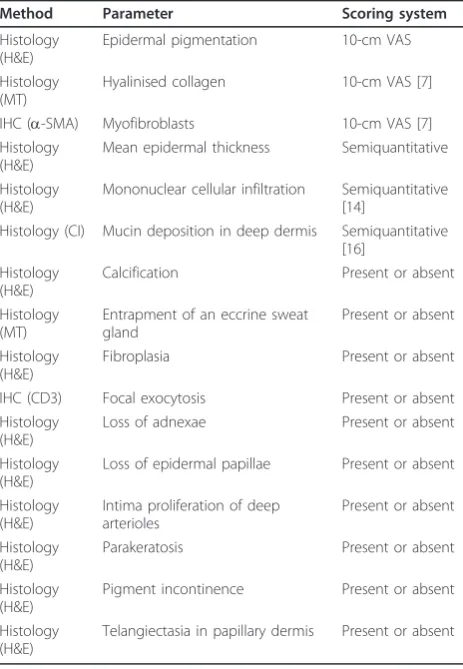
Studies of SSc skin histopathology have described altera-tions in different skin compartments, including atrophy and increased pigmentation of the epidermis [11], loss of the epidermal papillae [11], increase of melanophages (’pigment incontinence’) [11], the presence of a mono-nuclear perivascular infiltrate and myofibroblasts [7,12-14], sclerosis [7], narrowing of arteriolar lumina in the deep vascular plexus (reticular dermis) [15] and dis-appearance and entrapment of dermal adnexae and cal-cification [11,12]. To this set of alterations, we added two key histopathological features of other scleroderma-like disorders, namely, mucin deposition and fibroplasia [16,17]. Because analysis of routine biopsies revealed the presence of telangiectasia, focal exocytosis (that is, the presence of lymphocytes in the epidermis) and parakera-tosis in some SSc biopsies, these items were also added to the set of scoring parameters [18]. Scoring systems for different parameters were obtained from previous publications as much as possible. An overview of the skin parameters that were scored is shown in Table 1.
random (0 = mean less than three layers, 1 = mean of three or four layers, 2 = mean of five or six layers, 3 = mean of more than six layers), a scoring system which has also been used for the synovial lining layer [14,19]. Mono-nuclear cellular infiltration was scored on a semiquantita-tive scale as 0 (few scattered cells), 1 (maximum number of cells per collection at least 10), 2 (maximum number of cells per collection between 10 and 50) or 3 (maximum number of cells per collection at least 50) [14]. Mucin deposition in the reticular dermis was evaluated by scoring the degree of acid mucopolysaccharide staining on a semi-quantitative scale as negative, very slight, slight, fair or abundant [16]. Entrapment of an eccrine sweat gland was defined as the absence of any surrounding fat tissue (Fig-ure 1A). Focal exocytosis was defined as the presence of T-lymphocytes in the epidermis at least at two distinct sites (Figure 1B). Intima proliferation in deep arterioles was considered pathologic if the vessel wall thickness exceeded the diameter of the vessel lumen (Figure 1C). Parakeratosis was defined as the presence of nuclei in the
stratum corneum (Figure 1D). Pigment incontinence was defined as the presence of melanin in papillary macrophages at least at two distinct sites (Figure 1E). Tel-angiectasia was defined as enlarged papillary capillaries (Figure 1F).
Stained slides were coded so that a blinded analysis could be performed. Slides from the dorsal forearm were analysed by two independent observers (MH and JTVP) who were uninformed of any clinical data. All scoring parameters were judged to be reliable, as the interobser-ver agreement was substantial (> 0.6 for all categorical parameters and intraclass correlation coefficient >0.7 for all continuous parameters). For continuous parameters, the mean of the two observers was used for analysis. In case of a discrepant score for a categorical parameter, a consensus was determined by the two observers. Slides from the upper inner arm were scored twice by one observer (JTVP). For continuous parameters, the mean of the two scores was used for analysis. In case of a discre-pant score for a categorical parameter, a consensus was determined by a third evaluation. Because fibroplasia and calcification were not observed in a single biopsy, these items were left out of all analyses.
Statistical analysis
In patients with lcSSc or dcSSc, associations of histo-pathological parameters with local clinical skin involve-ment were determined by generalised estimation equation (GEE) modelling with a correction for subject level and biopsy site. Local skin involvement at the dorsal forearm was defined as a local clinical score of at least 1. Since the upper inner arm local skin score is not included in the mRSS, all patients with dcSSc were considered to have local skin involvement at this site. In case a signifi-cant interaction between biopsy site and local clinical skin involvement was found in the GEE model, statistical testing for the effect of skin involvement was separately performed for the dorsal forearm and the upper inner arm. For normally distributed parameters, the Pearson correlation coefficient was used to determine the correla-tion with the dorsal forearm score. Otherwise, the Spear-man correlation coefficient was used. For categorical parameters, Fisher’s exact test was used to analyse the association with the dorsal forearm score. Mann-Whitney Utest (continuous data) and Fisher’s exact test (categori-cal data) were used to compare lSSc biopsies with normal controls.P≤0.05 was considered statistically significant. All analyses were performed using PASW 18.0 software (SPSS, Inc., Chicago, IL, USA).
Results
Clinical characteristics of the patients
[image:3.595.57.289.111.446.2]Skin biopsies from the dorsal forearm and the upper inner arm were obtained from 53 consecutive SSc patients Table 1 Overview of the skin histology parameters and
scoring systema
Method Parameter Scoring system
Histology (H&E)
Epidermal pigmentation 10-cm VAS
Histology (MT)
Hyalinised collagen 10-cm VAS [7]
IHC (a-SMA) Myofibroblasts 10-cm VAS [7]
Histology (H&E)
Mean epidermal thickness Semiquantitative
Histology (H&E)
Mononuclear cellular infiltration Semiquantitative [14]
Histology (CI) Mucin deposition in deep dermis Semiquantitative [16]
Histology (H&E)
Calcification Present or absent
Histology (MT)
Entrapment of an eccrine sweat gland
Present or absent
Histology (H&E)
Fibroplasia Present or absent
IHC (CD3) Focal exocytosis Present or absent
Histology (H&E)
Loss of adnexae Present or absent
Histology (H&E)
Loss of epidermal papillae Present or absent
Histology (H&E)
Intima proliferation of deep arterioles
Present or absent
Histology (H&E)
Parakeratosis Present or absent
Histology (H&E)
Pigment incontinence Present or absent
Histology (H&E)
Telangiectasia in papillary dermis Present or absent
a
(17 males and 36 females; mean age ± SD, 52 ± 12 years). Seven SSc patients (13%) had no clinical skin involvement (lSSc), and 46 SSc patients (87%) had skin involvement (29 lcSSc and 17 dcSSc patients). Twenty-five patients used methotrexate, and 11 patients used low-dose corticoster-oids (< 15 mg prednisolone/day). Table 2 summarizes the features of the different SSc patient subsets. Normal skin samples taken from the upper inner arm were included as a reference set (n= 18 comprising 7 males and 11 females; mean age ± SD, 44 ± 17 years).
Associations of local clinical skin involvement and histological alterations
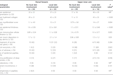
To examine associations of local skin disease with histolo-gical parameters, we analysed the biopsies from patients with lcSSc or dcSSc (n= 46). We found that, independently
of the anatomical site of the biopsy, the hyalinised collagen score, the myofibroblast score, the mean epidermal thick-ness, the mononuclear cellular infiltration and the fre-quency of focal exocytosis differed significantly between biopsies with and without local skin involvement (a= 0.05; GEE) (Table 3). For the continuous parameters, only the epidermal thickness (r= 0.553;P< 0.001), the myofibro-blast score (r= 0.507;P< 0.001) and the hyalinised col-lagen score (r= 0.572;P< 0.001) correlated with the local clinical skin score. No association was found between the local clinical score and the frequency of focal exocytosis (P = 0.06).
Sensitivity and specificity of histological alterations
To determine the specificity of the studied histological parameters, we analysed 18 upper inner arm biopsies D
A
E B
[image:4.595.61.539.87.488.2]C DF
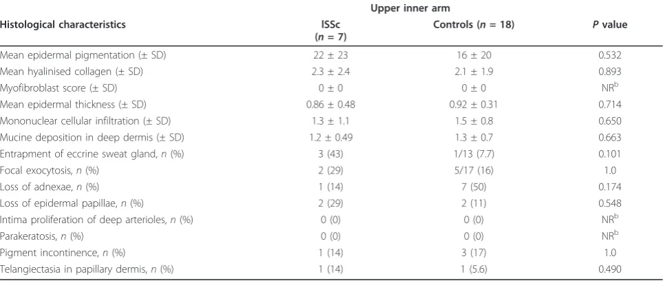
from patients who were referred for a lupus band test and in whom further evaluation excluded any specified autoimmune disease. Myofibroblasts, intima prolifera-tion of deep arterioles and parakeratosis were not seen in these biopsies (Table 4). Comparison with the biop-sies of the patients with lSSc (n = 7) revealed no
[image:5.595.56.544.99.247.2]statistically significant differences (Table 4). Analysis of all SSc biopsies showed that myofibroblasts, intima pro-liferation of deep arterioles and parakeratosis were pre-sent in, respectively, 22%, 19% and 5.8% of the dorsal forearm biopsies and in 14%, 14% and 0% of the upper inner arm biopsies.
Table 2 Clinical data of patients with lSSc, lcSSc and dcSSca
Subset lSSc (n= 7) lcSSc (n= 29) dcSSc (n= 17)
Mean age, yr (± SD) 51 ± 16 51 ± 13 55 ± 8,0
Female/male,n 6/1 21/8 9/8
Median disease durationb(rangec) 2 (2 to 21) 6 (0 to 35) 2 (0 to 12)
Median mRSS (range) 0 (0 to 0) 4 (0 to 14) 21 (4 to 27)
Median local skin score dorsal forearm (range) 0 (0 to 0) 0 (0 to 2) 1 (0 to 3)
ACR criteria,n 0 14 17
ANA,n
Topoisomerase I 0 5 8c
Centromere 6 14 3c
RNA polymerase III 0 1 3
U1-RNP 0 3 0
a
ACR, American College of Rheumatology; ANA, antinuclear antibodies; dcSSc, diffuse cutaneous systemic sclerosis; lSSc, limited systemic sclerosis; lcSSc, limited cutaneous systemic sclerosis; mRSS, modified Rodnan skin score; U1RNP, U1-ribonucleic protein;b
disease duration from first non-Raynaud’s phenomenon symptom;cone patient had both anticentromere and anti-topoisomerase I antibodies.crange denotes the full interval between the smallest and largest values.
Table 3 Histological characteristics of patients with lcSSc or dcSSca
Dorsal forearm Upper inner arm Histological
characteristics
No local skin involvement
(n= 24)
Local skin involvement
(n= 22)
No local skin involvement
(n= 29)
Local skin involvement
(n= 16)
P
valueb
Mean epidermal pigmentation (± SD)
33 ± 22 37 ± 23 17 ± 18 40 ± 20 NRc
Mean hyalinised collagen (± SD)
20 ± 21 42 ± 29 11 ± 13 43 ± 33 < 0.001
Mean myofibroblast score (± SD)
1.1 ± 4.9 12 ± 21 0.79 ± 3.8 14 ± 27 0.004
Mean epidermal thickness (± SD)
1.6 ± 0.58 2.3 ± 0.61 1.1 ± 0.51 1.5 ± 0.48 < 0.001
Mean mononuclear cellular infiltration (± SD)
0.89 ± 0.34 1.1 ± 0.38 1.4 ± 0.79 1.9 ± 0.77 0.005
Mean mucin deposition in deep dermis (± SD)
1.7 ± 1.3 2.1 ± 1.4 0.96 ± 0.83 1.5 ± 1.2 0.09
Entrapment of eccrine sweat gland,n(%)
10 (42) 11/21 (52) 8 (28) 6/15 (40) 0.284
Focal exocytosis,n(%) 1 (4.2) 5 (23) 14 (48) 11 (69) 0.043
Loss of adnexae,n(%) 10 (42) 12 (55) 9 (31) 3/15 (20) 0.94
Loss of epidermal papillae,
n(%)
10 (42) 5 (23) 1 (3,4) 0 (0) 0.122
Intima proliferation of deep arterioles,n(%)
3 (13) 6 (27) 5 (17) 2/15 (13) 0.596
Parakeratosis,n(%) 0 (0) 3 (14) 0 (0) 0 (0) NRd
Pigment incontinence,n (%)
9 (38) 13 (59) 16 (55) 9 (56) 0.141
Telangiectasia in papillary dermis,n(%)
2 (8,3) 0 (0) 0 (0) 0 (0) NRd
a
lcSSc, limited cutaneous systemic sclerosis; dcSSc, diffuse cutaneous systemic sclerosis; SD, standard deviation; NR, not reported;b
generalised estimation equation (GEE) modelling was used to determine the effect of local skin involvement for each variable, correcting for subject level and biopsy site;c
in the GEE model, the effect of this parameter differed significantly between the skin sites (Student’st-test was used to evaluate the upper inner arm,P< 0.001, and the dorsal forearm,P= 0.580);d
[image:5.595.65.536.383.697.2]Discussion
Because of the rarity of the disease, few studies have sys-temically addressed the skin histopathology of SSc. Pre-vious studies may have been hampered by bias due to the inclusion of only dcSSc patients [7], by failure to perform biopsies at the same anatomical site in all patients [15] or by failure to include a normal control group or to link alterations to clinical scoring [11,13]. The present study was designed to overcome these issues and to identify histopathological alterations in the skin of SSc patients which are linked to clinical skin scoring or might have diagnostic relevance.
The results of this study show that independent of the biopsy site (dorsal forearm or inner upper arm), the hya-linised collagen score, the myofibroblast score and the mean epidermal thickness are associated with the pre-sence of local clinical skin involvement. Furthermore, these three parameters correlated well with the local clinical skin score at the dorsal forearm. In agreement with our data, Kissinet al.[7] reported a good correla-tion of the hyalinised collagen score and the myofibro-blast score with clinical scoring in patients with dcSSc. The link between histopathological alterations and clini-cal assessment at two independent skin sites in a large set of SSc biopsies, including patients from different dis-ease subsets and with early and late disdis-ease, suggests these parameters might be potential candidates as out-come measures in clinical trials on skin disease. How-ever, longitudinal studies should address their sensitivity to change before they can be considered validated mea-sures [20]. Given that the link with clinical scoring was independent of the skin site, our data indicate that in
patients with dcSSc, biopsies from the upper inner arm may be used to study histological parameters.
One interesting finding of our study is the link between clinical skin scoring and epidermal changes. Apart from the increased mean epidermal thickness in biopsies with local clinical skin involvement, we also found parakerato-sis in a minority of clinically involved skin biopsies from the dorsal forearm, which points to a disturbance of epi-dermal differentiation [21]. Consistent with these results, Aden et al.[22] demonstrated that the epidermis in involved SSc skin shows thickening and altered differen-tiation, mimicking an active wound-healing phenotype. In contrast, older literature reported atrophy of the epi-dermis in SSc skin biopsies [11]. Also, we found that local clinical skin involvement was associated with a higher epidermal pigmentation at the upper inner arm but not at the dorsal forearm, which is probably related to the different sun exposure of the two biopsy sites.
[image:6.595.57.539.101.305.2]A second research aim of this study was to determine the sensitivity of SSc-specific histological alterations, focusing on SSc patients without clinical skin involve-ment (lSSc). These patients have Raynaud’s phenom-enon and SSc-associated antinuclear antibodies and/or nailfold capillaroscopic alterations without skin involve-ment. At the upper inner arm, we found no differences between lSSc and control biopsies. Concerning the spe-cificity of histological parameters for SSc, we could not detect parakeratosis, myofibroblasts or intima prolifera-tion of the deep arterioles in controls. However, these alterations were present in only a minority of the SSc biopsies. Thus, SSc-specific histological alterations have a low diagnostic sensitivity.
Table 4 Histological characteristics of patients with lSSc and controlsa
Upper inner arm Histological characteristics lSSc
(n= 7)
Controls (n= 18) Pvalue
Mean epidermal pigmentation (± SD) 22 ± 23 16 ± 20 0.532
Mean hyalinised collagen (± SD) 2.3 ± 2.4 2.1 ± 1.9 0.893
Myofibroblast score (± SD) 0 ± 0 0 ± 0 NRb
Mean epidermal thickness (± SD) 0.86 ± 0.48 0.92 ± 0.31 0.714
Mononuclear cellular infiltration (± SD) 1.3 ± 1.1 1.5 ± 0.8 0.650
Mucine deposition in deep dermis (± SD) 1.2 ± 0.49 1.3 ± 0.7 0.663
Entrapment of eccrine sweat gland,n(%) 3 (43) 1/13 (7.7) 0.101
Focal exocytosis,n(%) 2 (29) 5/17 (16) 1.0
Loss of adnexae,n(%) 1 (14) 7 (50) 0.174
Loss of epidermal papillae,n(%) 2 (29) 2 (11) 0.548
Intima proliferation of deep arterioles,n(%) 0 (0) 0 (0) NRb
Parakeratosis,n(%) 0 (0) 0 (0) NRb
Pigment incontinence,n(%) 1 (14) 3 (17) 1.0
Telangiectasia in papillary dermis,n(%) 1 (14) 1 (5.6) 0.490
a
lSSc, limited systemic sclerosis; NR, not reported;b
Conclusions
In conclusion, the systematic analysis of skin biopsies from 53 consecutive SSc patients and 18 normal con-trols revealed that the mean epidermal thickness, the hyalinised collagen score and the myofibroblast score are linked to local clinical skin involvement and are cor-related with the local skin score. Concerning histological alterations in lSSc, we found no significant differences with control skin at the upper inner arm. Finally, myofi-broblasts, intima proliferation of the deep arterioles or parakeratosis in a skin biopsy are useful diagnostic mar-kers for SSc, although they have a low sensitivity.
Abbreviations
dcSSc: diffuse cutaneous systemic sclerosis; GEE: generalised estimation equation; lcSSc: limited cutaneous systemic sclerosis; lSSc: limited systemic sclerosis; mRSS: modified Rodnan Skin Score; SSc: systemic sclerosis.
Acknowledgements
The authors thank Rita Heyse, Sofie D’hont and Dorothea Van Limbergen for excellent technical assistance. JTVP is supported by a research grant from the Fund for Scientific Research-Flanders.
Author details
1
Department of Rheumatology, Ghent University Hospital, De Pintelaan 185, BE-9000 Gent, Belgium.2Department of Dermatology, Ghent University
Hospital, De Pintelaan 185, BE-9000 Gent, Belgium.3Department of Pathology, Stedelijk Ziekenhuis Roeselare, Brugsesteenweg 90, BE-8800 Roeselare, Belgium.
Authors’contributions
JTVP, VS, MH and FDK designed the study. VS acquired the capillaroscopic and clinical data. JTVP and FDK acquired the serological data. JTVP, MH and ND acquired the histological data. JTVP, VS, MH, DE and FDK participated in the manuscript preparation and finalisation. All authors read and approved the final manuscript.
Competing interests
The authors declare that they have no competing interests.
Received: 16 October 2010 Revised: 19 January 2011 Accepted: 28 February 2011 Published: 28 February 2011
References
1. Denton CP, Black CM, Abraham DJ:Mechanisms and consequences of fibrosis in systemic sclerosis.Nat Clin Pract Rheumatol2006,2:134-144. 2. Akesson A, Fiori G, Krieg T, van den Hoogen FH, Seibold JR:Assessment of
skin, joint, tendon and muscle involvement.Clin Exp Rheumatol2003,21: S5-S8.
3. LeRoy EC, Medsger TA Jr:Criteria for the classification of early systemic sclerosis.J Rheumatol2001,28:1573-1576.
4. Koenig M, Joyal F, Fritzler MJ, Roussin A, Abrahamowicz M, Boire G, Goulet JR, Rich E, Grodzicky T, Raymond Y, Senécal JL:Autoantibodies and microvascular damage are independent predictive factors for the progression of Raynaud’s phenomenon to systemic sclerosis: a twenty-year prospective study of 586 patients, with validation of proposed criteria for early systemic sclerosis.Arthritis Rheum2008,58:3902-3912. 5. Smith V, Van Praet JT, Vandooren B, Van der Cruyssen B, Naeyaert JM,
Decuman S, Elewaut D, De Keyser F:Rituximab in diffuse cutaneous systemic sclerosis: an open-label clinical and histopathological study.
Ann Rheum Dis2010,69:193-197.
6. Nash RA, McSweeney PA, Crofford LJ, Abidi M, Chen CS, Godwin JD, Gooley TA, Holmberg L, Henstorf G, LeMaistre CF, Mayes MD, McDonagh KT, McLaughlin B, Molitor JA, Nelson JL, Shulman H, Storb R, Viganego F, Wener MH, Seibold JR, Sullivan KM, Furst DE:High-dose immunosuppressive therapy and autologous hematopoietic cell
transplantation for severe systemic sclerosis: long-term follow-up of the US multicenter pilot study.Blood2007,110:1388-1396.
7. Kissin EY, Merkel PA, Lafyatis R:Myofibroblasts and hyalinized collagen as markers of skin disease in systemic sclerosis.Arthritis Rheum2006,
54:3655-3660.
8. Smith V, Pizzorni C, De Keyser F, Decuman S, Van Praet JT, Deschepper E, Sulli A, Cutolo M:Reliability of the qualitative and semiquantitative nailfold videocapillaroscopy assessment in a systemic sclerosis cohort: a two-centre study.Ann Rheum Dis2010,69:1092-1096.
9. LeRoy EC, Black C, Fleischmajer R, Jablonska S, Krieg T, Medsger TA Jr, Rowell N, Wollheim F:Scleroderma (systemic sclerosis): classification, subsets and pathogenesis.J Rheumatol1988,15:202-205.
10. Clements PJ, Lachenbruch PA, Seibold JR, Zee B, Steen VD, Brennan P, Silman AJ, Allegar N, Varga J, Massa M,et al:Skin thickness score in systemic sclerosis: an assessment of interobserver variability in 3 independent studies.J Rheumatol1993,20:1892-1896.
11. Montgomery H, O’Leary PA, Ragsdale WE Jr:Dermatohistopathology of various types of scleroderma.AMA Arch Derm1957,75:78-87. 12. Prescott RJ, Freemont AJ, Jones CJ, Hoyland J, Fielding P:Sequential
dermal microvascular and perivascular changes in the development of scleroderma.J Pathol1992,166:255-263.
13. Torres JE, Sanchez JL:Histopathologic differentiation between localized and systemic scleroderma.Am J Dermatopathol1998,20:242-245. 14. Roumm AD, Whiteside TL, Medsger TA Jr, Rodnan GP:Lymphocytes in the
skin of patients with progressive systemic sclerosis: quantification, subtyping, and clinical correlations.Arthritis Rheum1984,27:645-653. 15. Fleming JN, Shulman HM, Nash RA, Johnson PY, Wight TN, Gown A,
Schwartz SM:Cutaneous chronic graft-versus-host disease does not have the abnormal endothelial phenotype or vascular rarefaction
characteristic of systemic sclerosis.PLoS One2009,4:e6203.
16. Rongioletti F, Gambini C, Micalizzi C, Pastorino A, Rebora A:Mucin deposits in morphea and systemic scleroderma.Dermatology1994,189:157-158. 17. Boin F, Hummers LK:Scleroderma-like fibrosing disorders.Rheum Dis Clin
North Am2008,34:199-220, ix.
18. Ackerman AB:Histologic Diagnosis of Inflammatory Skin Diseases: An
Algorithmic Method Based on Pattern Analysis.2 edition. Baltimore: Williams
& Wilkins; 1997.
19. Baeten D, Demetter P, Cuvelier C, Van Den Bosch F, Kruithof E, Van Damme N, Verbruggen G, Mielants H, Veys EM, De Keyser F:Comparative study of the synovial histology in rheumatoid arthritis,
spondyloarthropathy, and osteoarthritis: influence of disease duration and activity.Ann Rheum Dis2000,59:945-953.
20. Furst D, Khanna D, Matucci-Cerinic M, Clements P, Steen V, Pope J, Merkel P, Foeldvari I, Seibold J, Pittrow D, Polisson R, Strand V:Systemic sclerosis: continuing progress in developing clinical measures of response.J Rheumatol2007,34:1194-1200.
21. Fitzpatrick TB, Wolff K, Access Medicine:Fitzpatrick’s Dermatology in General
Medicine.7 edition. New York: McGraw-Hill Medical; 2008.
22. Aden N, Shiwen X, Aden D, Black C, Nuttall A, Denton CP, Leask A, Abraham D, Stratton R:Proteomic analysis of scleroderma lesional skin reveals activated wound healing phenotype of epidermal cell layer.
Rheumatology (Oxford)2008,47:1754-1760.
doi:10.1186/ar3267
Cite this article as:Van Praetet al.:Histopathological cutaneous