N A N O E X P R E S S
Open Access
Enhancing oral bioavailability of quercetin using
novel soluplus polymeric micelles
Linghui Dian
1,2, Enjiang Yu
1, Xiaona Chen
2, Xinguo Wen
2, Zhengzan Zhang
2, Lingzhen Qin
2, Qingqing Wang
2,
Ge Li
3and Chuanbin Wu
2,3*Abstract
To improve its poor aqueous solubility and stability, the potential chemotherapeutic drug quercetin was encapsulated in soluplus polymeric micelles by a modified film dispersion method. With the encapsulation efficiency over 90%, the quercetin-loaded polymeric micelles (Qu-PMs) with drug loading of 6.7% had a narrow size distribution around mean size of 79.00 ± 2.24 nm, suggesting the complete dispersibility of quercetin in water. X-ray diffraction (XRD) patterns illustrated that quercetin was in amorphous or molecular form within PMs. Fourier transform infrared spectroscopy (FTIR) indicated that quercetin formed intermolecular hydrogen bonding with carriers. Anin vitrodialysis test showed the Qu-PMs possessed significant sustained-release property, and the formulation was stable for at least 6 months under accelerated conditions. The pharmacokinetic study in beagle dogs showed that absorption of quercetin after oral administration of Qu-PMs was improved significantly, with a half-life 2.19-fold longer and a relative oral bioavailability of 286% as compared to free quercetin. Therefore, these novel soluplus polymeric micelles can be applied to encapsulate various poorly water-soluble drugs towards a development of more applicable therapeutic formulations.
Keywords:Soluplus; Polymeric micelles; Oral bioavailability; Quercetin
Background
Oral administration is by far the easiest and most ac-ceptable route of drug delivery, especially for the long-term medication of patients [1]. But about 40% of the approved active molecules have low solubility, resulting in poor oral bioavailability. Many efforts have been de-voted to the development of oral sustained-release sys-tems that can not only improve drug bioavailability leading to better efficacy and less administration frequen-cies but also decrease the fluctuation of plasma drug con-centration to lower side effects [2]. In recent decades, emerging nanotechnology provides a novel platform to solve the solubility problem of drugs [3,4]. Especially, polymeric micelles as a promising drug delivery system is a new research hotspot [5], most current studies concen-trated on developing polymeric micelles for injection drug delivery [6]. Drug-loaded micelles in the systemic circula-tion characterizes long retencircula-tion time and excellent tissue
permeability and can gather in the diseased tissue to gain passive targeting [7,8]. Furthermore, polymeric micelles with stable, biocompatible, and solubilizing properties have drawn considerable attention for oral administration. Polymeric micelles with inner‘core’and outer‘shell’are formed by amphiphilic copolymers composed of hydro-philic and hydrophobic chains that can self-assemble in water above the critical micelle concentration (CMC) [9]. A polymeric micelle has the ability to encapsulate a hydro-phobic drug into their cores and deliver the drug to the desired site at the concentration exceeding the intrinsic solubility of the drug. Moreover, the encapsulated drug can be not only protected from contact with the GI con-tents which likely induce degradation and metabolism but also conferred with the characteristics of sustained-release and direct uptake by cells. Many studies have proven that nanoparticles can transport across the intestinal mem-brane through paracellular or trancellular routes [10],
* Correspondence:[email protected]
2
School of Pharmaceutical Sciences, Sun Yat-Sen University, Waihuan Road 132, Guangzhou, Guangdong 510006, People’s Republic of China 3
R&D Center of Pharmaceutical Engineering, Sun Yat-sen University, Waihuan Road 132, Guangzhou 510006, Guangdong, People’s Republic of China Full list of author information is available at the end of the article
while maintaining their integrity [11]. Therefore, the oral formulation based on nanosized polymeric micelles was expected to achieve the advantages of nanoparticles, such as enhanced permeability and retention (EPR) effects.
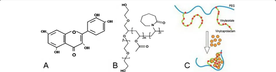
Quercetin (3, 3′, 4′, 5, 7-pentahydroxy flavones, Figure 1A) is a flavonoid compound widely present in flower, leaf, and fruit of plants such asSophora japonica L.,Dendranthema morifolium(Ramat.)Tzvel, and Cratae-gus pinnatifida bunge, with a variety of biological activity and high medical value [12]. Literature indicates that quercetin can inhibit the growth and proliferation of a var-iety of cancer cell lines (human ovarian cancer, breast cancer, lung cancer, human colon cancer, etc.) [13-16]. Quercetin can also lower the multidrug-resistance in cancer cells [17,18] and enhance the antitumor effects of drugs [19,20].
But clinical use of quercetin is limited due to its poor water solubility and instability in physiological media [21], leading to poor bioavailability [22]. Polymeric mi-celles are regarded as excellent candidates for anticancer drug delivery, and several anticancer drugs delivered by amphiphilic polymer micelles have already been pre-ceded to clinical study or market [23,24]. Recently, polymer micelles have been utilized for quercetin for-mulation too [25].
Soluplus, an amphiphilic polyvinyl caprolactam-polyvinyl acetate-polyethylene glycol graft copolymer (Figure 1B), was introduced by BASF. This graft copolymer has a polyethylene glycol (PEG) backbone as hydrophilic part and vinylcaprolactam/vinyl acetate side chains as lipo-philic structure. So, micelles can be formed in aqueous so-lution above the CMC of 7.6 mg · L−1(Figure 1C) [26].
The objective of this study was to develop a nanomi-celle delivery system by using soluplus and poloxamer 407(F127), which could solubilize quercetin in aqueous
media, reaching the clinically relevant concentration and delivering quercetin in a controlled manner. The previ-ously reported preparation method was slightly modified to produce soluplus micelles with suitable size, charge, and stable properties. Powder X-ray diffraction (XRD)
was employed to identify the physical state of quercetin in the polymeric micelles. Taking advantage of their permeation-enhancing effect, the soluplus nanomicelles were evaluated in vivo as potential vehicles, and the pharmacokinetic profile of orally administered quer-cetin encapsulated in micelles was investigated.
Methods
Materials
Soluplus® was friendly supplied by BASF Auxiliary Chem. Co., Ltd. (Shanghai, China). Poloxamer 407 (PEO
98-POP67PEO98) was obtained from BASF (Ludwigshafen,
Germany). Quercetin (Qu) was purchased from Shanxi Sciphar Biotech. Co. Ltd. (Shanxi, China). Methanol (high-performance liquid chromatography (HPLC) grade) was purchased from Fisher Scientific, Waltham, MA, USA. Milli-Q grade water purified through a Millipore system (ELGA LabWater, Sartorius, UK) was used throughout this study. All solvents were used without further purification.
Animals
Beagle dogs were obtained from Experimental Animal Center of Sun Yat-sen University (Guangzhou, China). Beagle dogs were provided with standard food and water at will and were exposed to alternating 12-h periods of light and darkness. Relative humidity and temperature were maintained at 50% and 25°C, respectively. All care and handling of animals were performed with the ap-proval of Institutional Authority for Laboratory Animal Care of Sun Yat-sen University.
Preparation of quercetin-loaded soluplus polymeric micelles
Quercetin-loaded polymeric micelles (Qu-PMs) were formed by a modified film dispersion method using solu-plus and F127 [27]. Briefly, soluplus (10 mg) and
[image:2.595.58.538.587.713.2]quer-cetin (7 mg) were dissolved together in organic solvent acetone, followed by evaporation under reduced pressure in a rotary evaporator at 35°C. The deionized water was
then added into the polymer and drug solution, allowing the self-assembly of soluplus and quercetin to form quercetin-encapsulated polymeric micelles with core-shell structure (Figure 1C) at 650 rpm. Finally, the pre-pared Qu-PMs were lyophilized for future application.
Determining the optimum concentration of F127
Then the optimum concentration of F127 required for
preparing the desirable Qu-PMs was determined based on particle size and encapsulation efficiency.
Determining the optimum stirring time
The optimum time of magnetic stirring for the prepar-ation of Qu-PMs was determined on the basis of particle size and encapsulation efficiency.
Optimization of drug loading
Qu-PMs were prepared using different theoretical Qu loading, i.e., 5%, 7%, and 9% of polymer on the basis of preliminary experiment, to determine the optimum per-centage of Qu in soluplus matrix and its effects on par-ticle size, polydispersity index (PDI), zeta potential, and encapsulation efficiency of Qu-PMs. The magnetic stir-ring time (2 h), stabilizer concentration (1% of F127), and
aqueous ratio were kept constant.
Characterization of Qu-PMs
Particle size and zeta potential measurements
The particle size and PDI of Qu-PMs were determined by using a Malvern Instruments Zetasizer Nano ZS90 (Malvern Instruments, Malvern, UK) on the basis of pho-ton correlation spectroscopy. The dispersion of Qu-PMs was diluted in double distilled water and measured at 25°C for analysis. The particle size and PDI were ob-tained by cumulate analysis using the MALVERN soft-ware. The Zeta potential of Qu-PMs also measured by using a Malvern Instruments Zetasizer Nano ZS90 (Malvern Instruments, Malvern, UK). All experiments were repeated three times.
Transmission electron microscopy
The surface morphology of Qu-PMs was examined by using a transmission electron microscope (TEM; H66009IV, Hitachi, Chiyoda-ku, Japan). The dispersion of Qu-PMs were placed on a copper grid covered with nitrocellulose, negatively stained with phosphotungstic acid, and allowed to dry at room temperature.
X-ray diffraction
The X-ray diffraction patterns of pure Qu, void PMs, physical mixture of void PMs and Qu, and Qu-PMs were obtained by using an X-ray powder diffractometer (Bruker AXS, Madison, WI, USA) at a voltage of 40 kV and
25 mA with a scanned angle from 5°≤2θ≤50°at a scan rate of 0.9 · min−1.
Fourier transform infrared spectrometer
The Fourier transform infrared spectroscopy (FTIR) spectra of Qu, void PMs, physical mixture of void PMs and Qu, and Qu-PMs were recorded on a Nicolet 5700 FTIR spectroscopy (Thermo, Waltham, MA, USA) using a Smart OMNI-sampler accessory. The samples were put on KBr plates. The FTIR spectra were recorded at 1 cm−1 resolution, with the range of 400 to 4,000 cm−1.
Encapsulation efficiency (EE)
The content of Qu encapsulated in PMs was determined by membrane filter method. 0.5 mL of Qu-PMs was fil-tered through the 0.22-μm membrane, while non-encapsulated Qu was retained on the membrane. The filtrate which contained Qu-PMs was demulsificated with methanol and analyzed for entrapped Qu content by high-performance liquid chromatography (HPLC). All experiments were repeated three times.
In vitro release
In vitro release of Qu from Qu-PMs was undertaken by the dialysis bag method [28]. The dialysis bags (MWCO 14000, Millipore, Boston, MA, USA) were immersed in double-distilled water for 24 h prior to loading with 2 mL of Qu-PMs dispersion or quercetin solution (equivalent to 4 mg of Qu). The loaded bags were putted into a conical flask and soaked in 100 mL of 35% (v/v) ethanol, and the flask was placed in a water bath at 37°C ± 0.5°C and stirred rate of 100 rpm. The release medium (5 mL) was taken out at time intervals of 0.5, 1.0, 2.0, 4.0, 6.0, 8.0, 12.0, 24.0, 48.0, 72.0, 96.0, 120.0, 144.0, 168.0, 192.0, 216.0, 240 h and added with the same volume of fresh medium to adjust a sink condition [29,30]. The content of Qu was determined by HPLC. Each test was carried out in triplicate.
The mechanism of Qu release from PMs was per-formed by fitting the release rate data into the following equations:
Zero‐order model equation: y¼k1tþa1 ð1Þ
First‐order model equation: ln 100ð −yÞ ¼k2tþa2 ð2Þ
Higuchi’s square‐root equation: y¼k3t0:5þa3 ð3Þ
Hereyrepresents the accumulative release percentage; tsampling time; k1, k2, andk3release rate constants for
Equations 1, 2, and 3, respectively; a1~ a3are constants
for Equations 1, 2, and 3.
Storage stability
caps and placed in an accelerated stability chamber with temperature of 30°C ± 2°C and RH of 65% ± 5% for 6 months. The formulations were evaluated for changes in particle size, PDI, and entrapment efficiency, besides phys-ical appearance and ease of reconstitution [31,32].
In vivo pharmacokinetics study after oral administration
To compare the pharmacokinetics of Qu-PMs with those of pure Qu after oral administration, an animal experi-ment was in favor of the Ethical Committee of the Sun Yat-sen University (Guangzhou, China) and performed in accordance with the National Institute of Health and Nutrition Guidelines for the Care and Use of Laboratory Animals.
Six beagle dogs (1.2 to 2.0 years of age) weighing 12 to 14 kg were acclimatized in an environmentally con-trolled breeding room for 1 week, before fasting over-night before the experiments, but allowed to drink water only. These dogs were randomly distributed into two groups each made up of three dogs. Dogs in one group were given pure Qu dispersed in Milli Q water contain-ing 0.3% (v/v) CMC-Na, while dogs in the other group were administered Qu-PMs dissolved in distilled water. All the formulations were administrated at an equivalent dose of Qu 16 mg · kg−1by oral gavage. Blood samples of 3 mL were collected from the hind leg vein and placed into heparinized tubes at time intervals of 0.5, 1.0, 1.5, 2.0, 3.0, 4.0, 5.0, 6.0, 8.0, 10.0, 12.0, 24.0, 36.0, and 48.0 h after oral administration. The plasma was segregated and the samples were kept at−20°C until analysis.
Qu was assayed using an HPLC method. In sample analysis, a 100 μL plasma sample aliquot was blended with protein-precipitating methanol agent (200 μL) and 0.25% HCL (100μL) were added and vortexed for 2 min, then heated at 50°C for 10 min. After centrifugation at 12,000 rpm for 10 min, the supernatant was analyzed by HPLC. Qu analysis was carried out by injecting an ali-quot (100 μL) of sample into the HPLC column (an odyssil C18 column, 4.6 × 250 mm, 5μm) with a
precol-umn (4.6 × 12.5 mm, 5 μm), using a mobile phase mak-ing up of methanol 0.2% phosphoric acid (60:40,v/v) at a flow rate of 1.0 mL · min−1. The detection wavelength was 375 nm [33].
Pharmacokinetic parameters were estimated with one compartmental model using the 3P87 software (pub-lished by the State Food and Drug Administration of China for pharmacokinetic study). The Cmax (highest
peak concentration), Tmax (the time at which Cmax
reached), and the AUC0−∞ (the total area under the
curve) were determined. The Mann-Whitney U-test was analyzed statistically. The results of the test were evaluated as mean values ± SD (standard deviation), and that ofp< 0.05 analysis was significant statistically difference.
Results and discussion
Preparation of Qu-PMs
Different process variables including stabilizer concen-tration, magnetic stirring time, and theoretical drug loading were optimized for the preparation of Qu-PMs.
Effect of stabilizer concentration
The effects of F127concentration on the size and
encap-sulation efficiency of Qu-PMs are shown in Figure 2. As the concentration of F127increased (1% to 3%), the
par-ticle size of Qu-PMs increased, but the encapsulation ef-ficiency remained almost constant at around 95%. The particle size with 3% F127was larger than 100 nm which
was unacceptable, and no significant difference was found in particle size and encapsulation efficiency with respect to 2% and 1% F127. So 1% F127 (w/v) was
opti-mized for Qu-PMs preparation.
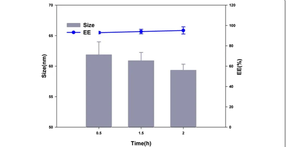
Effect of stirring time
Qu-PMs were prepared through magnetic stirring for particle size reduction. Figure 3 shows the decrease in particle size upon increasing the stirring time up to 2 h. Magnetic stirring of 2 h brought about the smaller par-ticle size (59.32 ± 1.01 nm) and larger encapsulation effi-ciency (95.12% ± 3.54%) compared with shorter stirring time. So, magnetic stirring of 2 h was selected for further research.
Optimum of drug loading
Qu-PMs were prepared with different theoretical drug loading, i.e., 5%, 7%, and 9% of polymer, to investigate the appropriate percentage of Qu in soluplus matrix. As shown in Table 1, initial drug loading affected the par-ticle size and drug encapsulation efficiency of Qu-PMs significantly (p< 0.05). The particle size became big with the increase of drug loading, and the encapsulation effi-ciency also increased as drug loading increased from 5% to 7% but decreased significantly as drug loading further increased to 9%. Drug loading of 7% resulted in stable PMs with higher encapsulation efficiency (95.91% ± 4.05%), optimum particle size (79.00 ± 2.24 nm), and PDI (0.154 ± 0.044), as well as the most negative zeta po-tential (−17.10 ± 2.30). Taking this into consideration, the initial drug loading of 7% (w/w) was selected for Qu-PMs formulation.
Properties of Qu-PMs
Particle size and morphology of Qu-PMs
to intestinal uptake and extension of circulation half-life, as well as being evaded by the reticuloendothelial system (RES).
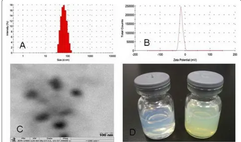
The average particle size and the PDI of Qu-PMs were studied by dynamic light scattering. The representative size distribution of Qu-PMs (Figure 4A) clearly shows a
[image:5.595.58.543.89.340.2]narrow size distribution with the average particle diameter of 79.00 ± 2.24 nm and PDI of 0.154 ± 0.044. This con-forms to the best particle size range for oral absorption. Zeta potential measurements possessed a negative surface charge of −17.10 ± 2.30 mV for Qu-PMs (Figure 4B), which certainly could increase the stability of Qu-PMs
Figure 2Effects of stabilizer F127concentration on size and EE of Qu-PMs (n= 3).
[image:5.595.58.540.467.714.2]in dispersion. Mono-disperse and spherical Qu-PMs with a diameter of approximately 80 nm was examined by TEM (Figure 4C), which is consistent with the above results of dynamic light scattering. One major purpose of encapsulating Qu in PMs was to enable Qu to be completely dispersible in aqueous media, and this was confirmed the uniform solution of Qu-PMs with an opalescence (Figure 4D).
X-ray diffraction
The physical status of Qu encapsulated in PMs was com-pared with that of pure Qu by XRD analysis. The XRD patterns of pure Qu, void PMs, physical mixture of void PMs and Qu, and Qu-PMs are shown in Figure 5. In the figure, pure Qu exhibit a lot of distinct peaks that are traits of a crystalline structure [35]. The physical mixture of Qu and void PMs also present a number of distinct peaks, indicating that Qu is crystalline in the mixture. In the case of Qu-PMs, here were some characteristic peaks
of Qu observed in 17°, which indicated that the drug was not completely amorphous in the PMs.
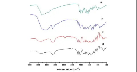
Fourier transform infrared spectrometer
[image:6.595.56.540.100.157.2]The molecular interactions within the solid matrix of the PMs were examined by FTIR method. On the basis of their structure, the possible interaction between quercetin and soluplus is hydrogen bonding, which may result in the shifting and peak broadening of the absorption bands at the interacting functional groups on the FTIR. The aro-matic -OH group in quercetin might come into a hydro-gen bond. FTIR of pure quercetin, void PMs, physical mixture of void PMs and quercetin, and Qu-PMs are pre-sented in Figure 6. Pure quercetin showed a number of characteristic bands representing O-H stretching (3,700 to 3,300 cm−1), C = O absorption (1,670 cm−1), C-C stretch-ing (1,612 cm−1), C-H bending (1,456, 1,383 and 866 cm−1), C-O stretching in the ring structure (1,272 cm−1), and C-O stretching (1,070 to 1,150 cm−1). The existence
Table 1 Effects of initial drug loading on size, PDI, zeta potential, and EE (mean ± SD,n= 3)
Drug loading (%) Size (nm) PDI Zeta potential EE (%)
5 59.97 ± 3.70 0.183 ± 0.023 −13.4 ± 0.20 93.24 ± 3.05
7 79.00 ± 2.24 0.154 ± 0.044 −17.10 ± 2.30 95.91 ± 4.05
9 111.2 ± 3.45 0.134 ± 0.082 −15.1 ± 1.60 75.06 ± 3.19
[image:6.595.53.538.428.713.2]PDI, polydispersity index; EE, encapsulation efficiency; SD, standard deviation.
of these bands is consistent with the report of past studies [36]. Void PMs also show a number of bands, including OH stretch (3,500 to 3,250 cm−1), sp3CH stretching (2,932 cm−1), ester carbonyl stretching (1,742 cm−1), and C = O stretching for tertiary amide (1,641 cm−1). On the spectra of Qu-PMs, the position of carbonyl absorption peaks in Qu-PMs was shifted to 1737.93 cm−1 and 1636.80 cm−1, respectively. However, no similar peak shift-ing was observed in the physical mixture. These results
illustrate that there might be some interactions between quercetin and soluplus in Qu-PMs.
Storage stability studies
The physicochemical properties of Qu-PMs containing 5% mannitol as a lyoprotectant were assessed after 6-month storage to establish their accelerated stability. Freeze dried Qu-PMs cakes were sealed in amber vials and stored in a stability chamber with temperature of 30 ± 0.5°C and relative humidity (RH) of 65% ± 5% for 6 months. The freeze-dried powder of Qu-PMs was stable without any shrinkage or collapse of the dried cake. The encapsulation efficiency, particle size, and PDI of the freeze dried Qu-PMs before and after storage were comparable (Table 2). Overall, these results sug-gested that the Qu-PMs had relatively good physical sta-bility in the presence of mannitol under accelerated conditions and were able to protect majority (>90%) of the encapsulated bioactive component.
In vitro release
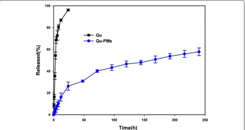
[image:7.595.57.291.88.287.2]As quercetin is insoluble in water, it simulated gastric fluid and intestinal fluid at room temperature (7.7, 5.4, and 28.87μg · mL−1, respectively) [37]. Ethanol (35% (v/v)) was used as a receptor medium to obtain a sink condition in the dynamic dialysis study [33]. Drug release from quer-cetin contained propylene glycol solution and Qu-PMs suspension through the dialysis membrane at 37°C was shown in Figure 7. The pure quercetin from the solution showed about 96.13% release for a period of 24 h, during
[image:7.595.59.540.464.717.2]Figure 6FTIR of Qu (a), physical mixture (b), void PMs (c), and Qu-PMs (d).
which no more than 26.22% of quercetin was released from Qu-PMs. Qu-PMs suspension exhibited sustained-release property and the accumulated sustained-release at 240 h is only 57.78%. The sustained release may be attributed to the diffusion of quercetin entrapped within the core of PMs.
In addition, the release data of Qu-PMs were fitted into different release mechanism models (Table 3). A linear relationship was established between the Qu re-lease rate and the square root of time (R2> 0.98), sug-gesting the release kinetics can be explicated by Higuchi’s equation. Namely, Qu is released from PMs by diffusion [38]. The reason for sustained drug release may be the formation of hydrogen bonds between drug and carrier molecules, which hinders the drug release. For the same reason, quercetin was entrapped in the polymeric micelle hydrophobic cores [39], and the
nanoencapsulation of quercetin in PMs may improve the bioavailability of this molecule significantly.
In vivo pharmacokinetics
[image:8.595.305.540.111.168.2]It has been reported that quercetin is found in plasma as conjugates of glucuronic acid and sulfate groups [40]. Quercetin is released from the binding complex by acid hydrolysis method, and total content of quercetin in plasma was determined by HPLC [41]. Calibration sam-ples were obtained by joining proper volumes of denom-inator Qu solution in methanol into blank plasma, gaining a calibration curve over the detected level range of 0.10 to 8.00 μg · mL−1 (R2> 0.99). The results of the method validation ascertained by assessing the precision, accuracy, recovery, and limit of quantification and proved that the method was reliable. The bioavailability of Qu-PMs was looked into by investigating beagle dogs and comparing with that of pure Qu. After oral adminis-tration of a single dose equivalent to 16 mg · kg−1of pure Qu or Qu-PMs, the mean quercetin concentrations in
Table 2 Characterization of freeze dried Qu-PMs with 5% mannitol after 6 months of storage at 30°C ± 0.5°C and 65% ± 5% RH (mean ± SD,n= 3)
Parameters Initial Final
Size(nm) 63.76 ± 2.35 65.63 ± 3.71
PDI 0.151 ± 0.023 0.183 ± 0.056
EE (%) 92.06 ± 2.41 90.36 ± 3.84
Physical appearance Intact cake Intact cake
Ease of redispersion By mere shaking By mere shaking
PDI, polydispersity index; SD, standard deviation; EE, encapsulation efficiency.
[image:8.595.57.293.123.206.2]Figure 7Release of quercetin from propylene glycol solution and Qu-PMs suspension.
Table 3 Fitting of Qu release data from Qu-PMs into various mechanism models
Model Equation R2
Zero-order y= 0.246t+ 8.291 0.8766
First-order ln(1−y) = 0.004t−16.129 0.9311
Higuchi y= 4.018t1/2−0.081 0.9894
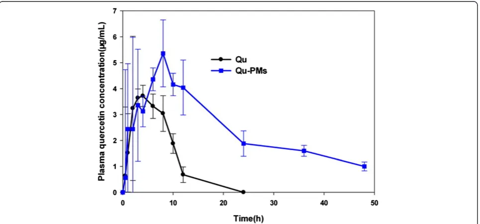
[image:8.595.58.540.456.712.2]dog serum at different time intervals are plotted in Figure 8 and the calculated key pharmacokinetic pa-rameters are summarized in Table 4. The plasma level of quercetin was detected only up to 24 h after admin-istration of free drug, with the Cmax of 5.24 μg · mL−1.
The drugs released from PMs were still detected in plasma 48 h after oral administration, with theCmax of
7.56μg · mL−1. As shown in Figure 8, after oral admin-istration, Qu-PMs were absorbed much slower than pure quercetin with Tmax of 7.02 ± 2.02 h and 5.31 ±
1.08 h (p< 0.05), respectively.
What is more, the half-life (T1/2) of Qu-PMs was
2.19-fold longer than that of pure quercetin (Table 4), indicat-ing a maximum residence time (MRT) of quercetin in the systemic circulation remarkably extended for Qu-PMs after oral administration. As the AUC0–∞ value of
Qu-PMs was significantly larger than that of pure quer-cetin, the relative oral bioavailability of Qu-PMs calcu-lated from AUC0–∞ values was about 286% (p< 0.05)
comparing with pure quercetin. These results implied an enhanced bioavailability of quercetin was achieved through incorporation of drug into PMs.
The main purpose of this study was to prepare an aqueous formulation which could improve the oral bio-availability of the hydrophobic quercetin. In this regard, a nanocarrier system based on soluplus PMs was devel-oped in this study and as anticipated, the pharmacoki-netic results indicated the bioavailability of the delivered quercetin was enhanced. When administered via oral route, PMs may be absorbed through specialized M-cells of the Peyer’s patches in the small intestine [1]. Though the PMs showed the potential to enhance the oral
bioavailability of poorly water-soluble drugs, the under-lying mechanisms of enhancement, however, are still un-clear and provoke future research interests.
Conclusions
[image:9.595.58.541.489.714.2]Quercetin was loaded into nanosized polymeric micelles based on amphiphilic polymers soluplus using a modified film dispersion method. The stable Qu-PMs showed sus-tained release of entrapped quercetin for up to 10 days in vitro, and more importantly, a sustained plasma level and enhanced systemic bioavailability of quercetinin vivo. Thus, the Qu-PMs provide a promising carrier candidate with efficient delivery of quercetin for therapeutic treat-ment in near future. Moreover, this study explores an in-teresting alternative approach for design and fabrication of novel polymeric micelles as delivery systems for bio-active compounds.
Figure 8Mean quercetin plasma concentration.
Table 4 Pharmacokinetic parameters of quercetin in serum after oral administration (mean ± SD,n= 3)
Parameter Pure Qu Qu-PMs
Cmax(μg/mL) 5.24 ± 1.32 7.56 ± 3.28
Tmax(h) 5.31 ± 1.08 7.02 ± 2.02
AUC0~∞(μg/h/mL) 37.68 ± 16.8 107.84 ± 54.4
T1/2(h) 4.94 ± 2.03 10.81 ± 3.7
MRT (h) 7.18 ± 2.25 27.09 ± 7.8
F(%) 286 ± 3.23
Competing interests
The authors declare that they have no competing interests.
Authors’contributions
LHD conceived and designed the study. XNC, LZQ, and QQW carried out the preparation, characterization, drug loading, and drug release studies of Qu-PMs. XGE and ZZZ performed the data analysis. LHD drafted the manuscript. GL and CBW touched up the manuscript. All authors read and approved the final manuscript.
Acknowledgements
The authors gratefully acknowledge the Medical Scientific Research Foundation of Guangdong Province, China (No A2013427), Administration of Traditional Chinese Medicine of Guangdong Province, China (No 20131261), Doctoral Fund of Guangdong Medical College (No XB13096), and International Science and Technology Cooperation Projects of Dongguan (No 2013508152013), Project Creative Young Talents in Colleges and Universities Guangdong Province (2014LYM0003) for the financial support.
Author details
1School of Pharmaceutical Sciences, Guangdong Medical College, Xincheng Road 1, Dongguan 523808, Guangdong, People’s Republic of China.2School of Pharmaceutical Sciences, Sun Yat-Sen University, Waihuan Road 132, Guangzhou, Guangdong 510006, People’s Republic of China.3R&D Center of Pharmaceutical Engineering, Sun Yat-sen University, Waihuan Road 132, Guangzhou 510006, Guangdong, People’s Republic of China.
Received: 28 October 2014 Accepted: 9 December 2014 Published: 18 December 2014
References
1. Lavellea EC, Sharif S, Thomas NW, Holland J, Davis SS:The importance of gastrointestinal uptake of particles in the design of oral delivery systems.Adv Drug Delivery Rev1995,18:5–22.
2. Deshpande AA, Rhodes CT, Shah NH, Malick AW:Controlled-release drug delivery systems for prolonged gastric residence: an overview.Drug Dev Ind Pharm1996,22:531–539.
3. Dian LH, Yang ZW, Li F, Wang ZH, Pan X, Peng XS, Huang XT, Guo ZF, Quan GL, Shi X, Chen B, Li G, Wu CB:Cubic phase nanoparticles for sustained release of ibuprofen: Formulation, characterization and enhanced bioavailability study.Int J Nanomedicine2013,8:845–854. 4. Johnston APR, Such GK, Ng SL, Caruso F:Challenges facing colloidal
delivery systems: FROM synthesis to the clinic.Curr Opin Colloid Interface Sci2011,16:171–181.
5. Bromberg L:Polymeric micelles in oral chemotherapy.J Control Release 2008,128:89–112.
6. Blanchette J, Peppas NA:Oral chemotherapeutic delivery design and cellular response.Annals Biomed Eng2005,33:142–149.
7. Dufresne MH, Garrec DL, Sant V, Leroux JC, Rangeret M:Preparation and characterization of water-soluble pH-sensitive nanocarriers for drug delivery.Int J Pharm2004,277:81–90.
8. Yamamoto Y, Xnagasaki Y, Kato Y, Sugiyama Y, Kataoka K:Long-circulating poly (ethylene glycol)-poly (L, D-lactide) block copolymer micelles with modulated surface charge.J Control Release2001,77:27–38.
9. Kataoka K, Harada A, Nagasaki Y:Block copolymer micelles for drug delivery: design, characterization and biological significance.Adv Drug Delivery Rev2012,64:37–48.
10. Jin Y, Song Y, Zhu X, Zhou D, Chen CH, Zhang ZR, Yuan H:Goblet cell-targeting nanoparticles for oral insulin delivery and the influence of mucus on insulin transport.Biomaterials2012,33:1573–1582. 11. Yao HJ, Ju RJ, Wang XX, Zhang Y, Li RJ, Yu Y, Zhang L, Lu WL:The antitumor
efficacy of functional paclitaxel nanomicelles in treating resistant breast cancers by oral delivery.Biomaterials2011,32:3285–3302.
12. Russo M, Spagnuolo C, Tedesco I, Bilotto S, Russo GL:The flavonoid quercetin in disease prevention and therapy: facts and fancies.Biochem Pharmacol 2012,83:6–15.
13. Scambia G, Panici PB, Ranelletti FO, Ferrandina G, De-Vincenzo R, Piantelli M, Masciullo V, Bonanno G, Isola G, Mancuso S:Quercetin enhances transforming growth factorβ1, secretion by human ovarian cancer cells.Int J Cancer1994,
57:211–215.
14. Choi JA, Kim JY, Lee JY, Kang CM, Kwon HJ, Yoo YD, Kim TW, Lee YS, Lee SJ:
Induction of cell cycle arrest and apoptosis in human breast cancer cells by quercetin.Int J Oncol2001,19:837–844.
15. Yang JH, Hsia TC, Kuo HM, Chao PD, Chou CC, Wei YH, Chung JG:Inhibition of lung cancer cell growth by quercetin glucuronides via G2/M arrest and induction of apoptosis.Drug Metab Dispos2006,34:296–304. 16. Kuo SM:Antiproliferative potency of structurally distinct dietary
flavonoids on human colon cancer cells.Cancer Lett1996,110:41–48. 17. Jakubowicz-Gil J, Paduch R, Piersiak T, Gowniak K, Gawron A,
Kandefer-SzerszeńM:The effect of quercetin on pro-apoptotic activity of cisplatin in HeLa cells.Biochem Pharmacol2005,69:1343–1350.
18. Asaum J, Matsuzaki H, Kawasak S, Kuroda M, Takeda Y, Kishi K, Hiraki Y:
Effects of quercetin on the cell growth and the intracellular accumulation and retention of adriamycin.Anticancer Res2000,20:2477–2483. 19. Čipák L, Rauko P, Miadoková E, Cipáková I, Novotný L:Effects of flavonoids
on cisplatin-induced apoptosis of HL-60 and L1210 leukemia cells.
Leuk Res2003,27:65–72.
20. Chan MM, Fong D, Soprano KJ, Holmes WF, Heverling H:Inhibition of growth and sensitization to cisplatin-mediated killing of ovarian cancer cells by polyphenolic chemopreventive agents.J Cell Physiol2003,
194:63–70.
21. Pralhad T, Rajendrakumar K:Study of freeze-dried quercetin-cyclodextrin binary systems by DSC, FT-IR, X-ray diffraction and SEM analysis.J Pharm Biomed Anal2004,34:333–339.
22. Ratnam DV, Ankola DD, Bhardwaj V, Sahana DK, Kumar MN:Role of antioxidants in prophylaxis and therapy: a pharmaceutical perspective.J Control Release 2006,113:189–207.
23. Hamaguchi T, Matsumura Y, Suzuki M, Shimizu K, Goda R, Nakamura I:
NK105, a paclitaxel-incorporating micellar nanoparticle formulation, can extend in vivo antitumour activity and reduce the neurotoxicity of paclitaxel.Brit J Cancer2005,92:1240–1246.
24. Nakanishi T, Fukushima S, Okamoto K, Suzuki M, Matsumura Y, Yokoyama M, Okano T, Sakurai Y, Kataoka K:Development of the polymer micelle carrier system for doxorubicin.J Control Release2001,74:295–302.
25. Gao X, Wang BL, Wei XW, Men K, Zheng FG:Anticancer effect and mechanism of polymer micelle-encapsulated quercetin on ovarian cancer.
Nanoscale2012,4:7021–7030.
26. Ali S, Langley N, Djuric D, Kolter K:Eye on excipients.http://www.kollidon. com/Documents/ENP/Articles/EN/EyeOnExcipients_1010TC.pdf. 27. Kim Y, Dalhaimer P, Christian DA, Discher DE:Polymeric worm micelles as
nano-carriers for drug delivery.Nanotechnology2005,2005(16):1–8. 28. Zeng N, Gao X, Hu Q, Song QX, Xia HM, Liu ZY, Gu GZ, Jiang MY, Pang ZQ,
Chen HZ, Chen J, Fang L:Lipid-based liquid crystalline nanoparticles as oral drug delivery vehicles for poorly water-soluble drugs: cellular interaction and in vivo absorption.Int J Nanomedicine2012,7:3703–3718.
29. Panwar P, Pandey B, Lakhera P, Singh KP:Preparation, characterization, and in vitro release study of albendazole-encapsulated nanosize liposomes.Int J Nanomedicine2010,5:101–108.
30. Jain S, Mittal A, Jain AK, Mahajan RR, Sing D:Cyclosporin A loaded PLGA nanoparticle: preparation, optimization, in vitro characterization and stability studies.Curr Nanoscience2010,6:422–431.
31. Abdelwahed W, Degobert G, Fessi H:Investigation of nanocapsules stabilization by amorphous excipients during freeze-drying and storage.
Eur J Pharm Biopharm2006,63:87–94.
32. Amit KJ, Nitin KS, Chandraiah G, Raman PS, Sanyog J:The effect of the oral administration of polymeric nanoparticles on the efficacy and toxicity of tamoxifen.Biomaterials2011,32:503–515.
33. Li HL, Zhao XB, Ma YK, Zhai GX, Li LB:Enhancement of gastrointestinal absorption of quercetin by solid lipid nanoparticles.J Control Release 2009,133:238–244.
34. Feng SS, Mei L, Anitha P, Gan CW, Zhou W:Poly(lactide)-vitamin E derivative/montmorillonite nanoparticle formulations for the oral delivery of docetaxe.Biomaterials2009,30:3297–3306.
35. Wu TH, Yen FL, Lin LT, Tsai TR, Lin CC, Cham TM:Preparation, physicochemical characterization, and antioxidant effects of quercetin nanoparticles.Int J Pharm2008,46:160–168.
36. Xie JX, Chang JB, Wang XM:Infrared spectroscopy application in organic chemistry and drug chemistry.Beijing: Science Press; 2001.
38. Derakhshandeh K, Soheili M, Dadashzadeh S, Saghiri R:Preparation and in vitro characterization of 9-nitrocamptothecin-loaded long circulating nanoparticles for delivery in cancer patients.Int J Nanomedicine2010,
5:463–471.
39. Kataoka K, Matsumoto T, Yokoyama M, Okano T, Sakurai Y, Fukushima S, Okamoto K, Kwon GS:Doxorubicin-loaded poly-(ethylene glycol)-poly (B-benzyl-L-aspartate) copolymer micelles: Their pharmaceutical characteristics and biological significance.J Control Release2000,64:143–153.
40. Erlund L, Kosonen T, Alfthan G, Menp J, Perttunen K, Kenraali J:
Pharmacokinetics of quercetin from quercetin aglycone and rutin in healthy volunteers.Eur J Clin Pharmacol2000,56:545–553.
41. Khaled KA, El-Sayed YM, Al-Hadiya BM:Disposition of the flavonoid quercetin in rats after single intravenous and oral doses.Drug Dev Ind Phann2003,
29:397–403.
doi:10.1186/1556-276X-9-684
Cite this article as:Dianet al.:Enhancing oral bioavailability of quercetin using novel soluplus polymeric micelles.Nanoscale Research Letters20149:684.
Submit your manuscript to a
journal and benefi t from:
7Convenient online submission
7Rigorous peer review
7Immediate publication on acceptance
7Open access: articles freely available online
7High visibility within the fi eld
7Retaining the copyright to your article