ORIGINAL RESEARCH ARTICLE
CERVICAL SPINE PAIN AND ITS ASSOCIATION WITH SHOULDER JOINT PROBLEMS
*Dr. Nabeel Ahmed Altaee, Dr. Wameedh Ramzi Al-Omari and Dr. Hamid Rija Hamid
M.B.ch.B, Diploma of Rheumatology, Mousl Health Directorate
ARTICLE INFO ABSTRACT
This study was conducted on 200 patients divided into 2 main groups. One group, consisting of 100 patients with neck pain, were kept as patient group and another group, consisting of 100 patients with low back pain but without neck pain, were kept as a control group.The aim of the study was to investigate the association between cervical spine pain and shoulder joint problems. A musculoskeletal history and examination form was done to both the first and second group concerning the cervical spine and shoulder.The parameters studied included age, sex, educational level, marital state, smoking, cervical and shoulder range of motion, neurological examination and special tests to both the neck and shoulder.The data obtained from the study revealed a high frequency of shoulder pain and limitation (51%) in the patients group compared with the control group (10%). Also, this study showed that the severity of neck pain (according to the visual analogue scale for neck pain) was associated with a higher number of shoulder pain and limitation. Females with shoulder pain (67.56%) were more than males (38.46%). Married patients (94%) were involved more than those who were single (6%) and educated patients (64%) were more affected by neck pain than those who were illiterate (36%).
Copyright © 2019, Nabeel Ahmed Altaee et al. This is an open access article distributed under the Creative Commons Attribution License, which permits unrestricted use, distribution, and reproduction in any medium, provided the original work is properly cited.
INTRODUCTION
Neck pain (NP) is a common musculoskeletal complaint (Ferrari, R., and Rassell, A., 2003) and is very common in the general population (Fejer R, et al., 2006). Once emerged, it will recur or continue in at least half of the cases (Carroll LJ, et al., 2008). Neck pain can be a substantial burden on the society, because it is related to work disability, unemployment and insurance claims. The majority of these costs are not related to health care, but are due to sick leave, disability and loss of productivity (Borghouts, J. A. J., et al., 1998). Epidemiological studies have demonstrated that neck pain is more common in women than in men (LeResche L. 2005). Biological and psychosocial factors have been suggested as an explanations of the sex-specific pain differences (Fillingim RB. 2000). The increased pain sensitivity and decreased pain tolerance in women point to biological factors as a possible explanation for the gender disparities (Rollman GB, et al., 2004). The pathological basis for neck pain is unclear in approximately 80% of the cases (Bongers, P. M., et al., 2002) and hence the term ‘‘non-specific’’ or idiopathic is applied to these cases (Loeser, J. D., and Melzack, R. 1999).
*Corresponding author: Dr. Nabeel Ahmed Altaee,
M.B.ch.B, Diploma of Rheumatology, Mousl Health Directorate
Neck pain is often concurrent with shoulder pain and limited shoulder/arm function and can affect the prevalence of neck pain (Soer, R., et al., 2006). A clear relationship has been demonstrated between the nature of occupation and neck pain, with manual workers having higher frequencies of neck pain than those with sedentary jobs. Work related neck pain (WRNP) is a multi-factorial disorder and is more commen among workers. It may in part be caused, aggravated, accelerated or exacerbated by occupational exposures, and may be related to impaired work capacity (Walker-Bone, K. and Cooper, C. 2005). Other positive associations with neck pain include self-reported heavy workload, level of education (Jacobsson L, et al., 1992) (probably a confounding factor with heavy workload), depression (Leino P and Magni G. 1993) and increasing age (Lawrence JS. 1969). It has been shown that prolonged extreme flexion of the neck will precipitate neck pain, most probably through strain of the posterior zygapophyseal joint capsule (Hams Ringdahl K and EkholmJ. 1986). Most studies of the aetiology of NP have focused on occupational risk factors either with regard to specific occupations (i.e. dentists, nurses, bus drivers, office workers, etc.) or to specific physical (Malchaire J, et al., 2001) and psychosocial risk factors (Ariëns GAM, et al., 2001) across a variety of different occupations or populations. Also comorbidities (Cote P, et al., 2000) and previous histories of
ISSN: 2230-9926
International Journal of Development Research
Vol. 09, Issue, 03, pp.26480-26496, March, 2019
Article History:
Received 19th December, 2018 Received in revised form 24th January, 2019
Accepted 11th February, 2019 Published online 31st March, 2019
Key Words:
Neck, Pain, Muscle.
Citation: Dr. Nabeel Ahmed Altaee, Dr. Wameedh Ramzi Al-Omari and Dr. Hamid Rija Hamid. 2019. “Cervical spine pain and its association with
shoulder joint problems”, International Journal of Development Research, 09, (03), 26480-26496.
neck injury (Croft PR, et al., 2001) have been associated with NP. The etiology of neck, shoulder and forearm/hands complaints in computer users is still not completely understood but several risk factors related to different physical exposures at work and psychosocial conditions have been identified as potential causes for neck, shoulder and forearm/hands complaints. These exposures can be physical exposures related to static neck and arm postures, repetitive tasks, workplace design (Andersen JH, et al., 2008) and also psychosocial factors related to job characteristics, high quantitative job demands, having little influence on one’s work situation, and limited support from coworkers or supervisors (Van den Heuvel SG, et al., 2005). In addition to physical risk factors, non-physical risk factors are also known to influence NP and sickness absence. Psychological and personality traits, health beliefs, environmental and social circumstances at work or at home, coping resources, mood, and psychopathology are potentially important in the development of NP (Feurerstein,
M., et al., 2004). The natural course of neck/shoulder pain
(NSP) is not well documented, but the onset of symptoms often takes place at a young age. In young populations, 7-15% suffer from weekly NSP (Mikkelsson et al., 1997b; Vikat et al., 2000), and the proportion of young population with symptoms increased during the past decade (Hakala et al., 2002). Neck shoulder pain in adolescence has also been shown to predict NSP in adulthood (Hertzberg 1985). Age, female gender (Viikari-Juntura et al., 2001), physical work loads (Ariëns et al., 2000), and certain psychosocial factors (Ariëns et al., 2001) have fairly consistently been shown to associate with NSP. Most of the studies in adult populations have concerned different occupational groups, and the main interest has focused on work-related risk factors. On the other hand, shoulder pain is a common clinical symptom and a notable cause of work disability and health care costs (Silverstein B, et al., 2002). Shoulder pain is as common as neck pain with a prevalence in the general population as high as 6-11% under the age of 50 years, increasing to 16-25% in the elderly people (Luime JJ, et al., 2004). Rotator cuff disease and impingement syndrome are terms used synonymously with shoulder pain and these conditions have an unfavourable outcome in many patients and may impose a burden on the individual and society (van der Windt DA, et al 1996). The prognosis of shoulder pain may be influenced by different factors or a combination of factors such as sociodemographics, genetics, psychological, personal traits, occupational factors, work status, characteristics of the shoulder pain, use of medication, and treatment (Kennedy CA, et al., 2006).
Potential risk factors related to physical load on the shoulder include heavy work load, awkward postures (for example, with trunk flexed forward), repetitive movements, vibration, work with elevated arms or working with arms above shoulder level, monotonous repetitive work, forceful exertions, pushing and pulling, carrying loads supported by the shoulder and duration of employment. Consistent findings were found for repetitive movements, vibration, and duration of employment (van der Windt DA, et al., 2000; Hoozemans MJM, et al., 2002). Nearly all studies that assessed psychosocial risk factors reported at least one positive association with shoulder pain, but the results were not consistent across studies for either high psychological demands, poor control at work, poor social support, or job dissatisfaction (van der Windt DA, et al., 2000). Shoulder pain may also reflect shoulder joint disorders such as adhesive capsulitis, synovitis, glenohumeral instability,
as well as, particularly in aging people, acromioclavicular and glenohumeral osteoarthritis (Burbank KM, et al., 2008).
Aim of the study
The aim of the present study is to investigate the association between cervical spine pain and shoulder joint problems.In the context of little knowledge about the mechanism of the frequently occurring neck shoulder pain, this study aimed at determining whether patients with neck pain do have an intrinsic shoulder problem. To our knowledge no large-scale studies had investigaed the true mechanism of shoulder related neck pains.
Review of Literatures
Neck Pain: Different types of definitions appeared in the
literatures based on anatomical location, etiology, severity, and duration of symptoms. The International Association for the Study of Pain (IASP) in its classification of chronic pain defines cervical spinal pain as pain perceived anywhere in the posterior region of the cervical spine, from the superior nuchal line to the first thoracic spinous process (Merskey and Bogduk, 1994). This is clearly a topographic definition, and it states that neck pain is usually perceived posteriorly. Pain only in the front of the neck may be related to the soft tissue such as the throat and not to the neck (Bogduk N. and McGuirk B., 2006). Neck pain may be subdivided into upper cervical spinal pain and lower cervical spinal pain, above or below an imaginary transverse line through the fourth cervical vertebra. From the upper cervical segments, pain can usually be referred to the head, whereas from the lower cervical segments, pain can be referred to the scapular region, anterior chest wall, shoulder, or upper limb (Victoria Misailidou, et al., 2010). Suboccipital pain is a pain located between the superior nuchal line and the seond cervical vertebra, an area that appears to be the source of cervicogenic headache. In that aspect, the division of neck pain into suboccipital, upper and lower cervical pain may be important for clinicians and researchers in recognizing the source of pain and trying to determine the possible causes (Bogduk N. and McGuirk B., 2006). The Bone and Joint Decade 2000-2010 Task Force on Neck Pain and its associated disorders describes neck pain as pain located in the anatomical region of the neck with or without radiation to the head, trunk, and upper limbs.(Guzman J. et al., 2008). The IASP definition limits the pain symptoms down to the first thoracic vertebra and does not include the various regions that neck pain can be referred to, whereas the Neck Pain Task Force includes in its definition the areas of referral destination (Ylinen J., 2007). Neck pain (NP) can be also defined as “pain, ache or discomfort” in the anatomical area between occiput and third thoracic vertebra and laterally between the medial margin of the scapulae (see figure 1) (Kuorinka I, 1987).
Anatomy of the cervical spine
The cervical spine consists of seven vertebrae. The articulation between the occiput and the first cervical vertebra (the atlantooccipital joint) allows for approximately one-third of flexion and extension and one-half of lateral bending of the neck (Monahan JJ and Waite RJ. 1999). The articulation between the first and second cervical vertebrae (the atlantoaxial joint) allows for 50 percent of the rotational range of motion.
Figure 1. Neck pain boundaries according to (Kuorinka I, 1987)
The articulations between the second to the seventh cervical vertebrae allow for approximately two-thirds of flexion and extension, 50 percent of rotation, and 50 percent of lateral bending (Monahan JJ and Waite RJ. 1999).The bony anatomy of the atlas (C1) and axis (C2) are unique, whereas C3 through C7 have fairly consistent anatomy. The atlas is a ring, consisting of anterior and posterior arches with two lateral masses and no vertebral body. The superior aspe
lateral masses articulate with the skull through the occipital condyles and form the atlantoccipital joints, which are supported further by the anterior and posterior occipital membranes (Daniels D.L. et al., 1983). The axis consists of two lamina, a spinous process, two lateral masses, two pedicles, a vertebral body, and the dens or odontoid peg, which projects upward and anteriorly to articulate with the posterior aspect of the anterior arch of the atlas. The principle stabilizer of the odontoid to the anterior arch of the atlas is the transverse ligament, with the alar and apical ligaments acting as secondary stabilizers. This is a true synovial joint and is susceptible to inflammatory processes, like rheumatoid arthritis. There is no intervertebral disk between the atlanto occipital joint and atlantoaxial joint, and without the stability conferred by a disk, the area is often involved by destructive inflammatory arthritides, which may result in instability (Kim D.H. and Hilibrand A.S., 2005).
The axis articulates with the vertebra above and below through the superior and inferior facets, also termed the zygapophyseal joints. Posteriorly, the axis has a large spinous process, which can be easily palpated just below the occiput. The atlantoaxial articulation also provides approximately 50% of rotatory motion of the cervical motion (Nachemson A.L.,
The subaxial cervical spine consists of C3 through C7 vertebrae, all with fairly similar anatomy. Each vertebra consists of a body, two interconnecting pedicles, two lateral masses, two transverse processes, two laminae, and a spinous process. The transverse and spinous processes project outward, providing attachment for ligaments and muscles and creating a moment arm to facilitate motion. The spinous processes of C3 through C6 are bifid, whereas the C7 spinous process is usually not. The C7 spinous process is large, however, and the next most prominent and easily palpable spinous process below C2 (Nachemson A.L., et al., 2000).
articulations between each vertebra from C2 through C7, including the intervertebral disk, two uncovertebral joints, and two facet or zygapophyseal joints. The facet joints are true apophyseal joints with hyaline cartilage articulations,
boundaries according to (Kuorinka I, 1987)
The articulations between the second to the seventh cervical thirds of flexion and extension, 50 percent of rotation, and 50 percent of lateral The bony anatomy of the atlas (C1) and axis (C2) are unique, whereas C3 through C7 have fairly consistent anatomy. The atlas is a ring, consisting of anterior and posterior arches with two lateral masses and no vertebral body. The superior aspects of the lateral masses articulate with the skull through the occipital condyles and form the atlantoccipital joints, which are supported further by the anterior and posterior occipital ., 1983). The axis consists of na, a spinous process, two lateral masses, two pedicles, a vertebral body, and the dens or odontoid peg, which projects upward and anteriorly to articulate with the posterior aspect of the anterior arch of the atlas. The principle stabilizer d to the anterior arch of the atlas is the transverse ligament, with the alar and apical ligaments acting as secondary stabilizers. This is a true synovial joint and is susceptible to inflammatory processes, like rheumatoid ebral disk between the atlanto-occipital joint and atlantoaxial joint, and without the stability conferred by a disk, the area is often involved by destructive inflammatory arthritides, which may result in instability (Kim
The axis articulates with the vertebra above and below through the superior and inferior facets, also termed the zygapophyseal joints. Posteriorly, the axis has a large spinous process, which can be easily palpated just below the occiput. The atlantoaxial articulation also provides approximately 50% of rotatory motion of the cervical motion (Nachemson A.L., et al., 2000). The subaxial cervical spine consists of C3 through C7 vertebrae, all with fairly similar anatomy. Each vertebra interconnecting pedicles, two lateral masses, two transverse processes, two laminae, and a spinous process. The transverse and spinous processes project outward, providing attachment for ligaments and muscles and creating a The spinous processes of C3 through C6 are bifid, whereas the C7 spinous process is usually not. The C7 spinous process is large, however, and the next most prominent and easily palpable spinous process 2000). There are five articulations between each vertebra from C2 through C7, including the intervertebral disk, two uncovertebral joints, and two facet or zygapophyseal joints. The facet joints are true apophyseal joints with hyaline cartilage articulations,
intervening menisci, synovial lining, and a joint capsule. This composition makes them susceptible to degenerative changes and systemic arthritides. Uncovertebral articulations (also known as joints of Luschka) are present in the C3
segments, located on the po
intervertebral disc, and in the anteromedial portion of the intervertebral foramen. These articulations are not true synovial joints, but can hypertrophy, associated with disc degeneration, and result in narrowing of the interver foramen. This foraminal narrowing is a common cause of cervical radiculopathy (Anderson BC. 2005).
wear and tear occurs between the C4 and C7 and the nerve roots passing through the intervertebral foraminal in these areas are C5, C6, and C7 (Monahan JJ and Waite RJ. 1999).
Etiopathogenesis of neck pain
Neck pain is believed to have a multifactorial etiology, with physical, psychosocial and individual factors interacting in the development of these disorders (
Some investigators conclude that psychosocial factors are of greater importance than physical factors (
Westgaard RH. 2002). When a pathoanatomical diagnosis of neck pain cannot be made, the IASP recommends the term cervical spinal pain of unknown origin to be applied (Merskey and Bogduk, 1994). Several theoretical models of how these factors relate to each other and their associations with musculoskeletal pain have been proposed (
2002). Some of the physical risk factors
Repetitive movements: repeated or cyclical neck movements, or repeated arm or shoulder motions that generate load to th neck/shoulder region e.g., trapezium muscle.
position: a combination of forceful and repetitive movements in an extreme position of the neck/shoulder region.
movements: loads to the neck and neck/shoulder, or described exposure as strenuous work involving the upper extremity that generates load to the neck/shoulder muscles.
Static contractions: long-term exposure or static posture that
generates load on the neck/shoulder muscles or other prolonged isometric contractions of the neck/shoul
(Bernard D. P. (Ed). 1997). Besides physical risk factors, also non-physical factors are known to influence NP and sickness absence. Psychological and personality traits, health beliefs, environmental and social circumstances at work or at home coping resources, mood, and psychopathology are potentially important in the development NP (Feurerstein, M., 2004).
Clinical manifestations of cervical spine disorders:
disorders affecting the cervical spine can be categorized as those that predominantly cause neck pain, and those that most often cause extremity pain and/or neurological dysfunction. Disorders that cause neck pain include cervical strain, internal disc disruption syndrome/discogenic pain, cervical facet mediated pain, cervical "whiplash" syndrome, and myofascial pain. Disorders that predominantly cause extremity symptoms and/or neurological dysfunction include cervical radiculopathy and cervical spondylotic myelopathy.
Cervical Sprain and Strain: Cervical sprain and strain is one
of the most common musculoskeletal problems encountered by generalists and neuromusculoskeletal specialists in the clinic. One cause of cervical strain is termed cervical acceleration deceleration injury; this is frequentl
nisci, synovial lining, and a joint capsule. This composition makes them susceptible to degenerative changes and systemic arthritides. Uncovertebral articulations (also known as joints of Luschka) are present in the C3-C7 spinal segments, located on the posterolateral border of the intervertebral disc, and in the anteromedial portion of the intervertebral foramen. These articulations are not true synovial joints, but can hypertrophy, associated with disc degeneration, and result in narrowing of the intervertebral foramen. This foraminal narrowing is a common cause of cervical radiculopathy (Anderson BC. 2005). The greatest wear and tear occurs between the C4 and C7 and the nerve roots passing through the intervertebral foraminal in these
d C7 (Monahan JJ and Waite RJ. 1999).
Etiopathogenesis of neck pain
Neck pain is believed to have a multifactorial etiology, with physical, psychosocial and individual factors interacting in the development of these disorders (Andersen JH, et al., 2002). Some investigators conclude that psychosocial factors are of greater importance than physical factors (Holte KA, and When a pathoanatomical diagnosis of neck pain cannot be made, the IASP recommends the term known origin to be applied (Merskey and Bogduk, 1994). Several theoretical models of how these factors relate to each other and their associations with musculoskeletal pain have been proposed (Huang GD, et al.,
). Some of the physical risk factors for NP include: Repetitive movements: repeated or cyclical neck movements,
or repeated arm or shoulder motions that generate load to the region e.g., trapezium muscle. Awkward position: a combination of forceful and repetitive movements in an extreme position of the neck/shoulder region. Forceful movements: loads to the neck and neck/shoulder, or described ous work involving the upper extremity that generates load to the neck/shoulder muscles.
term exposure or static posture that generates load on the neck/shoulder muscles or other prolonged isometric contractions of the neck/shoulder muscles (Bernard D. P. (Ed). 1997). Besides physical risk factors, also physical factors are known to influence NP and sickness absence. Psychological and personality traits, health beliefs, environmental and social circumstances at work or at home, coping resources, mood, and psychopathology are potentially important in the development NP (Feurerstein, M., et al.,
Clinical manifestations of cervical spine disorders: Clinical
disorders affecting the cervical spine can be categorized as that predominantly cause neck pain, and those that most often cause extremity pain and/or neurological dysfunction. Disorders that cause neck pain include cervical strain, internal disc disruption syndrome/discogenic pain, cervical
facet-rvical "whiplash" syndrome, and myofascial pain. Disorders that predominantly cause extremity symptoms and/or neurological dysfunction include cervical radiculopathy and cervical spondylotic myelopathy.
(Riley LH 3rd, et al., 1995).A history of previous neck injury is a significant risk factor for chronic neck pain (Croft PR, et al., 2001). The Quebec Taskforce on Whiplash-Associated Disorders has suggested the following system for classifying the severity of cervical strains and sprains (Spitzer WO, et al., 1995):
0 - No neck pain complaints, no physical signs
1 - Neck pain complaints, only stiffness or tenderness, no other physical signs
2 - Neck complaints and musculoskeletal signs (decreased range of motion (ROM) and point tenderness)
3 - Neck complaints and neurologic signs (weakness, sensory and reflex changes)
4 - Neck complaints with fracture and/or dislocation
Injuries to bony, articular (disks and facets), nerve (including root and spinal cord), and soft tissues of the cervical spine (ligament, tendon, muscle) are the most likely sources of dysfunction and pain. Cervical strain is produced by an overload injury to the muscle-tendon unit because of excessive forces on the cervical spine. The cause is thought to be the elongation and tearing of muscles or ligaments. Secondary edema, hemorrhage, and inflammation may occur (Siegmund
GP, et al., 2001) . At the time of accident, neck pain may be
minimal, with an onset of symptoms occurring during the subsequent 12-72 hours. Nonspecific neck and shoulder pain (a variety of cervical radiculopathies) may indicate an injury to a disk in the upper cervical spine (Chen TY. 2000). The most common symptoms of cervical disorders are suboccipital headache and/or ongoing or motion-induced neck pain. Headache is a frequent symptom of cervical strain (Haldeman S and Dagenais S. 2001). Facet joints and intervertebral disk damage have been implicated in the pathology of headaches due to neck injury (Anderson AV. 2001). Dizziness may result from injury to facet joints that are supplied with proprioceptive fibers. These fibers can cause confused vestibular and visual input to the brain (Anderson AV. 2001). Scientific evidence for the physiotherapeutic management of whiplash is sparse. An early, active strategy is recommended to improve functions, increase activity, and prevent chronicity (Scholten-Peeters GG, et al. 2002). Stretching and fitness training are commonly advised for patients with chronic neck pain, but stretching and aerobic exercising alone are less effective than strength training (Ylinen J, et al., 2003).
Cervical Radiculopathy: Cervical radiculopathy is a
dysfunction of a nerve root of the cervical spine. The seventh (C7; 60%) and sixth (C6; 25%) cervical nerve roots are the most commonly affected (Malanga GA. 1997). In the younger population, cervical radiculopathy is a result of a disc herniation or an acute injury causing foraminal impingement of an exiting nerve (Murphey F, et al., 1973). Factors associated with increased risk for cervical radiculopathy include heavy manual labor requiring the lifting of more than 25 pounds, smoking, and driving or operating vibrating equipment. Other, less frequent causes include tumors of the spine, an expanding cervical synovial cyst, synovial chondromatosis in the cervical facet joint, giant cell arteritis of the cervical radicular vessels, and spinal infections (Soubrier
M, et al., 2002). The most common cause of cervical
radiculopathy (in 70 to 75 percent of cases) is foraminal encroachment of the spinal nerve due to a combination of factors, including decreased disc height and degenerative
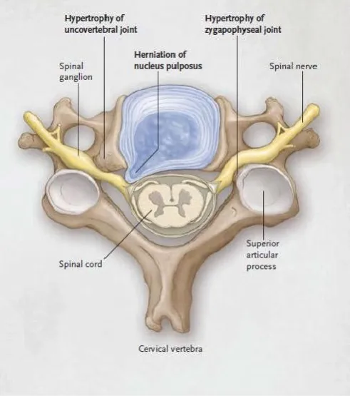
[image:4.595.311.557.100.378.2]changes of the uncovertebral joints anteriorly and zygapophyseal joints posteriorly (cervical spondylosis) (Fig. 2) (Viikari-Juntura E, et al., 1989).
Figure 2. Showing cervical spondylosis
Little is known about the natural history of cervical radiculopathy. A study in patients with cervical disc disease found that compression of a nerve root produced limb pain, whereas pressure on the disc of the lower segment produced pain in the neck and medial border of the scapula (Viikari-Juntura E, et al., 1989). Cervical radiculopathy is characterised by neck pain radiating to the arm and fingers corresponding to the dermatome involved. On examination, diminished muscle tendon reflexes, sensory disturbances, or motor weakness with dermatomal/myotomal distribution can be found (Nordin M, et al., 2008). The foraminal compression test, or Spurling test, is probably the best test for confirming the diagnosis of cervical radiculopathy. It is performed by positioning the patient with the neck extended and the head rotated, and then applying downward pressure on the head (approximately 7 kg). The test is considered positive if pain radiates into the limb ipsilateral to the side to which the head is rotated. The Spurling test has been found to be very specific (93%), but not sensitive (30%), in diagnosing acute radiculopathy (Tong HC, et al., 2002). Therefore, it is not useful as a screening test, but it is clinically useful in helping to confirm cervical radiculopathy (Nordin M,
et al., 2008). There are no universally accepted criteria for the
diagnosis of cervical radiculopathy (Wainner RS and Gill H. 2000). In most cases, the patient's history and physical examination are sufficient to make the diagnosis (Honet JC and Ellenberg MR. 2003). Magnetic resonance imaging of the cervical spine usually shows the cause of the radiculopathy which is usually spondylarthrosis or a herniated disc (Kuijper
B, et al., 2009). Radiography of the cervical spine is usually
study in all patients with chronic neck pain. Lateral, anteroposterior, and oblique views should be ordered (American College of Radiology. 2009). MRI has become the method of choice for imaging the neck to detect significant soft-tissue pathology, such as disc herniation. The American College of Radiology recommends routine MRI as the most appropriate imaging study in patients with chronic neck pain who have neurologic signs or symptoms but normal radiographs (American College of Radiology. 2009). patients in the early phase of cervical radiculopathy, can gain benefit from the use of a semi-hard cervical collar, rest for 3-6 weeks and physiotherapy accompanied by home exercises for 6 weeks. (Kuijper B, et al., 2009).
Cervical Disc Herniation: Cervical disc disorders include
herniated nucleus pulposus (HNP), degenerative disc disease (DDD), and internal disc disruption (IDD). HNP implies extension of disc material beyond the posterior margin of the vertebral body. Most of the herniation is made up of the annulus fibrosus. DDD involves degenerative annular tears and loss of disc height. IDD describes annular fissuring of the disc without external disc deformation.Clinically, cervical disc disorders frequently cause cervical axial pain, radiculopathy, and myelopathy. This kind of pain from the intervertebral disc is known to be caused by mechanical compression from extruded disc material, accompanying inflammatory response, and released chemical mediators (Nardi PV, et al., 2005). Intervertebral disc herniation in the cervical spine causes radicular pain (brachialgia) that radiates from the shoulder to the forearm to the hand (Carette S and Fehlings MG., 2005).
Cervical Spondylosis: Cervical spondylosis is a chronic
degenerative condition of the cervical spine that affects the vertebral bodies and intervertebral disks of the neck (in the form of, for example, disk herniation and spur formation), as well as the contents of the spinal canal (nerve roots and/or spinal cord). Some authors also include the degenerative changes in the facet joints, longitudinal ligaments, and ligamentum flavum. Spondylosis progresses with age and often develops at multiple interspaces. Chronic cervical degeneration is the most common cause of progressive spinal cord and nerve root compression. Spondylotic changes can result in stenosis of the spinal canal, lateral recess, and foramina. (Binder AI. 2007). It is widely assumed, that neck discomfort in the older population is related to the radiographic findings of facet joint osteoarthritis. Few patients with cervical OA have symptoms arising primarily from C1 through C2 facet joints, however, patients tend to be older women complaining primarily of occipital pain with crepitus in the upper cervical spine and occipital tender points. These patients usually respond to conservative therapy, but surgical fusion is occasionally indicated for intractable pain (Halla JT and Hardin JG. 1987).
X- ray changes in neck pain patients: cervical degenerative
spinal changes (CDSC) include reduced disc height, osteophytes, sclerosis of the vertebral endplates, anular tear, disc bulging, disc herniation (prolapsed disc), spondylolisthesis, bony changes of the vertebral bodies and facet joint osteoarthrosis (Svend Lings, et al., 2008).
Shoulder pain: Defining shoulder symptoms for
epidemiological studies of occurrence presents a number of difficulties. The complex interrelations between the shoulder
and adjacent areas and the frequent occurrence of referred pain make clinical case definition problematic (Bjelle A. 1989).
Anatomy of the shoulder: The shoulder refers to the
glenohumeral joint, but in fact the four joints of the shoulder girdle move synchronously to allow effective hand placement, and all four joints are potential sites of pain or dysfunction. These joints are the sternoclavicular, the acromioclavicular, the glenohumeral, and the scapulothoracic joints. The shoulder joint is the most mobile joint of the body, although mobility is gained at the sacrifice of stability. Only 25% of the humeral head surface has contact with the glenoid at any time. The labrum increases the contact area of the articular surface and confers stability to the joint (O'Brien S.J et al., 1990). The sternoclavicular joint is the shoulder girdle's only bony attachment to the axial skeleton. It is an incongruous joint, formed by a sternal articular surface that is concave from above downward and slightly convex front to back, a fairly flat clavicular articular surface, and the cartilage of the first rib. An articular disc between the sternum and clavicle improves the fit of the joint. The disc is attached to the clavicle above and to the first costal cartilage below, making the sternoclavicular joint very stable (Warwick R, and Williams PL. 1973). The costoclavicular ligament is just lateral to the sternoclavicular joint, and prevents excessive elevation or protraction of the clavicle. The acromioclavicular joint lies at the lateral end of the clavicle and allows the acromion (and scapula) to rotate on the clavicle. The sternoclavicular joint and acromioclavicular joint permit shrugging (elevation and depression of the shoulder girdle). In elevation of the arm above the head, movement at the sternoclavicular joint is seen in the first 90 degrees, with 4-degree elevation of the clavicle for every 10-degree elevation of the humerus. Movement at the acromioclavicular joint is important in the latter phase of abduction, above 135 degrees (O'Brien S.J et al., 1990).
deltoid and supraspinatus work together throughout abduction, with maximal deltoid activity at 120-degree abduction and peak supraspinatus activity near 100-degree abduction. The deltoid can initiate abduction in a shoulder whose supraspinatus muscle is paralyzed (Howell SM, et al., 1986). The glenohumeral joint and the scapulothoracic joint move synchronously in what is termed scapulohumeral rhythm. In the plane of the scapula (a plane tilted 30 degrees anterior from the coronal plane), glenohumeral abduction accounts for 103 degrees and scapular movement 65 degrees during full abduction. The glenohumeral joint moves approximately 3 degrees for every 2 degrees of scapular motion. If the scapulohumeral rhythm is abnormal and this ratio does not apply, usually the shoulder girdle has been injured (Kelly B.T., et al., 1996). The subacromial bursa allows the rotator cuff tendons to glide freely beneath the coracoacromial arch, without friction. The subdeltoid bursa, subcoracoid bursa, and supraspinatus bursa are extensions of the subacromial bursa, which serves as a secondary scapulohumeral joint or suprahumeral joint, separating the humerus from the fibro-osseous coracoacromial arch (Cailliet R. 1991).
Epidemiology of shoulder pain: Shoulder complaints are
poorly understood in relation to their epidemiology, impact upon the individual and society, appropriate management, and prognosis. Research has been limited by a lack of adequate randomization and blinding, the use of heterogeneous study populations, and a broad spectrum of treatment approaches, outcome measures, and follow-up intervals (Speed, C.A. and Hazleman, B.L. 2001).
Etiopathogenesis of shoulder pain: Some Risk factors have
been implicated in the development of shoulder pain. These include:
Individual factors: as sex, age, body mass index, mental
stress, smoking (non, previous and current smoker) and previous shoulder injuries.
Work related factors: like repetitive work, hand-arm
vibration, daily lifting of loads, amount of twisting movements of the trunk during a workday, working with the trunk flexed forward, working with a hand above shoulder level, working with rotated neck, working in sitting position, physical strenuousness of work, overload at work (difficulty at work, hurry at work), risk of accident at work due to tripping, slipping, climbing stairs, etc., frequency of physical exercise (times/week) and sports activity (Miranda H, et al., 2001).
Examination of the shoulder:- shoulder pain can be referred
from the neck, so the cervical range of motion should be ascertained. A careful neurologic exam of the upper extremities, particularly in the patient with weakness or paresthesia should be performed. Cervical radicular pain may radiate below the elbow to the hand, and may be completely relieved by elevating the arm above 120 degrees (Kozin F. 1997).
Clinical features of shoulder disorders:
Shoulder Impingement and Rotator Cuff Tendinopathy:
Shoulder impingement syndrome (SIS) refers to a combination of shoulder symptoms, examination findings, and radiologic signs attributable to non traumatic compression of the structures around the glenohumeral joint that occur with
shoulder elevation. Such compression causes persistent pain and dysfunction. Studies suggest that shoulder impingement syndrome (SIS) is the most common cause of shoulder pain (Faber E, et al., 2006). Impingement may be defined as the encroachment of the acromion, coracoacromial ligament, coracoid process, or AC joint on the rotator cuff as it passes beneath them during glenohumeral motion. The function of the posterior rotator cuff is to abduct and externally rotate the humerus. The cuff with the biceps tendon serves as a humeral head depressor to maintain the head centered within the glenoid fossa as the cuff and deltoid elevate the arm (Altchek
DW et al., 1990).Rotator cuff tendinitis is clinically defined as
shoulder pain exacerbated by movement against resistance when the shoulder is abducted (supraspinatus tendinitis), externally rotated (infraspinatus tendinitis), or internally rotated (subscapularis tendinitis). Active range of motion may be limited by pain, but passive range of motion is full (Chard
MD, et al., 1988).Repetitive activity at or above the shoulder
during work or sports represents the main risk factor for SIS. As with many shoulder disorders, increasing age also predisposes to SIS (Ardic F, et al., 2006). SIS is common among athletes who participate in overhead sports (Meister K. 2000). These sports may include swimming, throwing, tennis, weightlifting, golf, volleyball, and gymnastics (Hutton Ks and Julin MJ. 2002). Overhead work activities that can increase risk for developing SIS include painting, stocking shelves, and mechanical repair (Faber E, et al., 2006). The first clue to shoulder impingement is an insidious development of shoulder pain with overhead activity. A patient will often describe pain at a certain point in abduction or flexion of the arm, beyond which the pain disappears again. This “painful arc of motion” occurs between 60 and 120 degrees, when an inflamed rotator cuff or subacromial bursa tries to squeeze beneath the coracoacromial arch, as the arm is raised or lowered. Once the inflamed structure fully passes beneath the arch, the pain disappears, until the arm is brought back to its prior position and the inflamed tendons or bursa must pass beneath the coracoacromial arch again (DeFerm A, et al., 1997). Two physical signs suggest impingement.
In the Neer maneuver, the patient's scapula is immobilized and the painful arm is passively flexed as far as it will go. The patient grimaces or complains (Neer CS. 1983). (This maneuver can be done actively also, with the patient flexing the arm to 180 degrees, and the scapula free.) Hawkins and Kennedy described pain when the arm is flexed to 90 degrees, then internally rotated. (Hawkins RJ and Kennedy JC. 1980).
in passive shoulder range of motion exercises (Cailliet R. 1991). Subacromial steroid injections should be given to those who do not respond to rest, physical therapy, and NSAIDs within several weeks (Adebajo AO, et al., 1990).
Calcific Tendinitis: Calcific tendinitis is a painful condition
around the rotator cuff and is associated with deposition of calcium salts, primarily hydroxyapatite (McKendry R.J.R. et al., 1982). The cause of calcific tendinitis is unknown. The commonly accepted cause is degeneration of the tendon, which leads to calcification through a dystrophic process (Sarkar K. and Uhthoff H.K., 1978).Calcium deposits are seen about the shoulder on routine radiographs in 2.7% to 8% of adult shoulders. All calcium deposits larger than 1.5cm became symptomatic, and 34.6% of all calcium deposits in the rotator cuff structures would be symptomatic at some time (Codman EA. 1934). The calcium deposits that cause clinical calcific tendinitis are intratendinous, often bilateral and multifocal (Simon WH. 1975). The supraspinatus tendon is affected more than 50% of the time, followed in frequency by infraspinatus, teres minor, and subscapularis. The clinical syndromes are illnesses of middle age, peaking in the fifth decade. Sexes appear to be equally affected (Kozin F. 1997); however, there may be a female predominance (McKendry RJR, et al., 1982). When unilateral, the dominant shoulder is more likely to be affected (Mavrikakis ME, et al., 1989). Four histologic patterns can be seen in calcific tendinitis, and all patterns often coexist in the patient: (a) Precalcific changes are those of focal fibrocartilaginous transformation within the tendon. (b) The calcific phase is one of intratendinous calcium deposits without any inflammatory response. (c) The resorptive phase is that associated clinically with acute excruciating pain. (McKendry RJR, et al., 1982). Acute calcific tendinitis is self-limited and colchicine often helps with the pain, just as in acute gout or pseudogout (Kozin F. 1997). Local steroid injections are avoided, for they abort the natural resorptive process and promote recurrences. There is also the fear of local tissue necrosis and lessened tendon strength after steroid injections. Surgical removal of an acutely inflamed calcific deposit is not a commonly offered option. (Ebenbichler GR, et al., 1999).
Adhesive capsulitis: Frozen shoulder is a descriptive term that
refers to a stiffened glenohumeral joint that has lost significant range of motion (abduction and rotation). It is a reversible contraction of the joint capsule in almost all cases (Anderson, BC. 2006).Pathologically, the glenohumeral joint capsule has lost its normal distensibility. In long-standing cases, adhesions may form between the joint capsule and the humeral head (Bunker TD and Anthony PP. 1995). Frozen shoulder is the name given to a painful stiff shoulder, whose glenohumeral motion is globally limited by a contracted, poorly compliant joint capsule (Harryman DT, et al., 1998). Frozen shoulder may occur as an idiopathic process or as a result of an underlying disorder that leads to disuse. Idiopathic frozen shoulder occurs most commonly in the fourth through sixth decades of life. Rotator cuff tendinopathy, acute subacromial bursitis, fractures about the humeral head and neck, and paralytic stroke are relatively common predisposing factors for the development of frozen shoulder. The most common cause is rotator cuff tendinopathy. Approximately 10 percent of patients with this disorder will develop a frozen shoulder. (Pal
B, et al., 1986). Adhesive capsulitis has been reported in the
wrist, the ankle, and the hip, though in much lower incidences than in the shoulder. Other synonyms for frozen shoulder
include check-rein shoulder, periarthritis of the shoulder, and scapulohumeral periarthritis (Mont MA, et al., 1999). Frozen shoulder is rare before age 40, and generally involves persons aged 50 to 70. Women are affected slightly more than men (Rizk TE and Pinals RS. 1982). It is thought that frozen shoulder will not recur in a shoulder that has been affected previously. (Wright V and Haq AMMM. 1976). The patient with a frozen shoulder experiences diffuse shoulder pain, often night pain, and a gradual loss of mobility. Active and passive mobility of the shoulder is limited. External rotation, abduction, and internal rotation are the motions most affected, but all motions of the glenohumeral joint are involved. The patient may have pain-free motion within the confines of his decreased range, with pain only at the extremes of his range. Scapulothoracic motion remains normal, and is used by the patient to try to compensate for lost glenohumeral mobility. Frozen shoulder is a diagnosis of exclusion, and must be differentiated from chronic posterior dislocations, rotator cuff disease, septic arthritis, avascular necrosis, fracture, bony or pulmonary neoplasm, osteoarthritis of the shoulder or cervical spine, and other shoulder arthropathies.
The shoulder with a torn or inflamed rotator cuff generally has limited motion in only one or two directions, and has full passive motion once Xylocaine is used to eliminate pain and muscle spasm (Shaffer B, et al., 1992).The natural history of frozen shoulder has been divided into three phases: freezing, frozen, and thawing (Reeves B. 1975). The freezing phase may last 10 to 36 weeks, with generalized pain and growing stiffness. The second phase of marked stiffness typically lasts 4 to 12 months, and is less painful. The thawing phase generally lasts 5 to 26 months, and is characterized by gradual return of glenohumeral motion and function. Spontaneous recovery is the usual course of frozen shoulder. The cause of the spontaneous recovery from frozen shoulder is unknown but perhaps is due to an unrecognized tear of the joint capsule or to mechanical attrition of the tightened capsule (Rizk TE and Pinals RS. 1982). Plain radiographs typically are normal in patients with frozen shoulder, or show disuse osteopenia. Periarticular calcifications are seen in 7% to 9%, which is the frequency in the general population. An axillary view of the shoulder should be done in addition to standard anteroposterior views, to rule out unrecognized dislocations, fractures, or osteophytes (Shaffer B, et al., 1992). Exercises form the basis of every treatment proposed for frozen shoulder and includes active and passive range of motion programs. During the freezing phase, the emphasis is on pain relief. This can be accomplished with a sling, moist heat, NSAIDs, analgesics, oral steroids and/or local steroids (Kozin F. 1997). Local steroid injections benefit pain more than they increase range of motion. (Zuckerman JD and Cuomo F. 1993).
Bicepital tendinitis: Ninety-five percent of bicipital tendinitis
is associated with rotator cuff disease and impingement (Neer CS. 1972). The patient has anterior shoulder pain, which may extend to the biceps muscle belly, but does not radiate to the neck or past the biceps insertion distally. Usually, there is no history of trauma, but rather a history of repetitive use, often overhead. The pain worsens with lifting, carrying, or any use of the biceps. It is chronic and may worsen at night. On exam, the patient has point tenderness over the biceps tendon (felt directly anterior and about 2 to 3 inches below the acromion when the arm is internally rotated 10 degrees) (Burbank KM,
et al., 2008).The area of point tenderness moves as the arm
underlying bone. Flexion against resistance with the elbow extended and the forearm supinated causes pain over the biceps tendon (Speed's test). Maneuvers that stretch the inflamed biceps tendon (such as backward extension and external rotation with an extended elbow) cause pain (Reneman, M. F., et al., 2005).
Glenohumeral osteoarthritis
Primary glenohumeral osteoarthritis is uncommon, perhaps because the joint is non–weight-bearing and not a target joint for generalized osteoarthritis. The joint is subjected to high forces across it, largely related to the weight of the arm and the forces needed to hold the humeral head against the glenoid during abduction (DePalma AF. 1983). Clinically, primary glenohumeral arthritis causes decreased motion, particularly decreased rotation and abduction. Crepitus is felt with joint motion. Severe pain is unusual, and if it is present, an alternative explanation for the pain should be sought in the tendons and ligaments (rotator cuff tendinitis or
tendinitis) or neighboring joints (referred pain from the acromioclavicular joint or the neck). Treatment of glenohumeral osteoarthritis includes NSAIDs, analgesics, and avoidance of painful movements. Occasionally, joint arthroplasty or arthrodesis is indicated, depending on the functional demands on the shoulder (DePalma AF. 1983).
Acromioclavicular Disease
The acromioclavicular (AC) joint is a common source of shoulder pain. The articular surfaces are small, usually incongruous, and variable. Most people have advanced degenerative arthritis of the AC joint by mid
AF. 1983).
Neck/shoulder pain
Neck/shoulder pain is defined as pain lasting for longer than a day in an anatomical distribution bounded by the occiput and the lower edges of the scapulae (Figure 3)
[image:8.595.77.257.534.767.2]2003).
Figure 3. Boundaries of neck/shoulder pain ( 2003)
26487 Nabeel Ahmed Altaee et al.
underlying bone. Flexion against resistance with the elbow extended and the forearm supinated causes pain over the biceps tendon (Speed's test). Maneuvers that stretch the inflamed biceps tendon (such as backward extension and rnal rotation with an extended elbow) cause pain
Primary glenohumeral osteoarthritis is uncommon, perhaps bearing and not a target joint hritis. The joint is subjected to high forces across it, largely related to the weight of the arm and the forces needed to hold the humeral head against the glenoid during abduction (DePalma AF. 1983). Clinically, primary eased motion, particularly decreased rotation and abduction. Crepitus is felt with joint motion. Severe pain is unusual, and if it is present, an alternative explanation for the pain should be sought in the tendons and ligaments (rotator cuff tendinitis or tear, calcific tendinitis) or neighboring joints (referred pain from the acromioclavicular joint or the neck). Treatment of glenohumeral osteoarthritis includes NSAIDs, analgesics, and avoidance of painful movements. Occasionally, joint throdesis is indicated, depending on the functional demands on the shoulder (DePalma AF. 1983).
The acromioclavicular (AC) joint is a common source of shoulder pain. The articular surfaces are small, usually riable. Most people have advanced degenerative arthritis of the AC joint by mid-life (DePalma
Neck/shoulder pain is defined as pain lasting for longer than a day in an anatomical distribution bounded by the occiput and ower edges of the scapulae (Figure 3) (Smedley et al.,
ndaries of neck/shoulder pain (Smedley et al,
Prevalence and incidence of neck and shoulder pain
There are many studies concerning the prevalence of NSP, of which most have been carried out in different occupational groups of adult populations. There is a fairly large variation in the prevalence rates of NSP, since the symptoms have been determined and measured quite differently, mostly with self administered questionnaires (Sari Siivola. 2003).
epidemiologic studies among adolescents, the prevalence of NSP has consistently been shown to increase with age and the trend seems to continue through young adulthood (Salminen 1984; Vikat A, et al., 2000).
Etiopathogenesis of neck /shoulder p
The most commonly used term for non specific NSP has been ’tension neck’. Valtonen defined it as a cluster of symptoms, related to continuous tension of the muscles of the neck and shoulder region, but with no pathological change
or soft tissues of the cervical spine (Valtonen E, 1968). Although muscle tension often coexists with neck pain, this term is based on the idea of neck pain originating from tension of the neck muscles. In the latest versions of commonly use classification of diseases,
ICD-has been omitted. In the literature on occupational neck and shoulder disorders, the terms ’occupational cervicobrachial disorder’ (Maeda 1977; Hagberg M,
upper limb disorder’ (Kuorinka and Viikari
’repetitive strain injury’ (Ferguson 1984) have been suggested. In the majority of individuals suffering from NSP, the pain is non specific and the etiology of pain remains unclear. Most frequently, NSP arises from functional disorders, and no tissue damage can be displayed. Diversified classifications of NSP have been presented, and they have been based on the location of pain (cervicobrachialgia), the etiology of pain (work related), the duration of pain (acute/chronic pain), the findings of status (tension neck), radiological findings (degenerative disc disease), or dysfunction of cervical facet joints (hypo hypermobility) (Spitzer et al., 1987). The classification of NSP in the current care guidelines for neck pain in Finland (2002) is based on data elicited in a clinical interview, symptoms, and clinical findings: 1) local neck pain 2) radiating neck pain 3) whiplash injury 4) myelopathy (compression of the spinal cord) 5) other neck pains, which ar
diseases, tumors, or status following fractures of cervical spine (Guzman J. Et al., 2008). NSP is assumed to be a multifactorial disease, and it has been suggested that there are several risk factors contributing to its development (A al., 2000). The contributing factors of neck and shoulder symptoms have been a fairly common topic of both cross sectional and longitudinal studies in adults, but particularly the progression of NSP into a chronic problem demands clarifying. Age, female gender (Viikari
physical work loads (Ariëns psychosocial factors (Ariëns
consistently been shown to associate with NSP. Most of the studies in adult populations have concern
occupational groups, and the main interest has focused on work-related risk factors. The risk factors for NSP can be classified as:
Physical factors: Like age, female, specific neck, arm or trunk
postures/ movements, previous head or neck inj
lifting, hand arm vibration, work place design and low pressure pain threshold.
et al. Cervical spine pain and its association with shoulder joint problems
Prevalence and incidence of neck and shoulder pain
There are many studies concerning the prevalence of NSP, of have been carried out in different occupational groups of adult populations. There is a fairly large variation in the prevalence rates of NSP, since the symptoms have been determined and measured quite differently, mostly with
self-res (Sari Siivola. 2003). In the epidemiologic studies among adolescents, the prevalence of NSP has consistently been shown to increase with age and the trend seems to continue through young adulthood (Salminen
is of neck /shoulder pain
The most commonly used term for non specific NSP has been ’tension neck’. Valtonen defined it as a cluster of symptoms, related to continuous tension of the muscles of the neck and shoulder region, but with no pathological changes in the bones or soft tissues of the cervical spine (Valtonen E, 1968). Although muscle tension often coexists with neck pain, this term is based on the idea of neck pain originating from tension of the neck muscles. In the latest versions of commonly used -10 (1999, 2003), ‘tension neck’ has been omitted. In the literature on occupational neck and shoulder disorders, the terms ’occupational cervicobrachial disorder’ (Maeda 1977; Hagberg M, et al., 1995), ’neck and disorder’ (Kuorinka and Viikari-Juntura 1982), and ’repetitive strain injury’ (Ferguson 1984) have been suggested. In the majority of individuals suffering from NSP, the pain is non specific and the etiology of pain remains unclear. Most rises from functional disorders, and no tissue damage can be displayed. Diversified classifications of NSP have been presented, and they have been based on the location of pain (cervicobrachialgia), the etiology of pain
(work-(acute/chronic pain), the findings of status (tension neck), radiological findings (degenerative disc disease), or dysfunction of cervical facet joints (hypo- or ., 1987). The classification of NSP es for neck pain in Finland (2002) is based on data elicited in a clinical interview, symptoms, and clinical findings: 1) local neck pain 2) radiating neck pain 3) whiplash injury 4) myelopathy (compression of the spinal cord) 5) other neck pains, which are related to general diseases, tumors, or status following fractures of cervical spine ., 2008). NSP is assumed to be a multifactorial disease, and it has been suggested that there are several risk factors contributing to its development (Ariëns et , 2000). The contributing factors of neck and shoulder symptoms have been a fairly common topic of both cross-sectional and longitudinal studies in adults, but particularly the progression of NSP into a chronic problem demands female gender (Viikari-Juntura et al., 2001), physical work loads (Ariëns et al., 2000), and certain psychosocial factors (Ariëns et al., 2001) have fairly consistently been shown to associate with NSP. Most of the studies in adult populations have concerned different occupational groups, and the main interest has focused on related risk factors. The risk factors for NSP can be
Like age, female, specific neck, arm or trunk movements, previous head or neck injury, sitting, lifting, hand arm vibration, work place design and low
Psychosocial factors: like high job demands, mental stress, depression and anxiety.
Socioeconomic factors: like smoking, obesity, three or more
children, lower educational level, living alone.
Co morbidities: as headache, shoulder pain and digestive
problems. (Carroll LJ, et al. 2004; Walker-Bone K, et al., 2004). Several structures have been shown to cause pain in the neck and shoulder area. Bones, nerves, discs, longitudinal ligaments, muscles, facet joints, and dura are all capable of evoking pain, when irritated or inflamed (Spitzer et al., 1987; Cailliet 1991). Muscle tension is thought to be one of the causes of NSP, and the NSP in adolescents is most likely to be muscle pain in origin. Isometric muscle contraction, in Particularly, has been suggested to create intramuscular pressure, which might lead to tears of muscle fibers (Cailliet 1991). The studies concerning increased muscle activation measured by EMG in NSP have produced evidence mainly by two different study designs. Firstly, there are studies in occupational settings, where the muscle activity pattern has been recorded with surface EMG and a clinical examination of the shoulder region has been performed, and secondly, there are experimental studies, where the muscle activity patterns and pain development have been recorded in situations involving mental stress and minimal physical activity (Westgaard 1999). Especially the development of work-related complaints in the neck and shoulder region is commonly attributed to excessive loading of the shoulder muscles (Winkel and Westgaard 1992). It has still remained unclear whether Disc degeneration (DD) is a cause of spinal pain. However, outer anular rupture was documented to relate to pain (Moneta et al. 1994). Although there are indications that anulus rupture and disc herniation may be manifestations of different states of DD, their role as causes of NSP are still under discussion (Yu et al., 1988). There are only a few studies concerning the mechanism between stress and NSP (Hasselhorn et al., 2001). Clinical observations show that chronic NSP can be accompanied by depressive symptoms. The mechanism between NSP and depressive symptoms is, however, still under discussion. It has been suggested that chronic pain represents the expression of an underlying depressive disturbance and can be traced back to masked depression (Magni 1987).
MATERIALS AND METHODS
The present study had approval from regional research committee of Mosul health administration, and the scientific research committee of collage of medicine, university of Mosul, Mosul, Iraq. Patient collection started at October 2010 and ended at December 2011 at the rheumatology outpatient department in Ebn Sena teaching hospital.
Study design: Case – control study.
Patients group: Hundred Patients with neck pain for one
month or more duration (74 females, 26 males) aging between 35 to 50 years old were collected.
Control group: Hundred Patients with low back pain but
without neck pain (75 females, 25 males) aging between 35 to 50 years old were kept as a control.
Inclusion criteria: Patients with neck pain for one month and
more duration.
Exclusion criteria
Patients with previous or recent neck trauma.
Diseases with Inflammatory arthritis involving the neck (as rheumatoid arthritis and spondyloarthropathies).
Patients with previous or recent cardiovascular diseases.
Patients with malignancies or infection.
Cervical myelopathy
Diabetic and hypertensive patients.
Data collection
Data were obtained directly by interviewing the studied subjects. A clinical assessment form was designed to record the subjects informations, which includes name, age, sex, duration of the disease, a complete history of symptoms related to neck pain and local examination of the cervical spine and shoulder (including inspection, palpation, range of motion and special tests). A standard musculoskeletal examination method were followed. (appendix 1). X-rays for the patients (66 patient) were taken in the hospital and it included an A-P and lateral film for the cervical spine and an A-P view only for the shoulder in neutral position.
Instruments : No special instruments have been used in this
study except for the use of a hammer for reflex evaluation.
Methods : The presented study involved 2 groups, group one
consisted of one hundred subjects complaining from neck pain kept as patient group, and group two, also consisted of one hundred subjects complaining from low back pain kept as control group . The age of the subjects ranged between 35-50 years old and was classified into 3 groups: 35-39, 40-45 and 46-50 years. Both groups were questioned and examined at the rheumatology outpatient department in Ebn Sena teaching hospital. The patient group subjects undergone a clinical examination and a questionnaire about their symptoms regarding neck pain and the presence or absence of any shoulder joint problem. Both groups were questioned about their demographic parameters including age, sex, marital state, physical activity, education and smoking. The marital status of the subjects was classified as whether they are single or married. Finally, education status was classified as illiterate and non illiterate, and smoking as non smoker, smoker and x-smoker. A complete clinical symptom evaluation for neck pain was done for the patients like intensity, frequency, timing, aggravating and reliving factors and associated symptoms.
The duration of neck pain was classified into 3 classes:
Class a- one month to one year
Class b- more than one year to 5 years
Class c- more than 5 years
The intensity of pain was measured by the Visual Analogue Scale (VAS) scoring pain intensity from (0) for no pain to (100) as the most severe pain experienced by the patient. The patient group, according to pain severity, was classified into 3 main groups; mild (1-30) VAS group, moderate (31-60) VAS group and severe (61-100) VAS group.
The frequency of pain was classified into 3 classes: