IJPSR (2012), Vol. 3, Issue 07 (Research Article)
Received on 06 March, 2012; received in revised form 24 May, 2012; accepted 23 June, 2012
EFFECT OF DIFFERENT EXTRACTS OF STEM BARK OF FICUS SP. ON MULTIDRUG RESISTANT PATHOGENIC BACTERIA
D.M. Manimozhi*1, S. Sankaranarayanan 2 and G. Sampath Kumar 3
Mother Teresa Women’s University 1, Kodaikanal-624 102, Tamil Nadu, India
Sri Sairam Siddha Medical College and Research Centre 2, Chennai 600 044, Tamil Nadu, India Dr. Ambedkar Government Arts College 3, Chennai-600 039, Tamil Nadu, India
ABSTRACT
Medicinal plants represent a rich source of antimicrobial agents. The traditional medicine involves the use of different plant extracts of bioactive constituents. Ficus is a huge tropical deciduous or evergreen tree with more than 800 species.
Purpose: Ficus benghalensis, Ficus religiosa, Ficus recemosa are important ingredients in many siddha, ayurveda and traditional formulations and used for the treatment of bacterial infections. An attempt was made to study the antibacterial effect of Ficus sp., by appropriate methods.
Method: The acetone, methanol, ethylacetate extracts of Ficus benghalensis, Ficus religiosa, Ficus recemosa were evaluated for antibacterial activity against medically important bacteria Pseudomonas aeruginosa, Escherichia coli, Proteus vulgaris, Bacillus subtilis, Staphylococcus aureus. Phyto constituents of three plants were screened. The in-vitro antibacterial activity was performed by agar disc diffusion assay.
Results: The plant extract that showed higher active antibacterial activity against tested bacterial strain was chosen analyse beta-lactamase inhibitory activity. Ficus sp. Showed more antibacterial activity on Escherichia coli, Staphylococcus aureus and Proteus vulgaris.
Conclusion: Amongst the plant species screened, Ficus benghalensis extracts showed best antibacterial activity. These findings suggest a new pathway in elucidating a potent antimicrobial agent from Ficus benghalensis.
INTRODUCTION: Medicinal plants have been used an exemplary source for centuries as an alternative remedy for treating human diseases because they contain numerous active constituents of therapeutic value 1. The potential of higher plants as source for new drugs is still largely unexplored. Thus, any phytochemical investigation of a given plant will reveal only a very narrow spectrum of its constituents. Plants are important source of potentially useful structures for the development of new chemotherapeutic agents.
The first step towards, this goal is the invitro
antibacterial assay 2.
Infectious diseases are the leading cause of death world-wide. Antibiotic resistance has become a global concern 3. The clinical efficacy of many existing antibiotics is being threatened by the emergence of multi-drug resistant pathogens 4. Staphylococcus aureus is one of the major pathogens found on the mucous membranes and the skin of around a third of
Keywords: Ficus, Phytoconstituents, Disc diffusion assay, Bacterial pathogens
Correspondence to Author:
D.M. Manimozhi
the population. It’s extremely adaptable to antibiotic pressure. It was the first bacterium in which penicillin resistance was found. MRSA was responsible for 37% of fatal cases of blood poisoning in the UK in 1999; up fro 4% in 1991, half of all S. aureus infections in the U.S are resistant to penicillin, methicillin, tetracyline and erythromycin. Considering the vast potentiality of plants as sources for antimicrobial drugs with reference to antibacterial agents, a systematic investigation was undertaken to screen the antibacterial activity of three different species from
Ficus genus.
The genus Ficus includes some 750 species of woody plants occurring in most tropical and subtropical forests throughout the world 5. The genus is remarkable for the large variation in the habits of its species 6.
[image:2.612.320.575.185.626.2]Ficus benghalensis: Ficus benghalensis belongs to the family Moraceae, which is commonly known as Banyan tree. The tree is a fast growing, evergreen tree found in monsoon and rain forests grow up to 30 meters, with spreading branches and many aerial roots. Figure 1 shows the fully grown banyan tree with aerial roots.
FIGURE 1: FICUS BENGHALENSIS
Kingdom : Plantae
Division : Magnoliophyta Class : Magnoliopsida Order : Urticales Family : Moraceae Genus : Ficus
Species : F. benghalensis
Binomial name: Ficus benghalensis
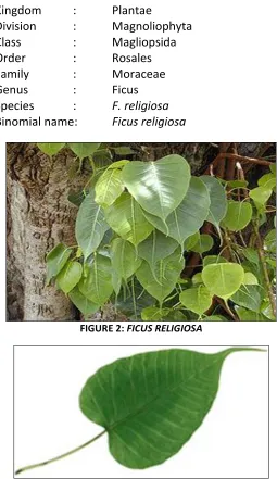
Ficus religiosa: Ficus religiosa Linn (Moraceae) commonly known as ‘Peepal tree’ is a large widely branched tree with leathery, heart‐shaped, leaves on long slender petioles and purple fruits growing in pairs. The tree is regarded as a sacred tree to both Hindus and Buddhists. It has got mythological, religious and medicinal importance 7. Figure 2 shows the branched peepal tree and figure 3 represents the typical leaf structure of the same tree.
Kingdom : Plantae
Division : Magnoliophyta Class : Magliopsida Order : Rosales Family : Moraceae Genus : Ficus Species : F. religiosa
Binomial name: Ficus religiosa
FIGURE 2: FICUS RELIGIOSA
FIGURE 3: TYPICAL LEAF STRUCTURE FICUS RELIGIOSA
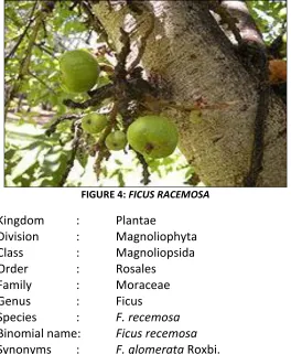
[image:2.612.31.282.410.724.2]issue from the stem and larger branches, muchcontracted at the base when young, subglobes, pyriform or subturbinate, 1.5 in. across, smooth or pubscent, red when ripe, with depressed umbilicus (edible but usually full of worms) ; basal bracts 3, ovate triangular ; male, female and gall flowers together in one receptacle, the male flower forming a zone near the mouth, the fertile female flowers forming a layers near the walls of the receptacle, and the gall flowers a more internal layer. Figure 4 shows the branched Ficus recemosa tree with figs.
FIGURE 4: FICUS RACEMOSA
Kingdom : Plantae
Division : Magnoliophyta Class : Magnoliopsida Order : Rosales
Family : Moraceae Genus : Ficus Species : F. recemosa
Binomial name: Ficus recemosa
Synonyms : F. glomerata Roxbi.
Ethnobotany of plants studied: Ficus plants are found throughout the world as moderate woody plants or trees. . Ficus species, namely, F. benghalensis and F. recemosa, F. religiosa are important ingredients in many ayurvedic and traditional formulations.Roots of
Ficus benghalensis shows antihelmintic activity. . Fruit extracts exhibits anti-tumour activity 8 .The fruit extracts of F. benghalensis exhibit antitumor activity and antibacterial activity, but no antifungal activity 9. In Ayurveda it is claimed that Ficus religiosa possesses anticonvulsant activity. Different parts of Ficus religiosa showed cholinesterase inhibitory activity and antianxiety activity. The methanolic extract of figs of
Ficus religiosa had anticonvulsant activity 10.
Malparmaram is an important group of ayurvedic formulation that constitutes the barks of Ficus recemosa, Ficus religiosa and Ficus benghalensis widely used in the treatment of skin diseases and also used in various ailments 11, 12.
Pharmacological studies of the plants: The bark of
Ficus benghalensis exhibited significant antibacterial activity against pathogenic bacterial like
Staphylococcus aureus, Pseudomonas aeruginosa and
Klebsiella pneumoniae 13. Ficus religiosa aqueous extract showed high antimicrobial activity. High activity was found against Bacillus subtilis and Pseudomonas aeruginosa, (multi-drug resistant) by Preeti et al14. Ethnomedicinal study and anti bacterial activities of F. recemosa was studied by Mahato et al.,15F. recemosa, country fig tree paste of bark is applied twice a day for 2-3 days to cure swellings of foot and hands . Stem bark of F. recemosa showed activity against Bacillus subtilis.
MATERIALS AND METHODS:
Plant material collection: The barks of Ficus benghalensis, Ficus religiosa, Ficus recemosa were collected from herbal garden of Gloris biomed Research center, Vadapalani. The plants authenticated identification done by Dr. S. Sankaranarayanan, Head, Department of Medicianl Botany, Sairam Siddha Medical College, Tambaram. The voucher specimens were submitted to Presidency College, Department of Botany. The voucher numbers are P.5123, P.5134, P.5137.
Test organisms: The screening for antibacterial activity was carried out in vitro condition using Pseudomonas aeruginosa (MTCC 27853), Escherichia coli (MTCC 25922), Proteus vulgaris (MTCC 13315), Bacillus subtilis (MTCC12567), and Staphylococcus aureus (MTCC 29213). All bacterial strains were were obtained from Micrbial Culture Collection Centre, Chandigarh, India.
Phytochemical analysis of the Plant Extract: The extracts were subjected to phytochemical tests for plant secondary metabolites, tannins, saponins, flavonoids, alkaloids and glycosides in accordance with Allan 16 and Harborne 17 with little modification (table 1).
1. Test for Tannins: About 0.5 g of the dried powdered samples was boiled in 20 ml of water in a test tube and then filtered. A few drops of 0.1% ferric chloride was added and observed for browrish green or a blue-black colouration.
2. Test for Saponin: About 2 g of the powdered sample was boiled in 20 ml of distilled water in a water bath and filtered. 10ml of the filtrate was mixed with 5 ml of distilled water and shaken vigorously for a stable persistent froth. The frothing was mixed with 3 drops of olive oil and shaken vigorously, then observed for the formation of emulsion.
3. Test for Flavonoids: Three methods were used to determine the presence of flavonoids in the plant sample 18, 19. 5 ml of dilute ammonia solution were added to a portion of the aqueous filtrate of each plant extract followed by addition of concentrated H2S04. A yellow colouration observed in each
extract indicated the presence of flavonoids. The yellow colouration disappeared on standing. Few drops of 1% aluminium solution were added to a portion of each filtrate. A yellow colouration was observed indicating the presence of flavonoids. A portion of the powdered plant sample was in each case heated with 10 ml of ethyl acetate over a steam bath for 3 min. The mixture was filtered and 4 ml of the filtrate was shaken with 1 ml of dilute ammonia solution. A yellow colouration was observed indicating a positive test for flavonoids. 4. Test for Terpenoids (Salkowski test): Five ml of
each extract was mixed in 2 ml of chloroform, and concentrated H2S04 (3 ml) was carefully added to
form a layer. A reddish brown colouration of the inter face was formed to show positive results for the presence of terpenoids.
5. Test for Glycosides (Keller-Killani test): Five ml of each extracts was treated with 2 ml of glacial acetic acid containing one drop of ferric chloride solution.
This was underlayed with 1 ml of concentrated sulphuric acid. A brown ring of the interface indicates a deoxysugar characteristic of cardenolides. A violet ring may appear below the brown ring, while in the acetic acid layer, a greenish ring may form just gradually throughout thin layer.
6. Test for Alkaloids:
(A) Dragendroffs reagent: 8g of bismuth nitrates Bi (NO3)3.5H2o was dissolve in 20ml of HNO3 and
2.72g of Potassium iodide in 50ml of H2O. These
were mixed and allowed to stand for deposition of KNo3 Crystals. The Supernatant was decanted off
and made up to 100ml with distilled water.
Procedure: To 0.5ml of leaf extract 2ml of HCl was added. To this acidic medium 1ml of dragendroffs reagent was added on, orange or red precipitate produced immediately indicate the presence of alkaloids.
(B) Mayer’s test: 1.36g of Mercuric chloride was dissolved in 60ml of distilled water and 5g of Potassium iodide in 10ml of water. These two solutions were mixed and diluted to 100 ml with distilled water.
Procedure: 1.2 ml of plant extract was taken in a test tube and to this 0.2 ml of dilute HCl and 0.1 ml of Mayer’s reagent was added. Formation of yellowish Puff coloured precipitate indicates the presence of alkaloid.
cloudy with 3-4 colonies of bacteria. After 30-60 min, 20 µl of sterile 1% starch solution and iodine solution were added.
If the color of iodine disappeared in 5 min, the isolate was considered beta-lactamase positive 21.
TABLE 1: PRELIMINARY PHYTOCHEMICAL ANALYSIS OF MEDICINAL PLANTS Tests
Medicinal Plant Species
Tannins Saponins Flavonoids Terpenoids Glycosides Anthraquinones
Alkaloids
Meyer’s test Dragendorff’s Test
Ficus Benghalensis + + + - - - - -
Ficus religiosa + + + + - - - -
Ficus recemosa + + + - + - - -
Antibacterial activity of different Extracts: The antibacterial activity was studied using the disc-diffusion Method 22. Bacteria were grown overnight on Mueller Hinton agar plates, five young colonies were suspended with 5ml of sterile saline (0.9%). The swab was used to inoculate the dried surface of MH agar plate by swabbing several times four times over the surface of the agar. The medium was allowed to dry for about 3 min before adding a sterile paper disc of 9mm diameter. Compounds (50µg) were weighed and dissolved in 1ml of 7% acetone. Twenty microlitres of the compounds were introduced on each disc (five replicates) and 7% acetone alone served as a normal control. The plates were incubated at 37 ◦C for 24 h; inhibition zones were measured and calculated.
RESULTS: Herbal extracts contain different phytochemicals with biological activity that can be of valuable therapeutics Index. Plants have the ability to produce a large variety of secondary metabolites (Phytochemicals) such as terpenoids, phenyl- propanoids, flavonoids and alkaloids which together account for over 2,00,000 compounds 23.
In the present study, three Ficus species namely, Ficus benghalensis Ficus religiosa and Ficus recemosa were analyzed for their phytochemical constituents. During the last three decades, the development of drug resistance and their undesirable side effects of certain antibiotics have led to the search of new antimicrobial agents mainly among plant extracts with the goal to discover new chemical structures. Preliminary screening of antibacterial activity was conducted for the three Ficus sp; against both gram positive and gram negative type of bacterial and two pathogenic fungal species. Among, those Ficus benghalensis
showed the highest inhibition of both Gram positive and Gram negative bacteria and fungi.
The antimicrobial activity was determined by measuring the diameter of the zone of inhibition, i.e. the mean of triplicates ±S.D. In recent years, secondary plant metabolites (phytochemicals) previously with unknown pharmacological activities have been extensively investigated as a source of medicinal agents 24. Phytochemicals with adequate, antibacterial effect will be used for the treatment of bacterial infections 25. It is time to examine more closely our natural resources, i.e., the plants, which contain compounds of potential medical use.
Phytochemistry: Phytochemical screening of aqeous and methanolic extract of Ficus benghalensis showed the presence of tannins, saponins, flavonoids. Aqueous and methanolic extracts of Ficus religiosa showed the presence of Tannins, saponins, flavonoids and terpenoids. Aqueous and methanolic extract of Ficus recemosa showed the presence of Tannins, saponins, flavonoids and glycosides.
Antibacterial activity: The dried bark powder of three
Ficus sp., namely Ficus benghalensis,Ficus religiosa Ficus recemosa were extracted with three organic solvents acetone, methanol and ethyl acetate. The antibacterial activity of these extracts was performed by disc diffusion assay. The results for antibacterial activity of Ficus beghalensis, Ficus religiosa and Ficus recemosa bythe agar disc diffusion assay are given in
tables 2, 3, 4 respectively.
Ficus benghalensis: Acetone extract of F. benghalensis
benghalensis was active against E. coli, S. aureus and B. subtilis.
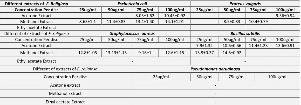
Ficus religiosa: The growth of Bacillus subtilis was significantly inhibited by acetone extract of Ficus religiosa. Higher concentrations of the same extract were required for the inhibition of E. coli. Methanol extract of the plant was very active against all the tested bacterial pathogens except P. aeruginosa. Ethyl acetate extract was not active against all the bacterial species.
Ficus recemosa: Acetone extract of Ficus recemosa was found to be active against E. coli, Proteus vulgaris and
Bacillus subtilis. It showed maximum activity against
Proteus vulgaris measuring 14.0mm of zone inhibition at 100µg/ml. Methanol extract of plant was found to be active against E. coli, P. vulgaris and B. subtilis and
P. aeruginosa. The ethyl acetate extract showed no anti bacterial activity against all the tested bacteria. The mean zone of inhibition on obtained using the agar disc diffusion assay ranged from 6mm (for
Pseudomonas aeruginosa) to 14mm (Proteus vulgaris, Bacillus subtilis, E. coli).
TABLE 2: ANTIBACTERIAL ACTIVITY OF DIFFERENT BARK EXTRACTS TESTED AGAINST PATHOGENIC BACTERIA BY DISC DIFFUSION ASSAY (FICUS BENGHALENSIS)
Different extracts of F.
benghalensis Escherichia Coli Proteus vulgaris
Concentration Per disc 25ug/ml 50ug/ml 75ug/ml 100ug/ml 25ug/ml 50ug/ml 75ug/ml 100ug/ml
Acetone extract 7.1±1.5 10.4±1.09 12.2±0.98 13.2±0.15 9.2±0.95 11.36±0.81 11.8±1.3 14.26±0.91
Methanol Extract 7.5±1.35 10.3±1.05 11.9±1.42 12.5±1.23 8.4±0.65 11.3±0.95 12.4±1.4 13.1±1.35
Ethyl acetate Extract 8.53±1.02 10.0±1.47 11.4±0.92 12.3±1.38 -
Different of extracts of F.
benghalensis Staphylococcus aureus Bacillus subtilis
Concentration Per disc 25ug/ml 50ug/ml 75ug/ml 100ug/ml 25ug/ml 50ug/ml 75ug/ml 100ug/ml
Acetone extract 8.16±1.25 9.53±0.55 11.2±1.06 13.3±1.30 7.3±0.90 10.3±0.95 11.66±1.56 13.3±1.21
Methanol Extract 9.1±1.05 11.13±1.05 12.2±1.05 13.5±0.60 7.4±1.02 10.5±1.1 12.4±0.80 13.5±0.66
Ethyl acetate Extract 7.2±0.98 9.1±0.9 11.5±0.8 12.8±0.3 8.46±0.76 11.3±1.12 12.86±0.75 13.6±1.25
Different of extracts of F. benghelensis Pseudomonas aeruginosa
Concentration Per disc 25ug/ml 50ug/ml 75ug/ml 100ug/ml
Acetone extract 8.3±0.61 10.46±1.29 11.96±1.05 13.9±1.76
Methanol Extract -
Ethylacelate Extract -
- No activity zone of inhibition in mm. The antimicrobial activity was determined by measuring the diameter of zone of inhibition that is the mean of triplicates ±SD.
TABLE 3: ANTIBACTERIAL ACTIVITY OF DIFFERENT EXTRACTS TESTED AGAINST PATHOGENIC BACTERIA BY DISC DIFFUSION ASSAY (FICUS RELIGIOSA)
Different extracts of F. Religiosa Escherichia coli Proteus vulgaris
Concentration Per disc 25ug/ml 50ug/ml 75ug/ml 100ug/ml 25ug/ml 50ug/ml 75ug/ml 100ug/ml
Acetone Extract - - 8.03±1.62 10.43±0.92 - - 9.36±0.94
Methanol Extract 8.63±1.1 11.4±0.83 13.4±1.40 14.1±1.01 - 8.5±0.83 10.4±0.79
Ethyl acetate Extract - -
Different of extracts of F. religiosa Staphylococcus aureus Bacillus subtilis
Concentration Per disc 25ug/ml 50ug/ml 75ug/ml 100ug/ml 25ug/ml 50ug/ml 75ug/ml 100ug/ml
Acetone Extract - 7.9±1.32 10.6±0.56 11.4±1.23 13.6±0.91
Methanol Extract 12.8±1.05 13.13±1.15 9.16±1 12.6±1.15 13.9±0.37 14.6±0.92
Ethyl acetate Extract - -
Different of extracts of F. religiosa Pseudomonas aeruginosa
Concentration Per disc 25ug/ml 50ug/ml 75ug/ml 100ug/ml
Acetone extract -
Methanol Extract -
Ethyl acetate Extract -
[image:6.612.13.607.257.451.2] [image:6.612.18.598.512.715.2]TABLE 4: ANTIBACTERIAL ACTIVITY OF DIFFERENT EXTRACTS OF FICUS RECEMOSA AGAINST BACTERIAL SPECIES TESTED BY DISC DIFFUSION ASSAY
Different extracts of F.
Recemosa Escherichia Coli Proteus Vulgaris
Concentration Per disc 25ug/ml 50ug/ml 75ug/ml 100ug/ml 25ug/ml 50ug/ml 75ug/ml 100ug/ml
Acetone extract 7.3±1.13 9.53±0.49 12.7±1.3 13.5±1.15 9.6±1.35 11.43±1.35 13.2±0.36 14.0±0.9
Methanol Extract 9.16±0.47 10.8±0.7 12.03±1 13.76±0.85 9.3±1.07 12.23±0.92 13.7±0.80 14.8±1.19
Ethyl acetate Extract - -
Different of extracts of F.
Recemosa Staphylococcus aureus Bacillus subtilis
Concentration Per disc 25ug/ml 50ug/ml 75ug/ml 100ug/ml 25ug/ml 50ug/ml 75ug/ml 100ug/ml
Acetone extract - 8±1.65 10.4±0.8 13.2±0.75 14.23±1.25
Methanol Extract - 8.2±1.23 10.6±1.07 12.36±1.02 13.8±0.85
Ethylacelate Extract - -
Different of extracts of F. recemosa Pseudomonas aeruginosa
Concentration Per disc 25ug/ml 50ug/ml 75ug/ml 100ug/ml
Acetone extract -
Methanol Extract 6.8±0.70 8.43±0.61 10.7±0.97 12.73±0.40
Ethyl acetate Extract F. recemosa -
- No activity zone of inhibition in mm. The antimicrobial activity was determined by measuring the diameter of zone of inhibition that is the mean of triplicates ±SD.
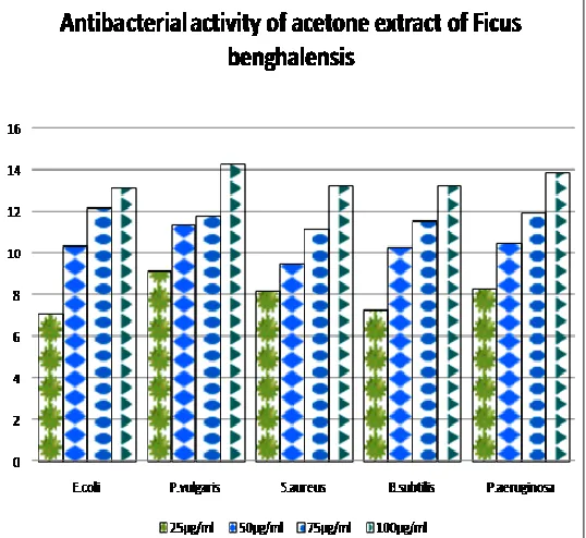
FIGURE 5: ANTIBACTERIAL ACTIVITY OF ACETONE EXTRACT OF
FICUS BENGHALENSIS
DISCUSSION: The previous studies on the phytochemical screening of Ficus benghalensis
revealed the presence of saponins, tannins and flavonoids in aqeous and methanolic extract 26. The preliminary phytochemical analysis of the methanol extract of Ficus religiosa bark studied by Uma et al., showed the presence of flavonoids, saponins and tannins 27.
The phytochemical screening of Ficus recemosa bark (various extracts) studied by Poongothai et al., showed the presence of alkaloids, flavonoids, glycosides, saponins, tannins and triterpenoids and the absence of anthraquinones 28.
The present study indicates strong antibacterial activity of bark extract of Ficus benghalensis. With the zone of inhibition more than 10mm against four of the five bacterial strains tested, the acetone extract of
Ficus beghalensis clearly possesses a strong and broad spectrum of antibacterial activity. The antibacterial activity against both gram positive and gram negative bacteria was in the order of Acetone>Methanol>ethyl acetate extract of F. benghalensis.
The methanol extract of Ficus religiosa and Ficus recemosa showed moderately good antibacterial activity against all tested bacterial strains. The ethyl acetate extract of these plants were less potent against most of the pathogens tested.
[image:7.612.18.599.68.291.2] [image:7.612.29.298.339.586.2]Such screening of various natural organic compounds and identification of active agents is the need of the hour because successful prediction of lead molecule at the onset of drug discovery will payoff later in drug discovery.
Lastly to conclude, the extracts were found to inhibit the growth of Gram positive bacteria as well as the Gram negative bacteria and also and the methanolic extract of the three plants was comparably more effective to inhibit the growth of microbes than acetone and ethyl acetate extracts.
ACKNOWLEDGEMENT: I am thankful to Mr. S. Shankaranarayanan for his guidance throughout the work. I also extend my thanks to Mr. G. Sampathkumar. The work is dedicated to all my teachers and students.
REFERENCES:
1. Nostro A, Germano MP, D'Angelo V, Marino A, Cannatelli MA. Extraction methods and bioautography for evaluation of medicinal plant antimicrobial activity. Letters in Appl. Microbiol. 2000, 30(5):379.
2. Tona, L., K. Kamlu, M. Ngimbi, K. cimanga and A.J. Vtientick. Anti amoebic and phytochemical screening of some congolose medicinal plants. J. Ethnopharmacol. 1998, 61:57-65.
3. Westh H, Zin C.S., Rosadahl V.T,et al. An international multicenter study of antimicrobial consumption and resistance in Staphylococcus aureus isolates from 15 hospitals in 14 countries. Microb drug resist. 2004, 10: 169-176
4. Bandew J.E, Brotz H, Leichrt LIO et al. Proteomic approach to understanding antibiotic action. Antimicrob agents chemotherapy. 2003, 47:948-955.
5. Berg CC, Classification and distribution of Ficus, experiential 1989, 45:605-611.
6. Jander EA, Machado KC, CA. Evolutionary Ecology of figs and their associates. Recent progress and outstanding puzzles. Ann. Rev Evol. Syst 2008, 39:439-458.
7. Sing.D. Goel R.K., Anticonvulasant effect of Ficus religiosa Role of serotonergic pathways, Journal of ethnopharmacology 2009, 123(2)330-4.
8. Rajib Ghosh, Sharath Chandra. Kh., Rita. S., Thokchem IS., Hypoglycaemic activity of Ficus hispidia (bark) in normal and diabetic abbine rats, Indian Journal of pharmacology, 2004, 36 (4), 222.
9. Mousa. O., Vuorela.P, KivirantaI, Abdul wahal.S, Hiltunen. R., Vuorela.H. Bioactivity of certain Egyptian Ficus species, journal of Ethnopharmacology, 1994; 41 (1-2), 71-76.
10. Daman. Preet Singh, Rajesh kumar Goel, Anticonvulsant effect of Ficus religiosa; Role of seratotergic pathways, Journal of Ethno pharmacology June 2009, 123 (2), 330-334.
11. Sivarajan VV & Balachandran I. Ayurvedi c Drugs and their sources, Oxford & IBH publishing co, pvt ltd; New Delhi 1994.
12. Joy PP, Thomas J, Mathew S & skaria BP. Medicinal plants In: Bopse TK, Kabir J, Das P & Joy OO (ed). Tropical Horticulture – vol 2. 449-632. 2001.
13. Gayathri. M., Kannabiran. K., Antim Harbone JB. Phytochemical Methods. A Guide to Modern Techniques of Plant Analysis. Chapman & Hall, London, 1998 182-190
14. Preeti. R., Vimal Devanathan V and M. Loganathan. Antimicrobial and antioxidant efficacy of some medicinal plants against food Borne pathogens advances in Biological Research 2010; 4 (2): 122-125.
15. Mahato RB., Chudary RP., Ethnomedicinal study and antibacterial activities of selected plants of palpa district Nepal. Scientific world 2005 volume 3. No: 3.
16. Allen ST. Chemical Analysis of Ecological Material. Blackwell Scientific Publication, New York, 1974, 313.
17. Harbone JB. Phytochemical Methods. A Guide to Modern Techniques of Plant Analysis. Chapman & Hall, London, 1976 p. 78 18. Sofowora A (1993). Medicinal Plants and Traditional Medicines in
Africa. Chichester John Wiley & Sons New York. 97- 145.
19. Harborne, A.J., 1973. Phytochemical Methods. Chapman and Hall, London, New York, Tokyo, 1-33.
20. Duma, RJ, Kinz LJ. Simple test for identifying penicillinase-producing staphylococci, Applied Microbiol, 1968 16(8), 1261-1262.
21. Oberhofer TR, Towle DW. Evaluation of the rapid penicillinase paper strip test for detection of betalactamase, J Clin Microbiol, 1982, 15(2), 196-199.
22. Bauer, A.W., Kirby, W.M., Sherris, J.C., Turck, M. Antibiotic susceptibility testing by a standardized single disk method. American Journal of Clinical Pathology 1996, 45, 493–496
23. Dixon, R.A., P.M. Dey, and C.J. Lamb. Phytoalexins: enzymology and molecular biology. Adv. Enzymol. 1983, 55:1-69.
24. Baladrin MF, Kjoke A.J., Wurtele E., et al. Natural plant chemicals: sources of Industrial and Mechanical materials science 1985, 228: 1154-1160.
25. Tanaka H, Sato m, Fujiwara S. Antibacterial activity of isoflavonoids isolated from Erythrina variegate against, methicillin resistant staphylococcus aureus. Let.Appl. Microbial, 2002, 35:494-498. 26. Manoj Aswar, Urmila Aswar, Akshaya wagh, Bhagyashri watkar,
Meenakshi vyas, Kishore M. Gujar. Antimicrobial activity of Ficus benghalensis. Pharmacology on line, 2008, 2: 715-725.
27. Uma. B., Prabaker. K., Rajendran. S., “Invitro antimicrobial activity and phytochemical analysis of Ficus religiosa and Ficus benghalensis L., against enterotoxigenic E. coli. “ Food chemical toxicology 2009 (11)2842-6.
28. Poongothai A., Sreena KP, Sreejith M, Uthairalingam, Annapoorani. S., 2011. Preliminary phytochemicals screening of Ficus remosa Linn. Bark. International Journal of Pharma and Biosciences vol 2, Issue 2, Apr-Jun 2011.
How to cite this article: