Copyright © 2001, American Society for Microbiology. All Rights Reserved.
Development of a Genomics-Based PCR Assay for Detection of
Mycoplasma pneumoniae
in a Large Outbreak
in New York State
ALFRED L. WARING, TANYA A. HALSE, CHARLES K. CSIZA, CYNTHIA J. CARLYN,†
KIMBERLEE ARRUDA MUSSER,ANDRONALD J. LIMBERGER*
David Axelrod Institute for Public Health, Wadsworth Center, New York State Department of Health, Albany, New York
Received 20 September 2000/Returned for modification 27 November 2000/Accepted 23 January 2001
A genomics-based PCR method was developed and used to test specimens from patients involved in a large
outbreak ofMycoplasma pneumoniaein a closed religious community in New York State. New P1 adhesin gene
primers were designed to bind to 9 of 10 target sequences in the repetitive-element sequences obtained from
the whole genome sequence ofM. pneumoniae. This PCR method had a sensitivity of 0.006 CFU and a specificity
of 100% forM. pneumoniae. The PCR was validated by testing a subset of patient samples by culture and
comparing the results to those obtained by PCR. Of the initial 280 samples tested, 73 were positive by PCR and 22 were positive by culture. All samples positive by culture were also positive by PCR. Follow-up testing of selected patients 3 to 6 weeks after antibiotic treatment revealed that eight samples remained positive by PCR and that three samples remained positive by culture. Additionally, no nonspecific PCR inhibition was detected as a result of the specimen type, transport medium, or sample preparation methodology. The study
demon-strates that the PCR described here is a rapid, sensitive, and specific method for the identification ofM.
pneumoniaeand was helpful for the detection and monitoring of the outbreak.
Mycoplasma pneumoniaeis primarily a respiratory pathogen that is responsible for approximately 15 to 20% of all commu-nity-acquired pneumonias (8). The clinical presentation of pa-tients withM. pneumoniaeinfection is not significantly differ-ent from that of patidiffer-ents with infections caused by other respiratory pathogens such asChlamydia pneumoniae(22), so diagnosis of infection relies primarily on laboratory testing.M. pneumoniae is, however, a fastidious organism, requiring a laborious effort and 21 days or more for growth in culture. Likewise, serology is lacking in sensitivity, has questionable specificity (22), and is dependent on specific collection times relative to the onset of illness (5). DNA probes have also been used, but cross-reactivity between M. pneumoniaeand Myco-plasma genitalium has been observed, and methods that use these probes also lack sensitivity (4).
Numerous PCR approaches have been developed to provide a rapid, sensitive method for the detection ofM. pneumoniae. PCR targets have included the P1 adhesin gene (3, 4, 5, 12, 17, 21), the ATPase operon (11), genomic clones (2, 7), or a combination of these (1). The P1 adhesin gene is responsible for cytadherence, which is necessary for colonization and in-fection of host cells byM. pneumoniae(22). This gene is also an intriguing target for PCR because of its repetitive nature within theM. pneumoniaegenome. Approximately 8% of this genome is composed of repetitive DNA elements with regions
homologous to the P1 adhesin gene (10). Use of the entire genomic DNA sequence enabled us to design primers that would theoretically amplify the maximum number of target molecules, resulting in an improved sensitivity of the PCR for
M. pneumoniae.
A large outbreak of respiratory illness occurred in a closed religious community in rural New York State in the summer of 1998, providing the opportunity to evaluate improved molec-ular biology-based methods for M. pneumoniaedetection. A study was designed to evaluate new, whole genome-based PCR primers and compare the detection capability of PCR to that of culture of throat swab specimens from community members. Additionally, the outbreak allowed us to determine whether PCR or culture of follow-up specimens could detectM. pneu-moniae after antibiotic treatment (azithromycin) of the in-fected individuals.
MATERIALS AND METHODS
Patients.Specimens were obtained from patients who were associated with an
outbreak of respiratory illness and whose clinical presentation was consistent with disease caused byM. pneumoniae. The outbreak occurred between 1 May 1998 and 11 August 1998 in a closed religious community in New York State. Throat swab specimens were initially obtained from 280 persons, consisting of symptomatic and asymptomatic persons. Five-day antimicrobial therapy with azithromycin (kindly donated by Pfizer Pharmaceuticals) was established for symptomatic individuals, and a 3-day prophylactic treatment was given to asymp-tomatic individuals following the collection of specimens. The total cumulative dose of azithromycin was identical (1,500 mg) for all treated individuals. After a 3- to 6-week period, follow-up throat swab specimens were collected from 69 of the initially tested individuals.
Specimen collection.Throat swabs were collected with a Dacron applicator
swab. The swabs were expressed in tubes containing 3 ml ofMycoplasma trans-port medium (Trypticase soy broth containing 0.25% glucose, 0.5% bovine serum albumin, 0.01% thallium acetate, 1,000 U of penicillin per ml), and the swabs were discarded. The specimen tubes were then transported on ice to the labo-ratory on the same day of collection and processed for culture and PCR or stored
* Corresponding author. Mailing address: David Axelrod Institute for Public Health, Wadsworth Center, New York State Department of Health, P.O. Box 22002, 120 New Scotland Ave., Albany, NY 12201-2002. Phone: (518) 474-4177. Fax: (518) 486-7971. E-mail: Ron [email protected].
† Present address: Infectious Disease Section, Stratton VA Medical Center, Albany, NY 12208.
1385
on May 15, 2020 by guest
http://jcm.asm.org/
at 4°C for 24 to 48 h. If culture was not performed at the time of PCR, the samples were stored at⫺70°C until culture was initiated.
Culture.The culturing ofM. pneumoniaewas accomplished by a method
adapted from that of Tully et al. (20) and presented recently (C. J. Carlyn, A. L. Waring, C. K. Csiza, T. A. Halse, S. J. Wong, and R. J. Limberger, Abstr. 99th Gen. Meet. Am. Soc. Microbiol., abstr. G-16, 1999). Briefly, the specimen tubes were vortexed, a volume of 100l was added to 3 ml of spiroplasma diphasic medium (SP4 agar; 2 ml of broth and 1 ml of solid media), and two serial 10-fold dilutions were made to dilute out potential inhibitors ofMycoplasmagrowth. These samples were then incubated at 37°C and examined twice weekly. When a color change occurred, 25l was subcultured onto SP4 agar. These subcultured samples were incubated in 5% CO2in a 37°C incubator and were then examined
twice weekly for the presence of typicalM. pneumoniaecolonies. Typical colonies were identified by guinea pig red blood cell hemadsorption and confirmed by a growth inhibition test (20).
PCR.A total of 500l of the original specimen was aliquoted into a micro-centrifuge tube for PCR analysis. This aliquot was concentrated by centrifugation at 13,000⫻gfor 10 min at room temperature, 480l of the medium was removed, and 30l of sterile water was added to the remaining 20l of medium and the pellet. The samples were then vortexed and heated to 95°C for 15 min. A 10-l aliquot of the sample lysate was then used directly for PCR amplifica-tion. AnM. pneumoniae-specific PCR was performed with a total reaction mix-ture volume of 100l. Each primer at a final concentration of 1M (primer MP-F [5⬘-CCCTCGACCAAGCCAACCTC-3⬘] and primer MP-R [5⬘-TGCGC GTTGTTCTTGTTGGTG-3⬘]), each deoxynucleoside triphosphate at a final concentration of 200M, and final concentrations of 10 mM Tris-HCl (pH 8.3), 50 mM KCl, 2 mM MgCl2, and 2.5 U of AmpliTaq Gold (Perkin-Elmer, Foster
City, Calif.) were used for PCR in a Perkin-Elmer 9600 thermocycler. Thermo-cycler conditions consisted of an initial incubation of 95°C for 9 min, followed by 40 cycles of 94°C for 30 s, 62°C for 30 s, and 72°C for 30 s. An additional incubation at 72°C for 7 min was added to complete the elongation. Negative controls with no template were included, as were controls known to be positive. To assess the overall effect of potential nonspecific inhibition of the amplifica-tion, an additional PCR analysis was performed with 41 randomly selected PCR-negative samples. Each specimen was spiked with 150 ng of HeLa cell DNA (laboratory stock), and a PCR specific for the human-globin gene was used to evaluate the patient samples. The PCR mixtures were spiked with this DNA to maintain a precise, consistent amount of template for analysis of nonspecific inhibition. The amplified products were analyzed on 2% GTG agarose gels containing ethidium bromide and were visualized at maximal levels of intensity with a Gel Doc 1000 gel analysis system (Bio-Rad Laboratories, Hercules, Calif.). All reagents and conditions for the PCR assay were optimized before the assays for sensitivity and specificity were performed. The oligonucleotides were synthe-sized in the Wadsworth Center Molecular Genetics Core Facility.
Dot blot hybridization of PCR products. In order to specifically confirm
amplification ofM. pneumoniaeDNA, a 26-bp probe specific for the internal region of the PCR product, MPP2 (5⬘-AATCCCGACTCGTTAAAGCAGGAT AA-3⬘), was used in all hybridization assays. A 30-l aliquot of PCR product was denatured at 37°C for 5 min with 0.1 volume of 1 N NaOH. Neutralization was obtained by adding 1 volume of 6⫻SSC (1⫻SSC is 0.15 M NaCl plus 0.015 M sodium citrate), and sterile water was then added to give a total volume of 400 l. The total volume was transferred onto a Hybond-N nylon membrane (Am-ersham Pharmacia Biotech, Piscataway, N.J.) with a dot blot apparatus (Schlei-cher & Schuell, Keene, N.H.). The membrane was UV cross-linked, prehybrid-ized overnight at 42°C, and then hybridprehybrid-ized at 42°C for 2 h with labeled probe (the hybridization solutions were specified by the manufacturer). The internal oligonucleotide probe was labeled according to the manufacturer’s protocol by using the ECL 3⬘oligolabeling and detection kit (Amersham Pharmacia Bio-tech). DNA hybridization was visualized by detection of a chemiluminescent signal when the blot was exposed to X-Omat AR film (Kodak, Rochester, N.Y.). Positive results by both PCR and hybridization assays were interpreted as a positive result for the specimen.
PCR sensitivity and specificity.The sensitivity of the PCR was determined by
using a viable suspension ofM. pneumoniaeATCC 29342. Tenfold serial dilu-tions were made in SP4 culture medium to 10⫺10. Identical volumes of each of
these dilutions were run in PCR assays and were plated onto SP4 agar. The concentration ofM. pneumoniaewas determined by plate counts, performed in triplicate.
A number of additionalMycoplasmaspp., respiratory organisms, and other common bacteria were tested to determine the specificity of the PCR assay. Concentrations of either 3⫻106CFU or 0.1 ng of DNA (equivalent to⬃1⫻106
copies of the genome) were used. The following organisms were tested by both PCR and probe hybridization assays to determine the specificities of the assays:
M. pneumoniaeATCC 29342, ATCC 15531, and NIH 1428;Mycoplasma arthri-tidisATCC 14152;Mycoplasma hominisATCC 23114; M. genitaliumATCC 33530; Mycoplasma salivarium ATCC 23064D; Mycoplasma orales ATCC 23714D;Mycoplasma fermentansATCC 19989D;Ureaplasma urealyticum; Bor-detella pertussis;Legionella pneumophila; group A streptococcus;Streptococcus pneumoniae;Haemophilus influenzae;Staphylococcus aureus;Neisseria meningi-tidis;Streptococcus sanguis;Branhamella catarrhalis;Corynebacterium aquaticum;
Klebsiella pneumoniae;Chlamydia pneumoniae;Chlamydia trachomatis; Trepo-nema pallidum;Escherichia coli; andHaemophilus ducreyi.
RESULTS
Primer and probe design.Analysis of the complete sequence
of the M. pneumoniae genome (found at the website http:// www.zmbh.uniheidelberg.de/M_pneumoniae/MP_Home.html) (10) enabled the selection of two new PCR primers. This sequence was manipulated by using the programs available in the Wisconsin Package (Wisconsin Package, version 10.1; Ge-netics Computer Group, Madison, Wis.), followed by manual sequencing analysis. Sequences within the repetitive element REPMP2/3 and the P1 adhesin gene (16, 19) were aligned, and the most highly conserved regions were selected to design primers for PCR. As shown in Fig. 1, nine amplicons could theoretically be synthesized in the first round of PCR with these primers, and thus, a significant increase in sensitivity over that of a single-target PCR assay would be achieved. The sizes of the expected products ranged from 309 to 339 bp and were visualized as one broad band upon gel electrophoresis (Fig. 2 and 3).
The internal probe, MPP2, was speculated to bind efficiently to seven of the amplified PCR products (sequence identities, 92 to 100%). The two remaining amplified PCR products had sequence identities to the probe of 61 and 69%, respectively, but were also capable of binding to the probe under our hy-bridization conditions (data not shown). The dot blot hybrid-ization procedure was designed to provide specificity for the assay to ensure that the PCR products were indeedM. pneu-moniae.
Sensitivity and specificity of PCR method.Under the
con-ditions used in this study, PCR produced an amplification product in the range of 309 to 339 bp, as expected by the design of the assay. The sensitivity of detection was 0.006 CFU, as determined by comparison of the PCR results with the culture results after plating of serial dilutions (Fig. 2). Negative con-trols with no template produced no PCR products, and positive controls forM. pneumoniaealways produced a product of 309 to 339 bp, as shown in Fig. 3.
The non-M. pneumoniaespecies and the unrelated respira-tory species tested as well as the additional bacterial species tested produced no detectable PCR products upon analysis by the PCR assay and no hybridization products by the hybrid-ization assay. Thus, the combined results of PCR and probing demonstrated 100% specificity.
PCR inhibition analysis. Forty-one of the specimens that
were negative by both culture and PCR were tested for non-specific PCR inhibition. After spiking of the samples with HeLa cell DNA, a region of the human -globin gene was amplified from all specimens by a PCR assay that is routinely used by our laboratory for inhibition testing. Because all of these specimens produced a band after the-globin PCR, they were determined to be devoid of any significant PCR inhibitory factors (data not shown). However, this does not preclude the
on May 15, 2020 by guest
http://jcm.asm.org/
potential presence of inhibitors in some of the patient samples that were not assayed by the-globin PCR.
Analysis of patient specimens. A total of 349 specimens
were tested by PCR. Of those, 280 specimens were from the original outbreak and 69 were received as follow-up specimens. Of the 280 specimens tested initially, 73 were positive and 207 were negative after PCR and hybridization. A typical ethidium bromide-stained 2% agarose gel, containing 11 patient speci-mens along with both positive and negative controls, is shown in Fig. 3. A total of 108 initial specimens were also tested by
culture. Of those initial specimens tested, 22 were culture pos-itive and 86 were culture negative.
PCR and culture results on posttherapeutic follow-up.The
[image:3.612.324.541.72.264.2]second follow-up specimen collected from 69 individuals was taken 3 to 6 weeks after antibiotic therapy. It was of interest to determine the effect that antibiotic treatment would have on the detection ofM. pneumoniaeby PCR and culture. Most of the individuals retested (53 of 69) were originally positive. Of those individuals retested, 8 remained positive by PCR and 61 were negative by PCR. All of those who were originally PCR negative remained PCR negative. The eight secondary speci-mens that remained positive by PCR were also tested by cul-ture. Of these, three specimens were positive by culture and had initially been positive by culture. The other five specimens
FIG. 1. Primer design strategy for the forward and reverse PCR primers (primers MP-F and MP-R, respectively) indicating the binding positions within theM. pneumoniae genome (10). The primer se-quences are shown at the bottom of each panel. Shaded areas indicate mismatches between the primer and the genome sequences within REPMP2/3 regions. The P1 adhesin gene sequence (P1) is indicated by the appropriate genome sequence. (A) Alignment of forward primer MP-F; (B) alignment of reverse primer MP-R.
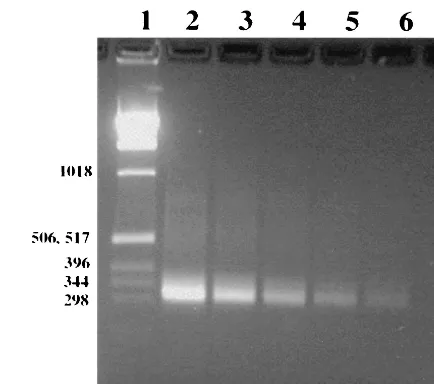
FIG. 2. Ethidium bromide-stained 2% agarose gel showing sensi-tivity of PCR after dilution of viableM. pneumoniae. Lane 1, 1-kb DNA ladder (numbers on the left are in base pairs); lanes 2, 3, and 4, amplifiedM. pneumoniae culture extracts containing 60, 6, and 0.6 CFU, respectively, in the total PCR mixture; lanes 5 and 6, extracts that did not produce any CFU (extrapolated to dilutions of 0.06 and 0.006 CFU, respectively); lane 7, negative control.
FIG. 3. PCR amplification of repetitive element REPMP2/3 within the P1 operon for direct detection ofM. pneumoniaein patient sam-ples. Samples were centrifuged, lysed, amplified by PCR, electropho-resed, and stained with ethidium bromide. Lane 1, 1-kb ladder (num-bers on the left are in base pairs); lanes 2 to 8, patient specimens; lane 9, negative control; lane 10, positive control.
on May 15, 2020 by guest
http://jcm.asm.org/
were negative by culture. Two of these were initially culture negative, one was initially culture positive, and two had not been previously tested by culture.
Dot blot hybridization analysis.Hybridization analysis was
carried out to confirm the specificity of the PCR. Figure 4 shows a typical blot with positive and negative controls, organ-isms used for testing of specificity (Treponema pallidumand
Chlamydia pneumoniae), and 65 patient specimens that were analyzed by PCR. A total of 131 patient specimens (initial and follow-up specimens) were tested by hybridization. Of those tested, 81 (73 initial specimens and 8 follow-up specimens) were positive by DNA hybridization and 50 were negative. These results correlated with the PCR results; thus, no PCR-negative specimens were determined to be positive by hybrid-ization and no PCR-positive samples were determined to be negative by hybridization. Thus, as expected, the hybridization assay provided sensitivity equivalent to those of PCR and Gel Doc gel analysis system for the 131 samples tested, and none of the observed PCR products from patient specimens had false-positive results.
Comparison of culture and PCR. A total of 116 patient
specimens (108 primary specimens and 8 follow-up specimens) were tested by both PCR and culture. A comparison of the results obtained by these methods with patient specimens is summarized in Table 1. All samples that were culture positive were also PCR positive. Of the 91 specimens found to be culture negative, 28 were PCR positive. The data in Table 1 demonstrate that PCR analysis was capable of markedly
in-creasing the level of detection ofM. pneumoniae. It should be noted that the first 87 outbreak-related specimens were tested by both culture and PCR; thereafter, the remaining specimens were tested only by culture if they were PCR positive. As a consequence of using PCR to prescreen samples for culture, the culture positivity rate was slightly higher than it would have been had all samples been cultured.
DISCUSSION
Pneumonia caused byM. pneumoniaehas long been a diffi-cult disease to diagnose because there are both clinical and laboratory diagnostic problems associated with its identifica-tion. It has been realized for quite some time that the detection ofM. pneumoniaeis greatly enhanced by the use of the PCR methodology. PCR methods have provided an advantage be-cause they are fast, specific, and sensitive: in this case 1 day is required for amplification and 1 day is required for the dot blot assay, whereas 1 to 3 weeks is required for traditional culture methods. We have designed a genomics-based PCR primer pair that targets multiple sites and that, we believe, on the basis of published CFU, provides an improvement over the current published methods.
An outbreak ofM. pneumoniaein New York State provided the opportunity to both validate our procedure and compare its sensitivity and specificity to those of culture. This outbreak was also the largest outbreak in New York State and one of the largest in the country to be evaluated by testing of specimens by PCR. Before development of this PCR assay, our New York State reference laboratory had gained significant experience with and expertise inMycoplasmaculture. The results of the present study indicate that our PCR method is at least twice as sensitive as the current culture methodology. Our PCR method was highly sensitive (0.006 CFU) and specific and displayed no nonspecific inhibition due to the transport or processing of throat swab specimens.
The present PCR method was designed to amplify the P1 adhesin gene ofM. pneumoniae. Numerous investigators have recognized the benefit of using the P1 adhesin gene as a target for PCR identification ofM. pneumoniae. A number of inves-tigators have published descriptions of PCR methods that tar-get sequences within this gene (3, 4, 5, 12, 17, 21). The P1 adhesin gene is an ideal amplification target for PCR because primers can be designed to target conserved regions within known repeats of theM. pneumoniaegenome. Portions of this gene are repeated up to 10 times within the genome, thus allowing an increase in the sensitivity of a PCR assay above that of a typical one-target PCR method. Our efforts were focused on taking full advantage of the repetitive elements in the genome by creating an assay that would be capable of amplifying the genome in the maximum number of regions. The newly designed PCR had the theoretical advantage of amplifying nine regions of theM. pneumoniaegenome (Fig. 1). The method was demonstrated to be further improved by in-cluding the addition of a reverse primer to bind to the 10th region (data not shown). Indeed, this method was highly sen-sitive (0.006 CFU). This detection limit is the equivalent to 3 serial dilutions below the 6 CFU concentration ofMycoplasma
determined by culture. In addition to the multiple-target ad-vantage of this PCR, it is also possible that nonviable bacterial
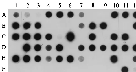
FIG. 4. Dot blot hybridization assay of amplified DNA from clini-cal specimens. Dots A1 to A3 represent hybridization to anM. pneu-moniaePCR product diluted 1:10, 1:40, and 1:160, respectively. Dot F1 is a negative control. Dots F2 and F3 indicateTreponema pallidumand
[image:4.612.57.292.71.195.2]Chlamydia pneumoniaeDNA samples, respectively, that were used as part of the specificity assessment. The remaining dots are patient specimens. Negative samples are dots A8, A9, B4 to B6, B10, B11, C7, C9, C12, D1, D6, E8, E9, E11, and F1 to F10; the rest of the samples are positive.
TABLE 1. Summary of results for paired outbreak-related specimens tested by both culture and PCR
Result No. of specimens
PCR negative PCR positive Total culture results
Culture negative 63 28 91
Culture positive 0 25 25
Total PCR results 63 53 116
on May 15, 2020 by guest
http://jcm.asm.org/
cells and extracellular DNA contribute to the discrepancy be-tween the high level of sensitivity of PCR compared to that of culture.
Other groups have also attempted to capitalize on this re-petitive aspect of the genome, but analysis suggests that those methods can potentially amplify only six or seven regions of the genome (3, 12, 21). From our sensitivity experiments, on the basis of serial dilution ofM. pneumoniae(ATCC 29342) and comparison of plate counts to PCR results, we found the level of detection of PCR to be 0.006 CFU. This was considerably more sensitive than the sensitivity reported for mostM. pneu-moniaePCR assays. One group (1) found a similar sensitivity (0.019 CFU), but a nested PCR was used to achieve its PCR sensitivity. Because nested PCR methods can be troubled by contamination problems (1, 6, 15), the simple, nonnested PCR assay described here, which has a sensitivity comparable to that of nested PCR, should provide an improvement over the cur-rent diagnostic tools used for laboratory detection ofM. pneu-moniaeby PCR.
Hybridization by a dot blot method and with an internal probe specific for our PCR product was used to add specificity to our level of detection. On occasion, after specimen ampli-fication we found secondary PCR products in addition to the expected 309- to 339-bp product. Additionally, we occasionally found nonspecific PCR products in the PCR-negative speci-mens. Both of these events usually occurred when specimens were tested on the same day that they were received or when recently obtained specimens were tested, and spurious bands often disappeared upon retesting. For these reasons, it was important to probe the PCR products with a specific oligonu-cleotide internal to the PCR primer set to ensure that any positive result was truly a positive result and any negative result was truly a negative result. This added test allowed confirma-tion that the specimen was indeedM. pneumoniae.
Amplicon contamination is an important issue that must be addressed with any PCR assay, especially one that is used frequently in a setting such as a large outbreak. Appropriate precautions were taken to maintain standard laboratory prac-tices to avoid contamination. At least one negative control, which contained no template, was run in every PCR, and this control was always negative. The hybridization reactions also contained multiple negative controls that were never positive. Finally, a second PCR was used to confirm the results for a number of specimens with PCR-positive, cultunegative re-sults. The PCR assay amplified a different portion of the P1 adhesin gene that was included in the REPMP4 portions of the
M. pneumoniaegenome (21).
As noted earlier in the report, there were twice as many PCR-confirmed cases of M. pneumoniae infection as there were culture-confirmed cases. We believe that these PCR-positive, culture-negative cases were true cases ofM. pneu-moniaeinfection for several reasons. There have been studies that suggest that PCR can detect mild cases of infection (8, 18), infection in patients who have previously been treated with antibiotics (8, 14), and infection in patients believed to be late in the course of disease (24). Moreover, PCR can detect non-viable bacteria, and in each of these instances culture would be negative. Epidemiological analysis suggested that the commu-nity members who were PCR positive had reported contact with patients in this closed community confirmed to be positive
by culture (data not shown). Conceivably, the significance of
M. pneumoniaeas a cause of infection is underestimated be-cause of the difficulties in the past of obtaining samples from patients with laboratory-confirmed cases of M. pneumoniae
infection.
Of the 172 primary specimens tested only by PCR and hy-bridization, a positivity rate of 30% was established within the community during the outbreak period. By comparison, a 20% positivity rate was demonstrated by culture. However, this number is probably the maximum culture positivity rate be-cause many of these specimens tested by culture were tested only after they were determined to be PCR positive to de-crease the amount of laborious culture manipulation during the outbreak. Because the sensitivity of this PCR assay is quite good, the likelihood of finding a culture-positive, PCR-nega-tive specimen was extremely unlikely. Most likely, the use of culture methods on all of the samples would have resulted in a lower culture positivity rate.
In the present study PCR provided a marked improvement over culture, as was expected. Table 1 illustrates the results for those paired samples that were tested by both PCR and cul-ture. These data demonstrate that, of the specimens tested, no PCR-negative specimen was found to be culture positive. This indicates the increased sensitivity of PCR over that of culture. Additionally, when those specimens that were PCR positive were observed for their rate of culture positivity, it was deter-mined that PCR was approximately twice as sensitive as cul-ture. Additionally, our sensitivity studies revealed that the level of detection by PCR was 2 log units above the level of detec-tion by culture and above the levels of detecdetec-tion by many of the published PCR assays.
An additional question addressed in the present outbreak study was what effect treatment with the antibiotic azithromy-cin would have on the ability of members of the community to harbor M. pneumoniae. To address this question, secondary specimens were taken from selected members of the commu-nity 3 to 6 weeks following the initial specimen collection from a group of 53 patient specimens that were initially positive by PCR, 8 specimens remained positive by PCR (including three specimens that remained culture positive). Because this group of specimens contained specimens from both symptomatic and asymptomatic patients, no clear link between the duration of therapy or the presentation of the infected individual and the persistence of the organism can be concluded from these re-sults. However, a marked decrease in the number of affected individuals was found after the 3- to 6-week period. Azithro-mycin therapy has previously been found to be effective at preventing outbreaks of infection with M. pneumoniae (9). Additionally, the combined use of the 1,500-mg cumulative dose of azithromycin and standard epidemic control measures has been associated with a significant reduction in the rate of transmission ofM. pneumoniae(13).
The results of the follow-up testing could be interpreted in several ways. First, the result can be interpreted as a 15% rate of carriage ofM. pneumoniaewithin the community following an outbreak, which would be consistent with past studies (23). One study (8) examined families in Seattle and found that a carrier state that lasted up to several months did exist, regard-less of symptoms or treatment with a broad-spectrum antibi-otic. That group also concluded that the rate of carriage ofM.
on May 15, 2020 by guest
http://jcm.asm.org/
pneumoniaemay increase during epidemic periods within com-munities. Second, the result can be interpreted as a failure of the antibiotic treatment to completely clear the infection be-cause of either the therapy or patient compliance. Third, the result can be interpreted as a possible reinfection of the patient due in part to the closeness of the community. Fourth, it is also possible that PCR testing is detecting nonviable organisms or residual cellular DNA that has not been cleared. However, it is possible that the very high sensitivity of PCR achieved the detection of infections that were not clinically relevant. This is a question relevant to a number of diseases that has not yet been fully addressed.
In conclusion, this report describes a genomics-based PCR that provided rapid detection ofM. pneumoniae in a closed community during a large outbreak. This PCR assay was de-veloped for the testing of throat swab specimens suspected to harbor M. pneumoniae and is highly sensitive (0.006 CFU) without the addition of a nested PCR component. This PCR is also 100% specific. In our hands this method was found to be twice as sensitive as culture, and as expected for a PCR pro-cedure, it is extremely rapid, especially when compared to the speed of culture.
ACKNOWLEDGMENTS
We thank Susan Wong, Wendy Archinal, Joel Ackelsberg, Stan Kondracki, Christopher Maendel, the Wadsworth Center Molecular Genetics Core Facility, and the Wadsworth Center Photography and Illustrations Department for valuable assistance.
REFERENCES
1.Abele-Horn, M., U. Busch, H. Nitschko, E. Jacobs, R. Bax, F. Pfaff, B.
Schaffer, and J. Heesemann.1998. Molecular approaches to diagnosis of
pulmonary diseases due toMycoplasma pneumoniae. J. Clin. Microbiol36:
548–551.
2.Bernet, C., M. Garret, B. De Barbeyrac, C. Bebear, and J. Bonnet.1989.
Detection ofMycoplasma pneumoniaeby using the polymerase chain reac-tion. J. Clin. Microbiol.27:2492–2496.
3.Buck, G. E., L. C. O’Hara, and J. T. Summersgill.1992. Rapid, sensitive
detection of Mycoplasma pneumoniaein simulated clinical specimens by DNA amplifications. J. Clin. Microbiol.30:3280–3283.
4.De Barbeyrac, B., C. Bernet-Poggi, F. Fe´brer, H. Renaudin, M. Dupont, and
C. Be´be´ar.1993. Detection of Mycoplasma pneumoniaeandMycoplasma
genitaliumin clinical samples by polymerase chain reaction. Clin. Infect. Dis.
17(Suppl. 1):S83–S89.
5.Dorigo-Zetsma, J. W., S. A. J. Zaat, P. M. E. Wertheim-Van Dillen, L.
Spanjaard, J. Rijntjes, G. Van Wavern, J. S. Jensen, A. F. Angulo, and J.
Dankert.1999. Comparison of PCR, culture, and serological tests for
diag-nosis of Mycoplasma pneumoniaerespiratory tract infection in children. J. Clin. Microbiol.37:14–17.
6.Ehrlich, G. D., and D. A. Sirko.1994. PCR and its role in clinical diagnostics,
p. 3–18.InG. D. Ehrlich and S. J. Greenberg (ed.), PCR-based diagnostics in infectious disease. Blackwell Scientific Publications, Boston. Mass.
7.Falguera, M., A. Nogues, A. Ruiz-Gonzalez, M. Garcia, and T. Puig.1996.
Detection ofMycoplasma pneumoniaeby polymerase chain reaction in lung aspirates from patients with community-acquired pneumonia. Chest110:
972–976.
8.Foy, H. M.1993. Infections caused byMycoplasma pneumoniaeand possible
carrier state in different populations of patients. Clin. Infect. Dis.17(Suppl. 1):S37–S46.
9.Gray, G. C., D. C. McPhate, M. Leinonen, G. H. Cassell, E. P. Deperalta,
S. D. Putnam, J. A. Karcher, M. H. Sawyer, A. Laurila, and J. D. Connor.
1998. Weekly oral azithromycin as prophylaxis for agents causing acute respiratory disease. Clin. Infect. Dis.26:103–110.
10. Himmelreich, R., H. Hilbert, H. Plagens, E. Pirkl, B.-L. Li, and R.
Herr-mann.1996. Complete sequence analysis of the genome of the bacterium
Mycoplasma pneumoniae. Nucleic Acids Res.24:4420–4449.
11. Honda, J., T. Yano, M. Kusaba, J. Yonemitsu, H. Kitajima, and M.
Ma-suoka.2000. Clinical use of capillary PCR to diagnoseMycoplasma
pneu-monia. J. Clin. Microbiol.38:1382–1384.
12. Ieven, M., D. Ursi, H. Van Bever, W. Quint, H. G. M. Niesters, and H.
Goossens.1996. Detection ofMycoplasma pneumoniaeby two polymerase
chain reactions and role ofM. pneumoniaein acute respiratory tract infec-tions in pediatric patients. J. Infect. Dis.173:1445–1452.
13. Klausner, J. D., D. Passaro, J. Rosenberg, W. L. Thacker, D. F. Talkington,
S. B. Werner, and D. J. Vugia.1998. Enhanced control of an outbreak of
Mycoplasma pneumoniaepneumonia with azithromycin prophylaxis. J. In-fect. Dis.177:161–166.
14. Kleemola, S. R. M., J. E. Karjalainen, and R. K. H. Ra¨ty.1990. Rapid
diagnosis ofMycoplasma pneumoniaeinfection: clinical evaluation of a com-mercial probe test. J. Infect. Dis.162:70–75.
15. Persing, D. H.1993. In vitro nucleic acid amplification techniques, p. 51–87.
InD. H. Persing, T. F. Smith, F. C. Tenover, and T. J. White (ed.), Diag-nostic molecular microbiology: principles and applications. American Soci-ety for Microbiology, Washington, D.C.
16. Ruland, K., R. Wenzel, and R. Herrmann.1990. Analysis of three different
repeated DNA elements present in the P1 operon ofMycoplasma pneu-moniae: size, number and distribution on the genome. Nucleic Acids Res.
18:6311–6317.
17. Sharma, S., R. Brousseau, and S. Kasatiya.1998. Detection and
confirma-tion ofMycoplasma pneumoniaein urogenital specimens by PCR. J. Clin. Microbiol.36:277–280.
18. Skakni, L., A. Sardet, J. Just, J. Landman-Parker, J. Costil, N. Moniot-Ville,
F. Bricout, and A. Garbarg-Chenon.1992. Detection ofMycoplasma
pneu-moniaein clinical samples from pediatric patients by polymerase chain re-action. J. Clin. Microbiol.30:2638–2643.
19. Su, C. J., V. V. Tryon, and J. B. Baseman.1987. Cloning and sequence
analysis of cytadhesin P1 gene fromMycoplasma pneumoniae. Infect. Im-mun.55:3023–3029.
20. Tully, J. G., D. L. Rose, R. F. Whitcomb, and R. P. Wenzel.1979. Enhanced
isolation ofMycoplasma pneumoniaefrom throat washing with a newly mod-ified culture medium. J. Infect. Dis.139:478–482.
21. Ursi, D., J.-P. Ursi, M. Ieven, M. Docx, P. Van Reempts, and S. R. Pattyn.
1995. Congenital pneumonia due toMycoplasma pneumoniae. Arch. Dis. Child.72:F118–F120.
22. Waites, K. B., and D. Taylor-Robinson.1999. Mycoplasma and ureaplasma,
p. 782–794.InP. R. Murray, E. J. Baron, M. A. Pfaller, F. C. Tenover, and R. H. Yolken (ed.). Manual of clinical microbiology, 7th ed. ASM Press, Washington, D.C.
23. Wenzel, R. P., R. B. Craven, J. A. Davies, J. O. Hendley, B. H. Hamory, and
J. M. Gwaltney, Jr.1977. Protective efficacy of an inactivatedMycoplasma
pneumoniaevaccine. J. Infect. Dis.136(Suppl.):S204–S207.
24. Williamson, J., B. P. Marmion, D. A. Worswick, T. W. Kok, G. Tannock, R.
Herd, and J. Harris.1992. Laboratory diagnosis ofMycoplasma pneumoniae
infection. Antigen capture and PCR-gene amplification for detection of the
Mycoplasma: problems of clinical correlation. Epidemiol. Infect.109:519– 537.