IFN-gamma action on pancreatic beta cells
causes class I MHC upregulation but not
diabetes.
H E Thomas, … , R D Schreiber, T W Kay
J Clin Invest.
1998;
102(6)
:1249-1257.
https://doi.org/10.1172/JCI2899
.
We have generated transgenic nonobese diabetic (NOD) mice expressing dominant
negative mutant IFN-gamma receptors on pancreatic beta cells to investigate whether the
direct effects of IFN-gamma on beta cells contribute to autoimmune diabetes. We have also
quantitated by flow cytometry the rise in class I MHC on beta cells of NOD mice with
increasing age and degree of islet inflammatory infiltrate. Class I MHC expression increases
gradually with age in wild-type NOD mice; however, no such increase is observed in the
transgenic beta cells. The transgenic mice develop diabetes at a similar rate to that of
wild-type animals. This study dissociates class I MHC upregulation from progression to diabetes,
shows that the rise in class I MHC is due to local IFN-gamma action, and eliminates beta
cells as the targets of IFN-gamma in autoimmune diabetes.
Research Article
J. Clin. Invest.
© The American Society for Clinical Investigation, Inc. 0021-9738/98/09/1249/09 $2.00
Volume 102, Number 6, September 1998, 1249–1257 http://www.jci.org
IFN-
g
Action on Pancreatic Beta Cells Causes Class I MHC Upregulation but
Not Diabetes
Helen E. Thomas,* Jennie L. Parker,* Robert D. Schreiber,‡ and Thomas W.H. Kay*
*Autoimmunity and Transplantation Division, Walter and Eliza Hall Institute of Medical Research, Victoria 3050, Australia; and
‡Department of Pathology, Washington University, St. Louis, Missouri 63110
Abstract
We have generated transgenic nonobese diabetic (NOD) mice expressing dominant negative mutant IFN-g receptors on pancreatic beta cells to investigate whether the direct ef-fects of IFN-g on beta cells contribute to autoimmune dia-betes. We have also quantitated by flow cytometry the rise in class I MHC on beta cells of NOD mice with increasing age and degree of islet inflammatory infiltrate. Class I MHC expression increases gradually with age in wild-type NOD mice; however, no such increase is observed in the trans-genic beta cells. The transtrans-genic mice develop diabetes at a similar rate to that of wild-type animals. This study dissoci-ates class I MHC upregulation from progression to diabetes, shows that the rise in class I MHC is due to local IFN-g ac-tion, and eliminates beta cells as the targets of IFN-g in au-toimmune diabetes. (J. Clin. Invest. 1998. 102:1249–1257.) Key words: nonobese diabetic mouse • cytokine •
autoim-mune disease • class I major histocompatibility antigen •
transgenic mice
Introduction
Insulin-dependent diabetes mellitus (IDDM)1 is characterized
by a mixed lymphocytic infiltrate in the pancreatic islets of Langerhans (termed insulitis), followed by beta cell
destruc-tion leading to hyperglycaemia. The pleiotropic cytokine IFN-g
is detected in the islets in increasing amounts during progres-sion to diabetes in the nonobese diabetic (NOD) mouse and biobreeding (BB) rat models of IDDM (1, 2). Recent data
with IFN-g receptor–deficient NOD mice, which do not
de-velop diabetes and have a reduced level of insulitis, confirms
that IFN-g has an important pathogenic role in the disease (3).
However, it is unclear whether the cellular target of IFN-g
rel-evant to diabetes is the beta cell or other cells such as
anti-gen-presenting cells. Treatment of NOD mice with anti-IFN-g
antibody or a nonimmunogenic soluble IFN-g receptor
pre-vents disease (4–6), and IFN-g–deficient mice crossed with the
lymphocytic choriomeningitis virus-glycoprotein (LCMV-GP) mouse model of diabetes are protected from disease after challenge with LCMV (7), which normally activates LCMV-GP–specific T cells (8, 9). Given the large amount of evidence
suggesting involvement of IFN-g, it is surprising that IFN-g–
deficient NOD mice develop insulitis and diabetes, albeit at a delayed and reduced rate (10).
Many direct effects of IFN-g on beta cells have been
dem-onstrated in vitro, including transcriptional regulation of genes implicated in beta cell dysfunction. The free radical nitric ox-ide (NO) is produced in beta cells by inducible NO synthase
(iNOS), which is induced by IFN-g in combination with IL-1
(for review see reference 11). Upregulation of iNOS by these cytokines leads to inhibition of glucostimulated insulin se-cretion by islets and DNA fragmentation in islet cells (12–14). Fas is a member of the TNF receptor family able to transduce an apoptotic signal (15). Its expression can be induced in vitro
by combinations of the cytokines IL-1, IFN-g,and TNF-a, and
once induced, the islets are susceptible to lysis by Fas anti-body, demonstrating that Fas-mediated cell death is possible in mouse islets (16). Adhesion molecules such as ICAM-1 are
also regulated by IFN-g (17), and may aid in the trafficking of
inflammatory mediators to the islets.
Class I major histocompatibility complex (MHC)
expres-sion is also upregulated by IFN-g, and is induced on beta cells
during destructive insulitis. Beta cell class I MHC overexpres-sion is observed in humans (18–20), NOD mice (21, 22), and BB rats (23), and is temporally associated with initiation of in-sulitis in the LCMV-GP model (24). This upregulation has
been proposed as a mechanism for beta cell–specific CD81 T
cell activation, either by increasing recognition of beta cells by
CD81 T cells, or indirectly by upregulating the
antigen-pro-cessing pathway (25). Insulitis and diabetes do not occur in
beta 2-microglobulin knockout NOD (NODb2mnull) mice (26–
29) that are deficient in cell surface class I MHC and CD81 T
cells (30). The absence of insulitis in NODb2mnull mice is
con-sistent with a role for CD81 T cells and class I MHC
expres-sion in the initiation of diabetes in NOD mice. When class I MHC was transgenically reconstituted on beta cells, insulitis returned, suggesting that initiation of autoimmune diabetes in the NOD mouse may involve a direct interaction between
CD81 T cells and peptide-class I MHC complexes on beta cells
(31). There is, however, also evidence that CD81 T cells can be
activated by cross-presentation of antigens derived from non-antigen-presenting cells (APCs) in association with class I MHC on professional APCs (32).
The effects of IFN-g on beta cells in vitro and the evidence
for its role in vivo have led to the hypothesis that IFN-g may
Address correspondence to Dr. Thomas Kay, Walter and Eliza Hall Institute of Medical Research, Post Office, Royal Melbourne Hospi-tal, Victoria 3050, Australia. Phone: 61-3-9345-2457; FAX: 61-3-9347-0852; E-mail: [email protected]
Received for publication 23 January 1998 and accepted in revised form 23 July 1998.
promote the course of diabetes in the NOD mouse by acting directly on the beta cell. We have tested this possibility by
ex-pressing a dominant negative mutant IFN-g receptor a chain
in transgenic mice to generate beta cells that are unresponsive
to IFN-g. This mutant receptor has been used previously in
transgenic mice under the control of T cell and
macrophage-specific promoters to study the effects of IFN-g on these
lin-eages (33). The IFN-g receptor a chain has been truncated
in-tracellularly so that when expressed at high levels, the mutant receptor is able to dimerize with endogenous receptors and bind ligand; however, it is unable to transduce a signal in
re-sponse to IFN-g (34). We have used this system to study the
di-rect effects of IFN-g on the beta cell while maintaining both an
intact immune system and the ability of other islet cells to
re-spond to IFN-g. We have found that while IFN-g response is
essential for upregulation of class I MHC on beta cells, it is not necessary for insulitis or progression to diabetes in the NOD mouse.
Methods
Generation of transgenic mice. The rat insulin promoter (RIP) was inserted in place of the human lysozyme promoter in the plasmid BSSK-hLP-myc-mgRDIC-hGH (33). The transgene sequence was verified and then excised with KpnI and NotI. C57Bl/6 3 SJL F2 em-bryos were microinjected with the transgene and transferred into pseudopregnant CBA 3 C57Bl/6 female fosters. Offspring were screened by Southern blot analysis of EcoRI-digested tail DNA using the RIP as a probe. Three founder lines obtained were crossed with NOD/Lt mice (the prevalence of diabetes of these mice at our institu-tion is 70–75% for females and 15% for males by 300 d) for seven generations with microsatellite analysis for NOD alleles at BC1 and BC2 generations. One line was intercrossed to generate mice ho-mozygous for the transgene. All mice were housed in the animal facil-ity at the Walter and Eliza Hall Institute of Medical Research.
Microsatellite analysis of back-crossed mice. At the first genera-tion back-cross (BC1), 96 transgene-positive males were tested by mi-crosatellite analysis for the presence of homozygous NOD alleles. A further 29 males were analyzed at the BC2 generation. PCR was per-formed on genomic DNA using 28 sets of simple sequence length polymorphic markers from the Whitehead Institute collection (35) purchased from Research Genetics (Huntsville, AL). Markers chosen were evenly spaced across the genome. Genotyping was performed as described (36, 37). The PCR products were pooled according to size, electrophoresed on a 7% polyacrylamide, 5.6 M urea, 32% formamide gel in 13 TBE, and were then transferred onto a positively charged nylon membrane (Boehringer Mannheim, Mannheim, Germany). Membranes were hybridized with biotinylated oligonucleotide probes and streptavidin-alkaline phosphatase, developed with CDPStar sub-strate (Boehringer Mannheim, Mannheim, Germany) and exposed to x-ray film for up to 3 h.
Islet isolation. Islets of Langerhans were isolated from mice as previously described (38). The common bile duct was cannulated, and the pancreas was distended with 3 ml of RPMI containing 1.3 U/ml collagenase P (Boehringer Mannheim). Pooled pancreata were di-gested at 378C for 20 min, and were then disrupted with shaking. Is-lets were purified on BSA density gradients (First Link, United King-dom), washed, and cultured at 378C in 10% CO2 in DME containing
2.5 mM glucose, penicillin, streptomycin, and 10% FCS. Murine IFN-g (Genentech Inc., South San Francisco, CA) was added to the medium as indicated at a final concentration of 100 U/ml unless stated other-wise. Islets were dispersed into single cells with 0.2% trypsin (Calbio-chem Corp., La Jolla, CA), 10 mM EDTA in HBSS, and were al-lowed to recover in culture medium for 1 h before staining with mAb. Antibodies. Biotinylated 9E10, which recognizes the myc epitope
at the 59 end of the transgene, was used for detection of the mutant IFN-g receptor (39), and GR20 rat mAb was used for detection of the murine IFN-g receptor a chain (40). To stain class I MHC, 34-1-2s, a mouse IgG2a mAb directed against H-2Kd and Dd, which cross-reacts
with H-2Kb and s, r, q, p H-2 haplotypes (41), was used. A2B5, a
mono-clonal IgM antiganglioside (42) obtained from American Type Cul-ture Collection (Rockville, MD), was used in some instances for de-tecting islet endocrine cells by flow cytometry.
Flow cytometry. Islet cell suspensions were incubated for 30 min on ice with mAb diluted in balanced salt solution (BSS) containing 2% FCS. They were then washed and incubated for 15 min with a sec-ondary Ab as necessary. Second antisera used were as follows: biotinylated anti-rat Ig (PharMingen, San Diego, CA) followed by phycoerythrin-conjugated streptavidin (Caltag Labs, San Francisco, CA) and phycoerythrin-conjugated anti-mouse Ig (Silenus Labs Pty., Ltd., Hawthorn, Australia). Cells were finally washed and resus-pended in BSS with 2% FCS and 1 mg/ml propidium iodide to stain dead cells. Analysis was performed on a FACScan (Becton Dickin-son, Mountain View, CA). Beta cells were sorted on a FACStarplus
(Becton Dickinson) on the basis of flavin adenine dinucleotide auto-fluorescence according the method described (43).
Immunohistochemistry. Pancreas and spleen were dissected, snap-frozen in OCT (Miles-Yeda Inc., Elkhart, IN) in a hexane/liquid ni-trogen bath, and stored at 2708C. Serial 5-mm cryostat sections were cut, fixed in acetone, air-dried, and stored at 2208C. Immunohis-tochemical staining was performed using an avidin–biotin immunoper-oxidase technique. Endogenous biotin present in pancreas exocrine tissue was blocked using an avidin/biotin blocking kit (Vector Labo-ratories, Inc., Burlingame, CA) and additional blocking with 10% mouse serum. Incubations were for 30 min followed by a 10-min wash in PBS. Insulin was detected by incubation with guinea pig anti-insu-lin Ab followed by incubation with horseradish peroxidase–conju-gated anti–guinea pig Ig (Dako Corp., Santa Barbara, CA). The DgR transgene was detected with biotinylated 9E10 followed by streptavi-din-conjugated horseradish peroxidase. Stains were developed for 4 min with 3,39-diaminobenzidine tetrahydrochloride (Sigma Chemical Co., St. Louis, MO) at 1 mg/ml in PBS containing 0.02% hydrogen peroxide and counterstained with hematoxylin.
Electrophoretic mobility shift assay (EMSA).Nuclear extracts were prepared by a method adapted from Schreiber (44). Cells were lysed in a buffer containing 10 mM Hepes, pH 7.9, 10 mM KCl, 1.5 mM MgCl2, protease inhibitors (0.5 mM PMSF, 10 mg/ml leupeptin), and
0.5 mM DTT. The lysate was vortexed with 25 ml 10% NP40, and was centrifuged. The pellet was incubated on ice in 420 mM NaCl, 20 mM Hepes, pH 7.9, 1.5 mM MgCl2, 0.2 mM EDTA, glycerol, protease
in-hibitors, and DTT, and was then centrifuged at 12,000 rpm for 5 min at 48C. Nuclear extracts were stored at 2708C. EMSA were carried out using oligonucleotides labeled with 32P. 1 pmol of annealed
double-stranded oligonucleotide was labeled by filling in the ends with Kle-now polymerase and [32P]a-dATP, and the labeled probe was
puri-fied on a spin column. The sequence used was the IFN-g–activated sequence (GAS; from the FcgR1 gene promoter [45]): 59 -GTAC-GAGATGTATTTCCCAGAAAA. For the EMSA, 4 ml of nuclear extract was incubated on ice in a reaction containing 13.3 mM Hepes, pH 7.6, 0.07 mM EDTA, 3.3 mM MgCl2, 34 mM KCl, 1 mM DTT,
10% glycerol, and 0.5 mg poly(dI-dC). After 15 min, 15,000 cpm–labeled probe was added. After a further 15 min, the DNA–protein com-plexes were analyzed by electrophoresis on a 5% nondenaturing polyacrylamide gel in 0.253 TBE and exposure to x-ray film (Hyper-film-MP; Amersham International, Little Chalfont, United Kingdom). Histological scoring and diabetes incidence. Pancreas and spleen were dissected from mice at 50 and 90 d of age and placed into Bouin’s fixative. 5-mm paraffin-embedded sections were cut and stained with hemotoxylin and eosin. Insulitis was scored on three sec-tions cut 40 mm apart on a 0–4 scale as described (46). Three mice were analyzed per group, with z100 islets scored in each group.
Splenocytes were suspended at 1 3 108 cells/ml in RPMI, and 200 ml
was injected intravenously into irradiated recipient mice. Eight male transgenic and eight nontransgenic mice aged 82 d were used as recip-ients. Blood glucose was measured every 5 d starting 2 wk after trans-fer. Mice were considered diabetic when blood glucose exceeded 15 mmol/liter.
Male mice (eight mice per group) . 100 d of age were used to as-sess the incidence of cyclophosphamide-accelerated diabetes. A single dose of 300 mg/kg body weight of cyclophosphamide (Cycloblastin Farmitalia; Carloerba, Hawthorn, Australia) was injected intraperito-neally, and blood glucose levels were monitored weekly starting 2 wk after cyclophosphamide.
Female transgenic and nontransgenic mice (15–30 per group) were set aside to assess the spontaneous incidence of diabetes. After 120 d of age, the mice were monitored weekly by urine glucose mea-surement with a follow-up blood glucose determination if required. Mice were considered diabetic with a blood glucose over 15 mmol/liter.
Results
Generation of dominant-negative IFN-g receptor transgenic mice. To generate mice with beta cells resistant to IFN-g, we
used a dominant negative form of the IFN-g receptor a chain.
The mutant IFN-g receptor a chain (DgR) was truncated
in-tracellularly so that it lacks the binding sites for Jak protein tyrosine kinases and Stat1, thus acting as a dominant negative receptor when expressed at high levels compared with endoge-nous receptors (34). Transgene expression was detected with an epitope from the myc proto-oncogene, recognized by the mAb 9E10. The gene was cloned under the control of the rat
insulin promoter (RIP-DgR), and was microinjected into
C57Bl/6 3 SJL F2 embryos. Three independent transgenic
lines were obtained, all expressing DgR protein in beta cells.
All three lines were crossed onto the NOD genetic back-ground.
One line (line 10) was chosen for accelerated back-cross
onto NOD, which was achieved by microsatellite analysis of
male mice at back-cross 1 (BC1) and BC2 generations. Mice heterozygous for the transgene were analyzed with 28 micro-satellite markers spread across the genome. Founder males at each generation were chosen as those with the greatest num-ber of homozygous NOD alleles so that the BC2 progenitor male was 93% homozygous for those alleles tested, including a marker within the MHC region, the major diabetes susceptibil-ity locus. The mice were then randomly back-crossed a further five times to obtain BC7 mice. Transgenic and nontransgenic animals were intercrossed to generate mice homozygous for the transgene and control animals with the same degree of NOD genetic background. Although it is possible with such a genome screen to identify the transgene location, we did not find any linkage in our study, probably due to the relatively small number of markers tested per chromosome.
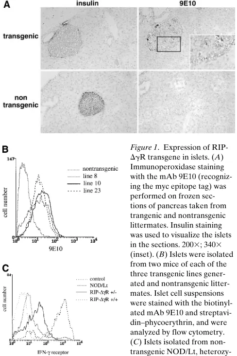
Expression of RIP-DgR in beta cells. The mAb 9E10 rec-ognizing the myc epitope tag (39) was used to detect
expres-sion of the DgR mutant receptor in pancreas sections from
transgenic mice by immunohistochemistry. Transgene expres-sion was detected in the islets of transgenic mice, but not in the surrounding exocrine pancreas, nor in the islets of littermate control animals (Fig. 1 A). The relative level of expression on the three lines of mice generated was compared by staining pu-rified islet cells with 9E10, and by analysis by flow cytometry. The three lines showed similar levels of transgene expression (Fig. 1 B).
The expression of IFN-g receptors on the beta cells of
RIP-DgR mice was also examined using the mAb GR20 that
rec-ognizes both the endogenous and transgenic murine IFN-g
receptors (40). Fig. 1 C shows levels of expression in mice wild-type, heterozygous, and homozygous for the mutant receptor. The level of GR20 staining is greatly increased in the beta cells
of mice homozygous for the RIP-DgR gene, possibly due to
the lack of turnover of the mutant receptor within the cell.
Stat1 is not activated in beta cells of RIP-DgR mice. As a
mea-sure of IFN-g responsiveness, we first looked at the ability of
the transcription factor Stat1 to bind DNA in response to IFN-g.
Stat1 normally binds to the IFN-g receptor a chain where it is
phosphorylated by members of the Janus (Jak) family of ty-rosine kinases. It then forms homodimers and translocates to the nucleus where it binds to the specific DNA sequence termed the gamma-activated sequence (GAS). When bound
to the GAS, Stat1 dimers are able to regulate IFN-g
-respon-sive genes transcriptionally (for review see reference 47). To
analyze the activation of Stat1 in the RIP-DgR mice, beta cells
were purified by FACS sorting of the A2B5-positive
[image:4.612.313.557.312.678.2]crine) and A2B5-negative (nonendocrine) cells. The cells were
treated with 100U/ml IFN-g for 15 min before nuclear extract
preparation. Nuclear extracts were analyzed by EMSA for the presence of Stat1 DNA-binding activity. While Stat1 is
acti-vated by IFN-g in the nontransgenic beta cells, the
heterozy-gous transgenic mice have a greatly reduced level of Stat1 DNA binding (Fig. 2). The small amount of Stat1 DNA bind-ing in the heterozygous mice could be due to insufficient levels of the transgene expression to achieve the dominant negative
effect, or due to the use of a very high level of IFN-g,causing a
small response (see below).
Class I MHC is not upregulated on RIP-DgR beta cells.
We next looked at the ability of IFN-g to upregulate a
respon-sive gene in the transgenic beta cells. It is well-documented that surface expression of class I MHC on beta cells increases
in response to IFN-g. We analyzed the expression of class I
MHC on beta cells by flow cytometry after incubation of
puri-fied islets in vitro for 48 h with 100 U/ml IFN-g. The
nontrans-genic beta cells showed a rise in cell surface class I MHC
ex-pression after incubation with IFN-g, which was reduced in the
mice homozygous for the transgene (Fig. 3 A). There was a slight but reproducible increase in class I MHC levels in the
transgenic mice after incubation with 100 U/ml IFN-g that
could be due to contaminating non-beta cells in the flow
cy-tometry analysis or to the high concentration of IFN-g during
the 48-h in vitro culture. To address the issue of IFN-g
concen-tration, we looked at class I MHC expression after incubation
with varying doses of IFN-g, from 0.1 U/ml to 1,000 U/ml.
Nontransgenic beta cells responded to as low as 1 U/ml IFN-g
with maximal upregulation of class I MHC after 10 U/ml. The
RIP-DgR beta cells did not upregulate class I MHC in
re-sponse to 10 U/ml IFN-g; however, they showed a slight
in-crease in class I MHC with 100 U/ml IFN-g (Fig. 3 A), which
was not further increased with up to 1,000 U/ml IFN-g (not
shown). Therefore, it is likely that with in vivo concentrations
of IFN-g, the RIP-DgR beta cells are not able to respond to
IFN-g, even though with high levels of cytokine in vitro class I
MHC is minimally upregulated (see results below also). Basal level of class I MHC on transgenic islets was not reduced as
compared with the wild-type mice, demonstrating that IFN-g
responsiveness is not required for basal class I MHC expres-sion in beta cells.
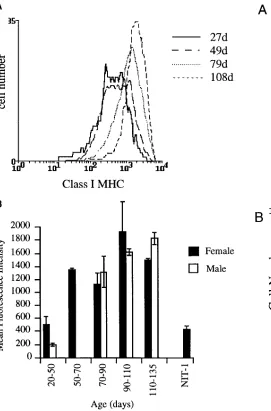
Quantitation of class I MHC expression increase with age on beta cells. Class I MHC surface protein levels were analyzed
on A2B5-positive islet endocrine cells isolated from NOD mice ranging in age from 27 d at the start of insulitis to 120 d when mice are developing diabetes. Class I MHC levels con-tinued to gradually increase from 50 d of age with no definite evidence of a plateau to this effect or step-wise change at the time that diabetes presents (Fig. 4 A). No difference was seen in female and male mice at any age tested despite the well-rec-ognized difference in risk of developing diabetes (Fig. 4 B). Staining on three consecutive days of islet cells isolated at the same time showed that class I MHC levels were stable in vitro for at least 48 h (not shown).
[image:5.612.59.304.60.242.2]NOD.scid mice do not develop insulitis or diabetes, but are congenic with NOD mice; that is, the genes controlling autoim-munity, including MHC genes, are the same as in NODs. D2.GD mice have the same class I MHC genes as NOD mice, but have different class II MHC and other genes, and are not prone to autoimmunity. We used these two strains of mice to ask whether the increase in class I MHC expression requires Figure 2. Stat1 is not activated
in purified beta cells from RIP-DgR mice. Islet cells from transgenic and nontransgenic littermates were sorted by flow cytometry into endocrine (FITC-A2B51) and nonendo-crine (FITC-A2B52) popula-tions. Cells were treated with 100U/ml IFN-g for 15 min, and nuclear extracts prepared and EMSA carried out with radio-labeled GAS oligonucleotide probe.
[image:5.612.58.426.527.737.2]the presence of insulitis. No increase in class I MHC expres-sion with age was seen in either NOD.scid or D2.GD islet cells (Fig. 5, A and B), and the levels seen were similar to those on islet cells from young NOD mice before or at the time of insu-litis. These results are consistent with overexpression of class I MHC being secondary to cytokines produced by cells in the in-sulitis lesion.
To determine if the rise in surface class I MHC expression seen in NOD mice of increasing age is due to the effect of local
IFN-g action on beta cells, we analyzed RIP-DgR mice at ages
after the onset of insulitis and compared beta cell class I MHC expression with that of NOD mice of similar age. NOD.scid
mice were used as an insulitis-free baseline class I MHC ex-pression level. The class I MHC level of beta cells from
RIP-DgR mice of ages 72–99 d remained unchanged at the level
seen in NOD.scid beta cells, whereas the class I MHC level on beta cells of 116-d-old NODs was elevated (Fig. 6). These data suggest that the class I MHC increase in NODs is due to
ef-fects of local IFN-g. This result further demonstrates that beta
cells from the transgenic mice are not responsive to IFN-g in
vivo. While it is conceivable that other IFN-g-responsive genes
may be regulated in the transgenic beta cells via an alternative signaling pathway, we have found no evidence for this.
Diabetic spleen cells transfer disease into RIP-DgR recipi-ents. We next looked at the ability of diabetogenic spleen cells
to transfer disease into RIP-DgR recipients. After transfer of
2 3 107 splenocytes from diabetic donors into irradiated
RIP-DgR(1/1) or NOD mice, diabetes incidence was assessed by
[image:6.612.62.333.58.469.2]regular blood glucose measurements. The incidence of diabe-tes was the same in the transgenics and control NOD mice Figure 4. Class I MHC expression increases with age on NOD/Lt
[image:6.612.296.550.63.461.2]beta cells. (A) Islet cells from NOD/Lt mice of 27, 49, 79, and 108 d of age were stained with 34-1-2s and phycoerythrin anti-mouse Ig for class I MHC expression. Beta cells are shown in this figure, identified with the mAb A2B5-FITC. (B) Class I MHC staining on A2B51 islet cells from female (solid bars) and male (open bars) NOD mice of the age groups indicated. The results represent the mean fluorescence in-tensity 1/2 SEM for three to four individual experiments, where is-lets from four to six mice of the same age have been pooled in each experiment. The NIT-1 insulinoma cell line (53) was included in each experiment to control for variability of class I MHC expression.
(Fig. 7). Thus, beta cell responsiveness to IFN-g is not neces-sary for the effector phase of beta cell destruction.
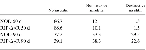
Insulitis and accelerated diabetes remain unchanged in
RIP-DgR mice. Insulitis scoring was done with homozygous mice
at 50 and 90 d of age. We found no significant difference be-tween the insulitis scores of the transgenic and nontransgenic
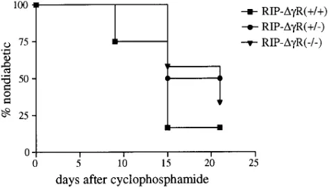
animals (Table I). RIP-DgR(1/1) and NOD controls were
given a single injection of 300 mg/kg cyclophosphamide to de-termine the incidence of accelerated diabetes. Blood glucose
levels were monitored weekly, and no difference (P 5 0.255,
logrank test) was found between cyclophosphamide-acceler-ated diabetes in the transgenic and nontransgenic animals (Fig. 8).
Spontaneous incidence of diabetes in RIP-DgR mice. Finally,
the effect of beta cell unresponsiveness to IFN-g on the
spon-taneous incidence of diabetes was determined. Fig. 9 shows the survival curve for transgenic vs. nontransgenic females, with a mean followup age of 185 d. We found no significant differ-ence in progression to diabetes between the wild-type and
transgenic mice, with a P 5 0.7 by the logrank test. We
con-clude that beta cell responsiveness to IFN-g is not required for
development of autoimmune diabetes in the NOD mouse.
Discussion
In this study we have described transgenic NOD mice with
beta cells that are unresponsive to IFN-g. These mice develop
insulitis and diabetes at the same rate as nontransgenic
litter-mates, showing that IFN-g does not promote the progression
of autoimmune diabetes in the NOD by directly affecting beta cell viability or gene expression, but must act indirectly via
an-other cell type. These mice also show that IFN-g is responsible
for increased beta cell class I MHC expression in NOD mice, a pathological feature of IDDM seen in affected humans and BB rats as well. Therefore, increased class I MHC expression on beta cells is not required for progression of diabetes.
We and others have linked in vitro effects on beta cells of
IFN-g and other cytokines to the pathogenesis of diabetes (for
reviews see references 48–50). While plausible circumstantial evidence for this link has existed, it has not previously been di-rectly tested. These in vitro effects remain valid, but they do
not explain the ability of IFN-g to promote diabetes. If direct
effects of IFN-g do play a role, it must be minor because
spon-taneous disease and diabetes development in two accelerated models of diabetes were not affected by transgene expression. This finding is important because of the emphasis placed on in
vitro effects of IFN-g on beta cells in diabetes research, which
we now believe to be of limited relevance.
Class I MHC expression on pancreatic beta cells and pro-gression to autoimmune diabetes have been linked in humans, BB rats, and NOD mice. In the current study they have been
dissociated for the first time because RIP-DgR mice developed
diabetes without beta cell class I overexpression. This result suggests that basal levels of class I expression are sufficient for
CD81 T cell recognition of beta cells, known to be required for
diabetes in the NOD mouse model (51).
Class I MHC overexpression on beta cells is closely associ-ated with insulitis, and is upregulassoci-ated very close to the time that insulitis begins. It is not found by immunohistology in is-lets without insulitis (21). In the current study, class I MHC overexpression on NOD beta cells has been quantitated for the first time by flow cytometry. Increased expression of class I MHC was first recognized at 50 d of age, and mean class I ex-pression then rose gradually with age until 90–110 d when hy-Figure 6. Class I MHC expression does not increase with age on
[image:7.612.58.295.56.239.2]RIP-DgR beta cells. Islet cells from NODscid, 116-d-old NOD/Lt, 72-, and 99-d-old RIP-DgR(1/1) mice were stained with the mAb 34-1-2s and phycoerythrin anti-mouse Ig for class I MHC expression. Shown is the class I MHC staining on the autofluorescent beta cell population.
[image:7.612.316.558.59.193.2]Figure 7. Adoptive transfer of diabetes into RIP-DgR recipients. Splenocytes from diabetic NOD/Lt female donors were injected in-travenously into irradiated NOD/Lt or RIP-DgR(1/1) recipients. The percentage of mice free from disease is shown up to 28 days post transfer. Eight mice were used per group.
Table I. Histological Scoring of Insulitis in Nondiabetic Wild-type NOD/Lt and RIP-DgR(1/1) Mice
No insulitis
Noninvasive insulitis
Destructive insulitis
NOD 50 d 86.7 12 1.3
RIP-DgR 50 d 88.6 10.1 1.3
NOD 90 d 37.2 33.3 29.5
RIP-DgR 90 d 39.1 38.3 22.6
[image:7.612.315.557.631.714.2]perglycaemia began to occur. We did not observe increased class I MHC expression before insulitis, and therefore it is likely to be a consequence of rather than required for its devel-opment. This conclusion was supported by the absence of in-creased class I expression with age in mice without insulitis,
and the development of insulitis in RIP-DgR mice in the
ab-sence of class I MHC overexpression. Similarly, increased class I expression is not likely to be due to the class I genes of the NOD since D2.GD mice with identical class I type do not show this phenotype. In contrast, total absence of class I MHC
ex-pression in the NODb2mnull completely protects NOD mice
from insulitis (for review see reference 51). We did not find any difference between class I MHC expression on beta cells from male and female NOD mice, making it also likely that class I overexpression is not sufficient for progression to diabe-tes since male mice in our colony rarely develop diabediabe-tes.
Our study shows that IFN-g is the major factor associated
with increased class I MHC expression on NOD beta cells. Other cytokines, endogenous or exogenous viruses, diet, or other environmental agents, although possibly contributing in-directly to this phenomenon, are unlikely to have a direct
ef-fect. This result is not surprising as, in vitro, IFN-g is the only
agent we have used that increases beta cell class I to the extent seen in vivo in NOD mice. An important implication of this re-sult is that beta cell class I can be regarded as a local indicator
of IFN-g action. If this is the case, our data do not support a
sudden shift to local IFN-g activity, but rather a gradual rise
with age.
The IFN-g knockout mice develop diabetes at a modestly
reduced and delayed rate (10). It is conceivable that global
IFN-g deficiency results in the development of compensatory
mechanisms that preserve immune homeostasis, and also
sub-stitute for IFN-g in the pathogenesis of diabetes. In contrast,
IFN-g receptor knockout NOD mice fail to develop diabetes,
and the results indicate that this is likely to be due to the
ab-sence of IFN-g–responsive macrophages or beta cells rather
than T cells (3). The discrepancy between these results is unex-plained, but both mice are known to have abnormal immune
responses that are not always identical to each other in other disease states (52). It is also possible that an as yet unidentified
second ligand for the IFN-g receptor exists that substitutes for
IFN-g in the IFN-g–deficient animals. Because of these
con-siderations and because we wished to test the relevance to
dia-betes of in vitro effects of IFN-g on beta cells, we chose an
ap-proach designed to produce IFN-g–unresponsive beta cells,
leaving the immune system intact. The approach of
tissue-spe-cific unresponsiveness to IFN-g allows elucidation of which
cell type is the target of IFN-g relevant to the pathogenesis of
IDDM. This cannot be achieved by transplantation of islets from knockout animals as there are many different cell types within the islet, including macrophages and endothelial cells,
that are potential targets of IFN-g action.
This study demonstrates the power of genetic manipulation of the NOD model to address clinically important questions directly. The use of class I–deficient mice back-crossed onto the NOD model dramatically changed our understanding of
the role of class I MHC expression and CD81 T cells in the
NOD, and the current data clarify that the requirement for class I expression is at basal levels only and have confirmed
that IFN-g is likely to play at most a minor role by its action on
beta cells. Clearly genetic manipulation of this sort is not appli-cable to IDDM in humans, in which virtually nothing defini-tive is known about pathogenesis beyond limited descripdefini-tive immunopathology. The challenge is to apply our understand-ing of diabetes pathogenesis in the NOD to human IDDM to enable rationally based treatment and prevention strategies.
Acknowledgments
We wish to thank Dr. Leigh A. Stephens, Dr. Jan Allison, Dr. Simon J. Foote, Ms. Vikki Marshall, and Ms. Rima Darwiche for expert ad-vice and assistance. We also thank Dr. Anne Kelso, Dr. Diane Mathis, and Dr. William R. Heath for critical reading of the manuscript.
This work was supported by the National Health and Medical Re-search Council of Australia and the Juvenile Diabetes Foundation as part of a Diabetes Interdisciplinary Research Program.
References
1. Rabinovitch, A., W. Suarez-Pinzon, A. El-Sheikh, O. Sorensen, and R.F. Power. 1996. Cytokine gene expression in pancreatic islet-infiltrating leuko-Figure 8. Cyclophosphamide induces diabetes in RIP-DgR mice.
[image:8.612.317.554.60.185.2]Nondiabetic male mice were given a single dose of cyclophosphamide injected intraperitoneally at 300 mg/kg body weight, and were ob-served for hyperglycemia development. The percentage of nondia-betic mice after cyclophosphamide injection of RIP-DgR(1/1), RIP-DgR(1/2), and nontransgenic littermates is shown. Eight mice were included in each group. The curves were not significantly different by the logrank test with a P 5 0.255.
[image:8.612.59.296.62.197.2]cytes of BB rats: expression of Th1 cytokines correlates with beta-cell destruc-tive insulitis and IDDM. Diabetes. 45:749–754.
2. Rabinovitch, A., W.L. Suarez-Pinzon, O. Sorensen, R.C. Bleackley, and R.F. Power. 1995. IFN-gamma gene expression in pancreatic islet-infiltrating mononuclear cells correlates with autoimmune diabetes in nonobese diabetic mice. J. Immunol. 154:4874–4882.
3. Wang, B., I. Andre, A. Gonzalez, J.D. Katz, M. Aguet, C. Benoist, and D. Mathis. 1997. Interferon-gamma impacts at multiple points during the progres-sion of autoimmune diabetes. Proc. Natl. Acad. Sci. USA. 94:13844–13849.
4. Campbell, I.L., T.W. Kay, L. Oxbrow, and L.C. Harrison. 1991. Essential role for interferon-gamma and interleukin-6 in autoimmune insulin-dependent diabetes in NOD/Wehi mice. J. Clin. Invest. 87:739–742.
5. Debray-Sachs, M., C. Carnaud, C. Boitard, H. Cohen, I. Gresser, P. Be-dossa, and J.F. Bach. 1991. Prevention of diabetes in NOD mice treated with antibody to murine IFN gamma. J. Autoimmun. 4:237–248.
6. Nicoletti, F., P. Zaccone, R. Di Marco, M. Di Mauro, G. Magro, S. Grasso, L. Mughini, P. Meroni, and G. Garotta. 1996. The effects of a nonim-munogenic form of murine soluble interferon-gamma receptor on the develop-ment of autoimmune diabetes in the NOD mouse. Endocrinology. 137:5567– 5575.
7. von Herrath, M.G., and M.B. Oldstone. 1997. Interferon-gamma is essen-tial for destruction of beta cells and development of insulin-dependent diabetes mellitus. J. Exp. Med. 185:531–539.
8. Oldstone, M.B., M. Nerenberg, P. Southern, J. Price, and H. Lewicki. 1991. Virus infection triggers insulin-dependent diabetes mellitus in a trans-genic model: role of anti-self (virus) immune response. Cell. 65:319–331.
9. Ohashi, P.S., S. Oehen, K. Buerki, H. Pircher, C.T. Ohashi, B. Odermatt, B. Malissen, R.M. Zinkernagel, and H. Hengartner. 1991. Ablation of “toler-ance” and induction of diabetes by virus infection in viral antigen transgenic mice. Cell. 65:305–317.
10. Hultgren, B., X. Huang, N. Dybdal, and T.A. Stewart. 1996. Genetic ab-sence of gamma-interferon delays but does not prevent diabetes in NOD mice.
Diabetes. 45:812–817.
11. Eizirik, D.L., M. Flodstrom, A.E. Karlsen, and N. Welsh. 1996. The har-mony of the spheres: inducible nitric oxide synthase and related genes in pan-creatic beta cells. Diabetologia. 39:875–890.
12. Kaneto, H., J. Fujii, H.G. Seo, K. Suzuki, T. Matsuoka, M. Nakamura, H. Tatsumi, Y. Yamasaki, T. Kamada, and N. Taniguchi. 1995. Apoptotic cell death triggered by nitric oxide in pancreatic beta-cells. Diabetes. 44:733–738.
13. Dunger, A., J.M. Cunningham, C.A. Delaney, J.E. Lowe, M.H. Green, A.J. Bone, and I.C. Green. 1996. Tumor necrosis factor-alpha and interferon-gamma inhibit insulin secretion and cause DNA damage in unweaned-rat islets. Extent of nitric oxide involvement. Diabetes. 45:183–189.
14. Corbett, J.A., and M.L. McDaniel. 1995. Intraislet release of interleukin 1 inhibits beta cell function by inducing beta cell expression of inducible nitric oxide synthase. J. Exp. Med. 181:559–568.
15. Nagata, S., and P. Golstein. 1995. The Fas death factor. Science. 267: 1449–1456.
16. Yamada, K., N. Takane-Gyotoku, X. Yuan, F. Ichikawa, C. Inada, and K. Nonaka. 1996. Mouse islet cell lysis mediated by interleukin-1-induced Fas.
Diabetologia. 39:1306–1312.
17. Yagi, N., K. Yokono, K. Amano, M. Nagata, K. Tsukamoto, Y. Hase-gawa, R. Yoneda, N. Okamoto, H. Moriyama, M. Miki, et al. 1995. Expression of intercellular adhesion molecule 1 on pancreatic cells accelerates beta-cell destruction by cytotoxic T beta-cells in murine autoimmune diabetes. Diabetes. 44:744–752.
18. Hanninen, A., S. Jalkanen, M. Salmi, S. Toikkanen, G. Nikolakaros, and O. Simell. 1992. Macrophages, T cell receptor usage, and endothelial cell activa-tion in the pancreas at the onset of insulin-dependent diabetes mellitus. J. Clin.
Invest. 90:1901–1910.
19. Itoh, N., T. Hanafusa, A. Miyazaki, J. Miyagawa, K. Yamagata, K. Yamamoto, M. Waguri, A. Imagawa, S. Tamura, M. Inada, et al. 1993. Mono-nuclear cell infiltration and its relation to the expression of major histocompati-bility complex antigens and adhesion molecules in pancreas biopsy specimens from newly diagnosed insulin-dependent diabetes mellitus patients. J. Clin.
In-vest. 92:2313–2322.
20. Somoza, N., F. Vargas, C. Roura-Mir, M. Vives-Pi, M.T. Fernandez-Figueras, A. Ariza, R. Gomis, R. Bragado, M. Marti, D. Jaraquemada, and R. Pujol-Borrell. 1994. Pancreas in recent onset insulin-dependent diabetes melli-tus. Changes in HLA, adhesion molecules and autoantigens, restricted T cell re-ceptor V beta usage, and cytokine profile. J. Immunol. 153:1360–1377.
21. Kay, T.W., I.L. Campbell, L. Oxbrow, and L.C. Harrison. 1991. Overex-pression of class I major histocompatibility complex accompanies insulitis in the non-obese diabetic mouse and is prevented by anti-interferon-gamma antibody.
Diabetologia. 34:779–785.
22. O’Reilly, L.A., P.R. Hutchings, P.R. Crocker, E. Simpson, T. Lund, D. Kioussis, F. Takei, J. Baird, and A. Cooke. 1991. Characterization of pancreatic islet cell infiltrates in NOD mice: effect of cell transfer and transgene expres-sion. Eur. J. Immunol. 21:1171–1180.
23. Ono, S.J., B. Issa-Chergui, E. Colle, R.D. Guttmann, T.A. Seemayer, and A. Fuks. 1988. IDDM in BB rats. Enhanced MHC class I heavy-chain gene expression in pancreatic islets. Diabetes. 37:1411–1418.
24. Ohashi, P.S., S. Oehen, P. Aichele, H. Pircher, B. Odermatt, P. Herrera, Y. Higuchi, K. Buerki, H. Hengartner, and R.M. Zinkernagel. 1993. Induction of diabetes is influenced by the infectious virus and local expression of MHC class I and tumor necrosis factor-alpha. J. Immunol. 150:5185–5194.
25. Vives-Pi, M., M.P. Armengol, L. Alcalde, M. Costa, N. Somoza, F. Var-gas, D. Jaraquemada, and R. Pujol-Borrell. 1996. Expression of transporter as-sociated with antigen processing-1 in the endocrine cells of human pancreatic islets: effect of cytokines and evidence of hyperexpression in IDDM. Diabetes. 45:779–788.
26. Wicker, L.S., E.H. Leiter, J.A. Todd, R.J. Renjilian, E. Peterson, P.A. Fischer, P.L. Podolin, M. Zijlstra, R. Jaenisch, and L.B. Peterson. 1994. Beta 2-microglobulin-deficient NOD mice do not develop insulitis or diabetes.
Dia-betes. 43:500–504.
27. Serreze, D.V., E.H. Leiter, G.J. Christianson, D. Greiner, and D.C. Roopenian. 1994. Major histocompatibility complex class I-deficient NOD-B2mnull mice are diabetes and insulitis resistant. Diabetes. 43:505–509.
28. Sumida, T., M. Furukawa, A. Sakamoto, T. Namekawa, T. Maeda, M. Zijlstra, I. Iwamoto, T. Koike, S. Yoshida, H. Tomioka, and M. Taniguchi. 1994. Prevention of insulitis and diabetes in beta 2-microglobulin-deficient non-obese diabetic mice. Int. Immunol. 6:1445–1449.
29. Katz, J., C. Benoist, and D. Mathis. 1993. Major histocompatibility com-plex class I molecules are required for the development of insulitis in non-obese diabetic mice.Eur. J. Immunol. 23:3358–3360.
30. Raulet, D.H. 1994. MHC class I-deficient mice. Adv. Immunol. 55:381–421. 31. Kay, T.W., J.L. Parker, L.A. Stephens, H.E. Thomas, and J. Allison. 1996. RIP-beta 2-microglobulin transgene expression restores insulitis, but not diabetes, in beta 2-microglobulin null nonobese diabetic mice. J. Immunol. 157: 3688–3693.
32. Kurts, C., W.R. Heath, F.R. Carbone, J. Allison, J.F. Miller, and H. Ko-saka. 1996. Constitutive class I-restricted exogenous presentation of self anti-gens in vivo. J. Exp. Med. 184:923–930.
33. Dighe, A.S., D. Campbell, C.S. Hsieh, S. Clarke, D.R. Greaves, S. Gor-don, K.M. Murphy, and R.D. Schreiber. 1995. Tissue-specific targeting of cy-tokine unresponsiveness in transgenic mice. Immunity. 3:657–666.
34. Dighe, A.S., M.A. Farrar, and R.D. Schreiber. 1993. Inhibition of cellu-lar responsiveness to interferon-gamma (IFN gamma) induced by overexpres-sion of inactive forms of the IFN gamma receptor. J. Biol. Chem. 268:10645– 10653.
35. Dietrich, W.F., J. Miller, R. Steen, M.A. Merchant, D. Damron-Boles, Z. Husain, R. Dredge, M.J. Daly, K.A. Ingalls, et al. 1996. A comprehensive ge-netic map of the mouse genome. Nature. 380:149–152.
36. Church, G.M., and S. Kieffer-Higgins. 1988. Multiplex DNA sequenc-ing. Science. 240:185–188.
37. Richterich, P., and G.M. Church. 1993. DNA sequencing with direct transfer electrophoresis and nonradioactive detection. Methods Enzymol. 218: 187–222.
38. Lake, S.P., J. Anderson, J. Chamberlain, S.J. Gardner, P.R. Bell, and R.F. James. 1987. Bovine serum albumin density gradient isolation of rat pan-creatic islets. Transplantation. 43:805–808.
39. Evan, G.I., G.K. Lewis, G. Ramsay, and J.M. Bishop. 1985. Isolation of monoclonal antibodies specific for human c-myc proto-oncogene product. Mol.
Cell. Biol. 5:3610–3616.
40. LeClaire, R.D., M. Basu, D.M. Pinson, M.L. Redick, J.S. Hunt, P.J. Zavodny, J.L. Pace, and S.W. Russell. 1992. Characterization and use of mono-clonal and polymono-clonal antibodies against the mouse interferon-gamma receptor.
J. Leukoc. Biol. 51:507–516.
41. Ozato, K., N.M. Mayer, and D.H. Sachs. 1982. Monoclonal antibodies to mouse major histocompatibility complex antigens. Transplantation. 34:113–120. 42. Eisenbarth, G.S., F.S. Walsh, and M. Nirenberg. 1979. Monoclonal anti-body to a plasma membrane antigen of neurons. Proc. Natl. Acad. Sci. USA. 76: 4913–4917.
43. Pipeleers, D.G., P.A. in’t Veld, M. Van de Winkel, E. Maes, F.C. Schuit, and W. Gepts. 1985. A new in vitro model for the study of pancreatic A and B cells. Endocrinology. 117:806–816.
44. Schreiber, E., P. Matthias, M.M. Muller, and W. Schaffner. 1989. Rapid detection of octamer binding proteins with mini-extracts, prepared from a small number of cells. Nucleic Acids Res. 17:6419.
45. Pearse, R.N., R. Feinman, K. Shuai, J. Darnell, Jr., and J.V. Ravetch. 1993. Interferon gamma-induced transcription of the high-affinity Fc receptor for IgG requires assembly of a complex that includes the 91-kDa subunit of transcription factor ISGF3. Proc. Natl. Acad. Sci. USA. 90:4314–4318.
46. Charlton, B., and T.E. Mandel. 1988. Progression from insulitis to beta-cell destruction in NOD mouse requires L3T41 T-lymphocytes. Diabetes. 37: 1108–1112.
47. Darnell, J., Jr., I.M. Kerr, and G.R. Stark. 1994. Jak-STAT pathways and transcriptional activation in response to IFNs and other extracellular sig-naling proteins. Science. 264:1415–1421.
48. Rabinovitch, A. 1996. Roles of cell-mediated immunity and cytokines in the pathogenesis of insulin-dependent diabetes mellitus. In Diabetes Mellitus-A Fundamental and Clinical Text. D. LeRoith, S.I. Taylor, and J.M. Olefsky, editors. Lippincott-Raven Publishers, Philadelphia. 314–325.
me-diators of autoimmune diabetes and diabetic complications. Endocr. Rev. 16: 164–176.
50. Harrison, L.C., I.L. Campbell, P.G. Colman, N. Chosich, T.W.H. Kay, B.D. Tait, R.K. Bartholomeusz, H.J. De Aizpurua, J.L. Joseph, S. Chu, and W.E. Kielczynski. 1990. Type 1 diabetes: immunopathology and immunother-apy. Adv. Endocrinol. Metab. 1:35–94.
51. Kay, T., H.L. Chaplin, J.L. Parker, L.A. Stephens, and H.E. Thomas. 1997. CD4(1) and CD8(1) T lymphocytes—clarification of their pathogenic roles in diabetes in the NOD mouse. Res. Immunol. 148:320–327.
52. Swihart, K., U. Fruth, N. Messmer, K. Hug, R. Behin, S. Huang, G. Del Giudice, M. Aguet, and J.A. Louis. 1995. Mice from a genetically resistant background lacking the interferon gamma receptor are susceptible to infection with Leishmania major but mount a polarized T helper cell 1-type CD41 T cell response. J. Exp. Med. 181:961–971.