Pdx1 restores
bb
cell function in Irs2 knockout
mice
Jake A. Kushner, … , Marc R. Montminy, Morris F. White
J Clin Invest.
2002;
109(9)
:1193-1201.
https://doi.org/10.1172/JCI14439
.
The homeodomain transcription factor Pdx1 is required for pancreas development,
including the differentiation and function of b cells. Mutations in Pdx1 or upstream
hepatocyte nuclear factors cause autosomal forms of early-onset diabetes (maturity-onset
diabetes of the young [MODY]). In mice, the Irs2 branch of the insulin/Igf signaling system
mediates peripheral insulin action and pancreatic b cell growth and function. To investigate
whether b cell failure in
Irs2
–/–mice might be related to dysfunction of MODY-related
transcription factors, we measured the expression of Pdx1 in islets from young
Irs2
–/–mice.
Before the onset of diabetes, Pdx1 was reduced in islets from
Irs2
–/–mice, whereas it was
expressed normally in islets from wild-type or
Irs1
–/–mice, which do not develop diabetes.
Whereas male
Irs2
–/–Pdx1
+/+mice developed diabetes between 8 and 10 weeks of age,
haploinsufficiency for
Pdx1
caused diabetes in newborn
Irs2
–/–mice. By contrast,
transgenic expression of Pdx1 restored b cell mass and function in
Irs2
–/–mice and
promoted glucose tolerance throughout life, as these mice survived for at least 20 months
without diabetes. Our results suggest that dysregulation of Pdx1 might represent a common
link between ordinary type 2 diabetes and MODY.
Article
Endocrinology
Introduction
The insulin receptor substrates (IRS proteins) coordi-nate many signals during insulin and IGF-1 stimula-tion, including activation of the phosphatidylinositol (PI) 3-kinase and ERK1/2 cascades (1). Although high-ly homologous and universalhigh-ly expressed, Irs1 and Irs2 display distinct biological function in mice (2). Irs1 strongly promotes somatic growth and mediates insulin action upon carbohydrate metabolism in skeletal mus-cle; however, Irs1–/–mice never develop diabetes, owing to islet hyperplasia and lifelong compensatory hyperin-sulinemia (2). By contrast, Irs2 plays a significant role in metabolic regulation, as hepatic gluconeogenesis and lipid metabolism are dysregulated in Irs2–/–mice (3). Moreover, Irs2–/–mice develop progressive βcell failure, which exacerbates the peripheral insulin resistance and leads to diabetes (2). The βcells in Irs2–/–mice display increased apoptosis, especially during weaning, which might contribute to the steady decline of βcell mass with increasing age (4). Although moderate hyperinsu-linemia compensates for peripheral insulin resistance in young Irs2–/–mice, diabetes occurs between 8 and 10 weeks of age, because the diminishing βcell content fails to secrete sufficient insulin (2).
Autosomal dominant forms of early-onset diabetes in young adults (maturity-onset diabetes of the young
[MODY]) are linked to mutations in glucokinase or tran-scription factors that promote normal βcell function, including HNF4α(MODY1), HNF1α(MODY3), PDX1
(MODY4), HNF1β (MODY5), and NeuroD1/BETA2
(MODY6); MODY2 is caused by mutations in glucokinase, whose expression is regulated by PDX1 (5). The regula-tion of these transcripregula-tion factors is complicated, but in certain cases it might be linked to insulin/IGF1 signaling though Foxo1or Hnf3β(6). Pdx1 plays an important role in islet development and βcell function. It is required for pancreas formation during the initial stages of gut devel-opment (7, 8). In adult mice, Pdx1 promotes the expres-sion of proinsulin, Glut2, and glucokinase, which mediates glucose-sensitive insulin secretion (9–11). Pdx1 also reg-ulates expression of FGFs and their receptors, which might promoteβcell growth and proinsulin processing via the prohormone processing enzyme PC1/3 (12). Con-sequently, disruption of Pdx1in murine βcells reduces insulin secretion and causes progressive βcell loss, which culminates in glucose intolerance and diabetes (13). Moreover, PDX1is required in adult humans to promote normal glucose sensing and insulin secretion, and muta-tions in PDX1represent a risk factor for type 2 diabetes (13–15). In this report, we reveal a close relation between Pdx1 and Irs2, which suggests that dysregulation of Pdx1 by genetic or functional mechanisms might be a
com-Pdx1 restores
β
cell function in Irs2 knockout mice
Jake A. Kushner,
1,2Jing Ye,
1Markus Schubert,
1Deborah J. Burks,
1Matthew A. Dow,
1Carrie L. Flint,
1Sanjoy Dutta,
3Christopher V.E. Wright,
4Marc R. Montminy,
5and Morris F. White
11Howard Hughes Medical Institute, Joslin Diabetes Center, Harvard Medical School, Boston, Massachusetts, USA 2Division of Endocrinology, Children’s Hospital, Boston, Massachusetts, USA
3Joslin Diabetes Center, Harvard Medical School, Boston, Massachusetts, USA 4Developmental Biology Program, Vanderbilt University, Nashville, Tennessee, USA 5Clayton Laboratories for Peptide Biology, The Salk Institute, La Jolla, California, USA
Address correspondence to: Morris F. White, Howard Hughes Medical Institute, Joslin Diabetes Center, 1 Joslin Place, Boston, Massachusetts 02215, USA. Phone: (617) 732-2578; Fax: (617) 732-2593; E-mail: [email protected].
Received for publication October 18, 2001, and accepted in revised form March 26, 2002.
The homeodomain transcription factor Pdx1 is required for pancreas development, including the dif-ferentiation and function of βcells. Mutations in Pdx1 or upstream hepatocyte nuclear factors cause autosomal forms of early-onset diabetes (maturity-onset diabetes of the young [MODY]). In mice, the Irs2 branch of the insulin/Igf signaling system mediates peripheral insulin action and pancreatic β cell growth and function. To investigate whether βcell failure in Irs2–/–mice might be related to
dys-function of MODY-related transcription factors, we measured the expression of Pdx1 in islets from young Irs2–/–mice. Before the onset of diabetes, Pdx1 was reduced in islets from Irs2–/–mice, whereas
it was expressed normally in islets from wild-type or Irs1–/–mice, which do not develop diabetes.
Where-as male Irs2–/–Pdx1+/+mice developed diabetes between 8 and 10 weeks of age, haploinsufficiency for Pdx1caused diabetes in newborn Irs2–/–mice. By contrast, transgenic expression of Pdx1 restored βcell
mass and function in Irs2–/–mice and promoted glucose tolerance throughout life, as these mice
sur-vived for at least 20 months without diabetes. Our results suggest that dysregulation of Pdx1 might represent a common link between ordinary type 2 diabetes and MODY.
mon element in autosomal early-onset (MODY) and common type 2 diabetes.
Methods
Mice. Irs1knockout (2), Irs2knockout (2), Pdx1knockout (8), and wild-type Pdx1transgene (16) mice were main-tained on a mixed C57BL/6 ×129Sv ×Black Swiss back-ground. Genotyping of animals was done by PCR as pre-viously described (4, 16). Male mice were used to characterize adult glucose homeostasis phenotypes and pancreatic pathology. Male and female Irs2+/–Pdx1+/– intercross newborn pups were grouped together, as no sexual dimorphism was apparent in neonatal glucose homeostasis (data not shown). Random-fed glucose and insulin measurements were performed as previously described (4). Intraperitoneal glucose tolerance tests were performed on mice fasted for 15–16 hours with 2 g
D-glucose per kg body weight as previously described (2).
Intraperitoneal insulin tolerance tests were performed on fed mice with 0.75 U human insulin per kg body weight as previously described (2).
Immunohistochemistry. Immunohistochemical localiza-tion of antigens and double-label immunohistochem-istry were performed similarly to previously described methods (4). Pancreas samples were dissected from fed mice and fixed with Bouin’s solution overnight. Five-micrometer longitudinal sections of paraffin blocks were rehydrated with xylene followed by decreasing concen-trations of ethanol, microwaved in 0.01 M sodium cit-rate (pH 6.0) for 20 minutes, and permeabilized with 1% Triton X-100 in PBS prior to primary antisera incuba-tion. Islet morphometry was performed similarly to the methods previously described (4). The primary antibod-ies were: guinea pig insulin and rabbit anti-glucagon antibodies (Zymed Laboratories Inc., South San Francisco, California, USA) rabbit N-terminal
anti-Pdx1 (from C.V.E. Wright), and mouse anti-BrdU (Roche Molecular Biochemicals, Indianapolis, Indiana, USA). Secondary antibodies were labeled with FITC or rhodamine (Jackson ImmunoResearch Laboratories Inc., West Grove, Pennsylvania, USA).
Islet morphometry. Newborn (postnatal day [P] 0.5–1.5) βcell area was quantified by acquiring adjacent ×10 images of the entire newborn pancreas from two anti-insulin–stained sections per animal, at least six animals per genotype (male and female were grouped together), with a Zeiss Axiovert microscope (Carl Zeiss MicroImag-ing, Thornwood, New York, USA). Images were analyzed for area with Improvision Open Lab software density slice software (Improvision Scientific Imaging, Lexing-ton, Massachusetts, USA). For each animal the ratio of βcell area to total pancreas area was calculated and reported as mean ± SEM. Adult βcell area was measured by acquiring images from two sets of eight to ten ran-dom nonoverlapping images at ×10 of insulin- and glucagon-stained sections from male mice, at least four animals per genotype. Results of βcell quantification
and size were calculated from captured insulin-stained images. Ratios of βto αcells were calculated from mean insulin- and glucagon-positive cell areas. Islet prolifera-tion and βcell size were examined by injecting 2- to 3-week-old male mice with 5-bromo-2-deoxyuridine (BrdU; Roche Molecular Biochemicals, Indianapolis, Indiana, USA; 100 µg/g body weight) and performing double-label insulin and BrdU immunohistochemistry on rehydrated Bouin’s-fixed paraffin-embedded sec-tions from the mice. Images of each islet per section were acquired at ×63. BrdU-positive βcell ratios were calculated as the mean ± SEM of BrdU-positive βcells over total βcells per section, two sections per animal, three to four animals per genotype. Size of βcells was determined by dividing total βcell area by the number of βcells examined per section.
RT-PCR and Western blotting. Islets were isolated from 6-week-old male wild-type, Irs1–/–, and Irs2–/–mice with Collagenase P (Liberase HI; Roche Molecular Bio-chemicals) digestion (17) and total RNA was extracted with Trizol (GIBCO BRL; Life Technologies Inc., Grand Island, New York, USA) and RNeasy (QIAGEN Inc., Valencia, California, USA) columns. cDNA syn-thesis was performed using the RETROscript kit (Ambion Inc., Austin, Texas, USA) and then TaqMan (Applied Biosystems, Foster City, California, USA) quantitative PCR (50°C for 2 minutes, 95°C for 10 minutes, followed by 40 cycles of 95°C for 15 seconds, 60°C for 1 minute) was performed with the ABI Prism 7700 PCR instrument (Applied Biosystems) to ampli-fy samples in triplicate for the Pdx1, Hnf1α, Hnf1β,
Hnf3β(Foxa2), Hnf4α, and cyclophillingenes. Primers were as follows: Pdx1 forward,
AGGAAAACAAGAGGAC-CCGTACT; Pdx1 probe, CCTACACCCGGGCGCAGCTG;
Pdx1 reverse, CGGGAGATGTATTTGTTAAATAAGAATTC; Hnf1αforward, TGCGTTCTACAACTGGTTTGC; Hnf1α probe, AGGCTTCCTCCTTGCGCCGG; Hnf1α reverse,
GGAGGTCCGTTATAGGTGTCCAT; Hnf1β forward,
GAGGAGTGTAACAGGGCAGAATGT; Hnf1β probe,
CCTTCCAAAGCCCACGGCCTAGG; Hnf1β reverse,
GGACCTCCGTGACCAAGTTG; Hnf3βforward,
GAACTC-CATCCGCCACTCTCT; Hnf3β probe,
TCAACGACT-GCTTTCTCAAGGTGCCC; Hnf3β reverse,
GCCCT-TGCCAGGCTTGT; Hnf4α forward,
ACGTGCTGC-TCCTAGGCAAT; Hnf4αprobe,
CACACGGCTCATCTC-CGCTAGCTCTG; Hnf4αreverse,
TCGAGGATGCGGATG-GA; cyclophillin forward, CAGACGCCACTGTCGCTTT; cyclophillin probe, CCTACACCCGGGCGCAGCTG; cyclophillin reverse, TGTCTTTGGAACTTTGTCTGCAA. For each sample, triplicate gene expression values were compared with wild-type islet cDNA dilution standard curves of log4dilutions (undiluted, 1:4, 1:16, 1:64) per-formed in triplicate (Pdx1threshold cycle (Ct) = 23,
r2 = 0.99; Hnf1α C
t= 28, r2 = 0.96; Hnf1β Ct= 29,
r2 = 0.88; Hnf3β C
t= 27, r2= 0.95; Hnf4α Ct= 29,
r2= 0.99; cyclophillin C
sev-Western blot analysis, SDS protein islet lysates were equalized for loading by total protein assay (Pierce Chemical Co., Rockford, Illinois, USA) run on 12% SDS-PAGE, transferred to a Protan nitrocellulose membrane (Schleicher & Schuell Inc., Keene, New Hampshire, USA), incubated with rabbit anti–N-ter-minal Pdx1 antisera (from C.V.E. Wright) and HRP-conjugated goat anti-rabbit antisera, and then treated for enhanced chemiluminescence (NEN Life Science Products Inc., Boston, Massachusetts, USA).
Results
Pdx1 mRNA levels were measured by RT-PCR in islet extracts from young male mice. Compared with wild-type, Pdx1 mRNA levels were reduced in Irs2–/–islets (Figure 1a); similar results were found with age-matched female Irs2–/–mice (data not shown). Hnf3β (also known as Foxa2) was also reduced in islets from
Irs2–/–mice, whereas the levels of Hnf1α, Hnf1β, and Hnf4αwere approximately normal in the male Irs2–/– mice (Figure 1a). Immunoblotting confirmed that Pdx1 protein levels were low in male and female Irs2–/–islets
(Figure 1b). By contrast, mRNA levels for Pdx1 and the various hepatocyte nuclear factors were nearly normal in Irs1–/–islets at 6 weeks of age, and immunoblotting confirmed that Pdx1 protein was normal in female
Irs1–/–mice (Figure 1b). The specific reduction of Pdx1 expression in Irs2–/–mice might be especially important for the progression to diabetes, as Pdx1 regulates com-ponents of the glucose sensing pathway, and patholog-ical processes that reduce Pdx1expression cause glucose intolerance (11, 15).
To test the role of Pdx1 in Irs2–/–mice, we intercrossed
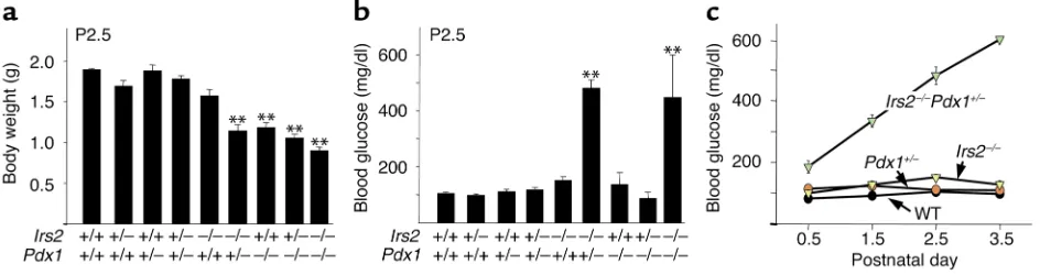
Irs2+/–mice with Pdx1+/–mice and backcrossed the com-pound heterozygotes to produce the nine expected genotypes at mendelian frequencies (8). On P2.5, hap-loinsufficiency for Pdx1or Irs2had no effect on body weight, whereas Irs2–/– mice were slightly smaller by comparison with littermates (Figure 2a). Irs2–/–Pdx1+/– mice and mice lacking Pdx1(Pdx1–/–, Irs2+/–Pdx1–/–, or
Irs2–/–Pdx1–/–) were about 40% smaller at P2.5 and failed to gain weight thereafter (Figure 2a and data not shown). During the first 5 postnatal days, most mice displayed normal blood glucose, including the Irs2–/– and Pdx1–/–mice (Figure 2b). However, Irs2–/–Pdx1+/– mice developed severe hyperglycemia and died by P4;
[image:4.576.67.278.55.255.2]Irs2–/–Pdx1–/–mice displayed a similar outcome (Fig-ure 2c and data not shown). No sexual dimorphism was seen in the Irs2–/–Pdx1+/–mouse phenotypes, as both male (n = 5) and female (n = 10) Irs2–/–Pdx1+/–pups devel-oped hyperglycemia and died by P4. By contrast, Pdx1–/– mice displayed normal blood glucose at P2.5 and sur-vived for up to 9 days, suggesting that Irs2 signaling might promote neonatal glucose homeostasis without insulin (Figure 2b). As shown previously, Irs2–/–mice Figure 1
Expression of hepatocyte nuclear factors (HNFs) and Pdx1in pancre-atic islets. (a) TaqMan RT-PCR of Hnf1α, Hnf1β, Hnf3β, Hnf4α, and Pdx1 in islets from wild-type (WT) (n = 4), Irs1–/–(n = 2), and Irs2–/– (n = 5) male mice at 6 weeks of age. Data are normalized to cyclophillin gene expression and expressed as mean ± SEM. (b) West-ern blots of Pdx1 in islets from male (top) and female (bottom) mice.
Figure 2
Characterization of progeny of Irs2+/–Pdx1+/–intercross. (a) Body weights of Irs2+/–Pdx1+/–intercross pups measured on P2.5. Results are reported as mean ± SEM of at least seven mice (except Irs2–/–Pdx1–/–mice, n = 3). **P < 0.01 compared with all other genotypes. (b) Blood glucose of Irs2+/–Pdx1+/–intercross pups measured on P2.5. Results are reported as mean ± SEM of at least seven mice, or three Irs2–/–Pdx1–/– mice. **P < 0.01 for Irs2–/–Pdx1+/–or Irs2–/–Pdx1–/–mice compared with all other genotypes. (c) Blood glucose values of wild-type, Irs2–/–,
[image:4.576.68.540.543.668.2]developed fasting hyperglycemia at 8 weeks and died between 12 and 15 weeks of age (2, 7).
Pancreas sections were immunostained with anti-bodies against insulin and glucagon to characterize islet histology and estimate pancreatic βcell content. As expected, the pancreas failed to develop in mice lack-ing Pdx1, including Irs2+/–Pdx1–/– and Irs2–/–Pdx1–/– mice; however, it developed in proportion to body size in the other mice (data not shown). At birth,
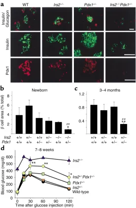
Irs2–/–Pdx+/–islets displayed few and small insulin-pos-itive βcell clusters, compared with the normal-sized islets in Irs2–/–, Pdx1+/–, and wild-type mice (Figure 3a). Pdx1 immunostaining was readily detected in wild-type islets but was consistently reduced in Pdx1+/–and in
Irs2–/– islets; and it was nearly undetected in
Irs2–/–Pdx1+/–islets, consistent with the severe hyper-glycemia that developed at birth (Figure 3a).
βCell content was quantified in the various mice by estimating the insulin-positive area in multiple
pan-β
and slightly decreased in Pdx1+/–, Pdx1+/–Irs2+/–, and
Irs2–/– mice; however, these small changes did not reach statistical significance (Figure 3b). By contrast, βcell content was significantly reduced in pancreatic slices from newborn Irs2–/–Pdx+/–mice (P < 0.05), con-sistent with the onset of fatal hyperglycemia 4 days after birth (Figure 3b). By 3–4 months of age, βcell content was similar in Irs2+/–, wild-type, and Pdx1+/– mice (Figure 3c); however, βcell content was reduced by 75% in compound heterozygous Irs2+/–Pdx1+/–mice, suggesting that both Irs2 and Pdx1 contribute to nor-mal islet function (Figure 3c). Consequently,
Irs2+/–Pdx1+/– mice displayed more severe glucose intolerance than did less affected littermates at 7–8 weeks of age (Figure 3d).
[image:5.576.58.334.50.471.2]To test whether Pdx1 promotes normal function of Irs2–/–βcells, we crossed Irs2+/–mice with Pdx1tgmice to increase the Pdx1gene dosage in the Irs2–/–mice. As pre-viously described, Pdx1tgmice have multiple copies of a
Figure 3
Islet morphology and quantification from Irs2+/–Pdx1+/– intercross mice and glucose tolerance tests of adult
Irs2+/–Pdx1+/–intercross mice. (a) Representative islet morphology from pancreas of newborn Irs2+/–Pdx1+/– intercross pups immunostained with antibodies against insulin (green) and glucagon (red, top panels), insulin (green, middle panels), and Pdx1 (red, bottom panels). Scale bars, 50 µm. (b) βCell area of newborn male and female progeny from Irs2+/–Pdx1+/– intercross pups (mean ± SEM relative to total pancreas area).
Irs2–/–Pdx1+/–pups have decreased βcell area compared with wild-type, Irs2–/–, or Pdx1+/–(*P < 0.05, ##P < 0.01). (c) Morphometric analysis of pancreas sections of 3- to 4-month-old male progeny from the Irs2+/–Pdx1+/– inter-cross. Results are reported as mean ± SEM of βcell area (percent relative to total pancreas area). At 3–4 months, Irs2+/–Pdx1+/–mice have decreased βcell area compared with wild-type (**P < 0.01) or Pdx1+/– (##P < 0.01). (d) Glucose tolerance tests of 7- to 8-week-old mice performed with 2 g D-glucose per kg body weight after a 15- to 16-hour fast (wild-type, n = 6;
Irs2+/–, n = 11; Pdx1+/–, n = 9; Irs2+/–Pdx1+/–, n = 11;
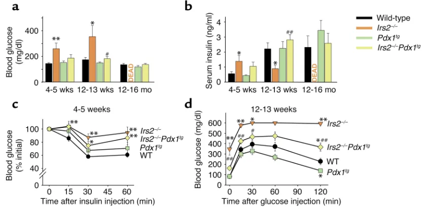
pression in the appropriate locations and developmen-tal stages (16). Random-fed glucose and insulin levels were identical in wild-type and Pdx1tgmice (Figure 4, a and b). Remarkably, Pdx1tgexpression prevented the hyperglycemia and relative hypoinsulinemia that devel-oped in Irs2–/–mice during the first 12–13 weeks of life (Figure 4, a and b). Moreover, Irs2–/–Pdx1tgmice survived with nearly normal glucose and insulin levels for up to 16 months; we now have Irs2–/–Pdx1tgmice that have sur-vived with mild glucose intolerance for up to 20 months. Based on these results, the Pdx1tgexpression restored βcell function in Irs2–/–mice and prevented the development of diabetes.
Although Pdx1tgexpression promoted βcell function in Irs2–/–mice, it had no significant effect on the sensi-tivity of wild-type mice to exogenous insulin injections and only slightly restored responsiveness of Irs2–/–Pdx1tg mice to insulin injections (Figure 4c). Glucose toler-ance at 12–13 weeks of age was significantly improved in Irs2–/–Pdx1tgmice compared with Irs2–/–mice; how-ever, tolerance was not entirely corrected, possibly owing to persistent peripheral insulin resistance (Fig-ure 4d). Irs2–/–mice develop various disorders, includ-ing impaired brain and retinal growth, and infertility in females, and these defects persisted in Irs2–/–Pdx1tg mice, suggesting that they are a result of dysregulated
Irs2signaling rather chronic hyperglycemia or diabetes (M. Schubert et al., manuscript submitted for publica-tion; and ref. 18).
Transgenic expression of Pdx1had profound effects on the histology of Irs2–/–islets. At 4–5 weeks of age, immunostaining of insulin and Pdx1 in Irs2–/–islets was
consistently reduced compared with that of wild-type mice (Figure 5). By contrast, insulin and Pdx1 immunostaining was consistently strong in Pdx1tgand
Irs2–/–Pdx1tgmice (Figure 5). Morphometric analysis of pancreatic sections at 4–5 weeks and at 3–4 months of age confirmed that βcell content (approximated by the mean cross-sectional β cell area) was significantly reduced in Irs2–/–mice (Table 1). Pdx1tgexpression did not change βcell content in wild-type pancreas sections, whereas it restored βcell content in the Irs2–/–Pdx1tg sec-tions (Table 1). Moreover, Pdx1tgexpression increased the βcell/αcell ratio in wild-type and Irs2–/–mice, espe-cially at 3–4 months of age (Table 1). Although βcell content in Irs2–/–sections decreased with age, the num-ber of islets detected was barely reduced, and Pdx1tg expression preserved or slightly increased the number of islets detected in the pancreas sections (Table 1). Thus, Pdx1tgexpression largely restores islet morpholo-gy in Irs2–/–mice.
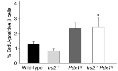
Since the size of βcells was barely affected by the absence of Irs2or the presence of Pdx1tg(Table 1), the increased βcell content in Pdx1tgmice pancreas might be related, at least in part, to increased mitogenesis. Mitogenesis was estimated by the incorporation dur-ing 6 hours of BrdU into βcells of 2- to 3-week-old male mice. BrdU labeling of Irs2–/–βcells was decreased about 40% compared with wild-type mice, consistent with decreased βcell content. By contrast, BrdU label-ing was increased twofold in both Pdx1tg and
[image:6.576.104.529.441.648.2]Irs2–/–Pdx1tg mice (Figure 6). Thus, Pdx1-mediated mitogenesis might contribute to increased βcell con-tent of Irs2–/–mice.
Figure 4
Characterization of male progeny of Irs2+/–Pdx1tgintercross. (aand b) Random-fed blood glucose (a) and serum insulin (b) of Irs2+/–Pdx1tg intercross mice at 4–5 weeks, 12–13 weeks, and 12–16 months. Results are mean ± SEM of six mice per genotype. *P < 0.05, Irs2–/–vs. wild-type; **P < 0.01, Irs2–/–vs. wild-type; #P < 0.05, Irs2–/–Pdx1tgvs. Irs2–/–; ##P < 0.01, Irs2–/–Pdx1tgvs. Irs2–/–. (c) Insulin tolerance test of 4- to 5-week-old fed mice performed with 0.75 U/kg human regular insulin on fed animals. Results are expressed as mean ± SEM of percent of ini-tial blood glucose value for at least eight animals per genotype. *P < 0.05 and **P < 0.01 vs. wild-type. (d) Glucose tolerance tests of 12- to 13-week-old mice performed with 2 g D-glucose per kg body weight after a 15- to 16-hour fast. Results reported as mean ± SEM for at least
Discussion
Our results show that the progressive loss of βcell func-tion and the onset of diabetes in Irs2–/–mice are associ-ated with decreased expression in pancreatic islets of the homeodomain transcription factor Pdx1(also called
Idx-1and Ipf1). PDX1 is critical for the development of the pancreas in mice and people, and pancreas agenesis occurs upon the complete disruption of PDX1(7, 19). Moreover, PDX1 is required in adult humans and mice to promote normal glucose sensing and insulin secretion (13, 15). Genetic defects in the PDX1gene occur in about 5% of people with type 2 diabetes. Inactivating muta-tions are associated with autosomal early-onset diabetes (MODY), whereas missense mutations predispose humans to late-onset type 2 diabetes (14, 20).
The functional association between Irs2 signaling and Pdx1 expression observed in mice might relate type 2 diabetes to autosomal forms of diabetes (MODY). Genetic reduction of Pdx1in Irs2–/–mice causes diabetes at birth owing to severe reduction of islet βcell content. By contrast, transgenic expression of Pdx1postpones βcell failure and prevents the progression of Irs2–/–mice to diabetes. Partial reduction of both genes in
Irs2+/–Pdx1+/–mice causes an intermediate phenotype of severe glucose intolerance. If insulin resistance in people includes an IRS2 component, diminished IRS2 signal-ing might reduce PDX1 function and eventually impair βcell compensation, resulting in type 2 diabetes. More-over, genetic defects in PDX1might exacerbate the effects of insulin resistance, even if the defects alone are
physi-PDX1 function might avoid diabetes even during chron-ic insulin resistance.
[image:7.576.58.354.53.338.2]While mutations in PDX1are associated with human diabetes, naturally occurring IRS2mutations are very rare (21, 22). However, physiological stress associated with acute injury, chronic obesity, inactivity, or aging promotes peripheral insulin resistance. Recent evi-dence suggests that serine phosphorylation or regu-lated degradation of Irs proteins might contribute to this insulin resistance (23, 24). If functional dysregu-lation of IRS2 extends to βcells, the ability to com-pensate for peripheral insulin resistance by increasing βcell mass and insulin secretion might be impaired and lead to glucose intolerance and diabetes. A related mechanism could contribute to type 1 diabetes, where proinflammatory cytokines produced by infiltrating
Figure 6
[image:7.576.323.512.571.686.2]BrdU incorporation analysis of male Irs2+/–Pdx1tgintercross progeny Figure 5
lymphocytes could inactivate Irs protein signaling and accelerate βcell death (25).
IRS-proteins coordinate multiple downstream signals through the PI 3-kinase and ERK1/2 cascades (26). Both pathways are regulated by Irs1 or Irs2, and both Irs pro-teins are expressed in βcells; however the Irs2 branch appears to be most important in mice (27). Irs proteins contain similar but not identical pleckstrin homology (PH) and phosphotyrosine-binding domains at their NH2-terminus that mediate coupling with activated membrane receptors. Cell-based experiments have not revealed a specificity that could explain the functional selectivity observed for Irs2 in βcells. However Irs2 inter-acts with the activated kinase regulatory loop of the insulin receptor, revealing a unique mechanism for cou-pling that is not shared with Irs1 (28). By contrast, the PH domain in Irs1 binds to PHIP, a protein that might contribute unique specificity (29).
The Irs2 → PI 3-kinase cascade controls many down-stream elements, including Akt/protein kinase B, p70s6k, BAD/Bcl2, and the Foxo subfamily of transcription fac-tors (30). Recently Akt1 and p70s6kwere shown to pro-mote βcell growth and function in mice, supporting the importance of signaling components regulated in β cells by the Irs2 →PI 3-kinase cascade (31, 32). One way that Akt promotes survival of cells is by phosphoryla-tion of BAD, which promotes the dissociaphosphoryla-tion of active Bcl2 (33). Since apoptosis of βcells might be crucial at several points during the onset of type 1 or type 2 dia-betes, BAD phosphorylation stimulated by the Irs2 → PI 3-kinase →Akt cascade might be an important sig-nal for βcell survival (34, 35). Previous reports show that Irs2–/–βcells undergo increased apoptosis at wean-ing, which might contribute to the early loss of βcell
mass (4); however, we did not investigate the effect of
Pdx1expression on apoptosis during weaning and did not detect apoptosis at later ages.
Pdx1 expression and function might be entirely inde-pendent of Irs2 signaling and reduced in Irs2–/–islets owing to βcell progressive failure. In this case, transgenic expression of Pdx1could overcome the negative effects of the Irs2knockout by strongly promoting the expression of genes that are essential for βcell function (10, 11). However, our results are consistent with the hypothesis that Irs2-signaling directly regulates expression and func-tion of Pdx1. Preliminary evidence suggests that Pdx1
[image:8.576.58.539.81.277.2] [image:8.576.326.494.559.699.2]expression in βcells is repressed through nuclear local-ization of the transcription factor Foxo1 (also called FKHR) (36). Foxo1 is ordinarily exported from the nucle-us and retained in the cytosol by Akt-mediated phos-phorylation (33). However, Foxo1 is largely nuclear in Table 1
Islet morphometry of Irs2+/–Pdx1tgintercross mice
Genotype βCell Area Islet Size Islet Density
(% total) (µm2 × 103/islet) (islets/µm2)
4–5 weeks 3–4 months 4–5 weeks 3–4 months 4–5 weeks 3–4 months
P P P P P P
Wild Type 0.51 ± 0.07 0.83 ± 0.19 3.1 ± 0.4 4.8 ± 0.5 1.3 ± 0.1 1.4 ± 0.1
Irs2–/– 0.26 ± 0.06 0.01 0.32 ± 0.04 0.01 2.5 ± 0.3 0.21 2.1 ± 0.2 < 0.01 0.8 ± 0.1 0.02 1.3 ± 0.1 0.22
Pdx1tg 0.60 ± 0.06 0.38 1.30 ± 0.25 0.28 2.8 ± 0.3 0.4 3.9 ± 0.3 0.11 1.8 ± 0.1 0.02 2.8 ± 0.4 < 0.01
Irs2–/–Pdx1tg 0.44 ± 0.05 0.04 0.80 ± 0.31 0.04 2.0 ± 0.2 0.2 4.6 ± 0.8 < 0.01 1.8 ± 0.3 0.01 1.4 ± 0.3 0.49
Genotype β/α-Cell Ratio βCell Size (µm2/islet)
4–5 weeks 3–4 months 2–3 weeks
P P P
Wild Type 4.9 ± 0.6 17.2 ± 2.1 108 ± 6
Irs2–/– 4.1 ± 1.0 0.53 5.6 ± 1.8 < 0.01 96 ± 7 0.29
Pdx1tg 8.5 ± 0.9 0.02 23.9 ± 2.2 0.07 107 ± 8 0.91
Irs2–/–Pdx1tg 8.3 ± 1.3 0.03 28.2 ± 10.8 0.08 118 ± 3 0.03
Morphometric βcell area analysis of male Irs2+/–Pdx1tgintercross progeny at 4–5 weeks and 3 months of age. βCell area was calculated from the mean cross-sectional βcell area of pancreas, reported as % of total pancreas area. Islet size was calculated from the mean cross-sectional βcell area per islet, reported as µm2×103/islet. Islet density was calculated from the mean islet density of pancreas, reported as islets/µm2. β/α-Cell ratio was calculated as the mean
cross-sectional β/α-cell area ratio. βCell size was calculated from the mean cross-sectional βcell size, reported as µm2/islet. Irs2–/–Pvalues against wild-type, Pdx1tg
Pvalues against wild-type, Irs2–/–Pdx1tgPvalues against Irs2–/–.
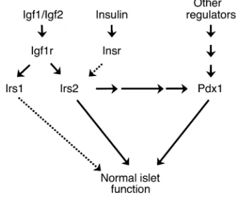
Figure 7
Irs2–/–βcells, consistent with the relative inactivity of the PI 3-kinase → Akt cascade (36). Interestingly, disruption of a single Foxo1allele reduces the level of nuclear Foxo1 in Irs2–/–βcells, which increases Pdx1expression and restores sufficient βcell function to normalize glucose homeostasis (36). More details about the integration of βcell transcription factors with the Irs2 branch of the insulin/Igf signaling cascade might reveal new strategies to promote βcell growth and function in both type 1 and type 2 diabetes (Figure 7).
PDX1is critical for the development of the pancreas, and pancreas agenesis occurs upon the complete dis-ruption of PDX1in mice and humans (7, 19). Although Pdx1 levels are low in Irs2–/–mice and lower in the
Irs2–/–Pdx1+/–mice, the exocrine pancreas develops nor-mally unless Pdx1is completely disrupted. During early development, Pdx1expression might be largely inde-pendent of Irs2 signaling, whereas after birth Pdx1
might be regulated by Irs2-dependent and -independ-ent mechanisms (Figure 7). The hepatocyte nuclear fac-tor Hnf3β promotes expression of Pdx1, so its reduced expression in Irs2–/–βcells might contribute to their failure (6, 37). However, genetic disruption of Hnf3βin βcells causes hyperinsulinemia at birth, possibly owing to the reduced expression of the ATP-sensitive potassi-um channel in βcells (38). So Hnf3βmight not be an essential element for Pdx1-mediated βcell function. The interaction of these related transcription factors and their integration with receptor signaling are an important direction of future work.
At the physiological level, the relation between Irs2 sig-naling and Pdx1 function is supported by the common development of diabetes between 10 and 15 weeks of age in Irs2–/–mice, or in mice lacking βcell Pdx1(2, 13). The progressive dysfunction of βcells specifically lack-ing Pdx1is related to the reduced expression of genes that promote glucose-sensitive insulin secretion, includ-ing proinsulin, glucose transporter-2, glucokinase, and components of the FGF receptor signaling system that control prohormone convertase 1/3 expression (10–12). Consistent with the relation between Irs2 and Pdx1, these gene products were always detected weakly by immunostaining Irs2–/– β cells, whereas they were strongly detected in Irs2–/–Pdx1tg β cells (data not shown). These preliminary results are consistent with the hypothesis that Pdx1mediates many of the effects of Irs2 on βcell function.
Irs2–/–Pdx1+/– and Irs2–/–Pdx1–/– mice were hyper-glycemic at birth, owing apparently to the near absence of βcells or the absence of a pancreas, respectively. However, Pdx1–/–mice were euglycemic until P4, sug-gesting that extrapancreatic ligands might regulate neonatal glucose homeostasis through Irs2. The importance of Irs2 at this stage is consistent with its role in hepatocytes to mediate the inhibitory effects of insulin on gluconeogenesis (3).
Based on our results we conclude that the Irs2 branch
effects of Irs2, including βcell growth and function are closely associated with Pdx1expression. Our results are consistent with a direct link between Irs2 and Pdx1 expression and function; however, additional experi-ments using cell-based strategies are necessary to prove this hypothesis. Our experiments provide a starting point to understand the relation between βcell failure and states of chronic insulin resistance. Moreover, dys-regulation of Pdx1by genetic or functional mechanisms might be one of the common links between early-onset (MODY) and ordinary type 2 diabetes.
Acknowledgments
This work was supported by grants from the NIH and the Howard Hughes Medical Institute funds. J.A. Kush-ner was supported by a Lawson Wilkins Pediatric Endocrine Society Fellowship, a National Institute of Diabetes and Digestive and Kidney Diseases training grant, and a Juvenile Diabetes Research Foundation Fel-lowship. J. Ye was supported by a Juvenile Diabetes Research Foundation Fellowship.
1. Yenush, L., and White, M.F. 1997. The IRS-signaling system during insulin and cytokine action. Bioessays.19:491–500.
2. Withers, D.J., et al. 1998. Disruption of IRS-2 causes type 2 diabetes in mice. Nature.391:900–904.
3. Previs, S.F., Withers, D.J., Ren, J.M., White, M.F., and Shulman, G.I. 2000. Contrasting effects of IRS-1 vs. IRS-2 gene disruption on carbohydrate and lipid metabolism in vivo. J. Biol. Chem.275:38990–38994. 4. Withers, D.J., et al. 1999. Irs-2 coordinates Igf-1 receptor-mediated
beta-cell development and peripheral insulin signalling. Nat. Genet.23:32–40. 5. Fajans, S.S., Bell, G.I., and Polonsky, K.S. 2001. Molecular mechanisms and clinical pathophysiology of maturity-onset diabetes of the young. N. Engl. J. Med.345:971–980.
6. Duncan, S.A., Navas, M.A., Dufort, D., Rossant, J., and Stoffel, M. 1998. Regulation of a transcription factor network required for differentiation and metabolism. Science.281:692–695.
7. Jonsson, J., Carlsson, L., Edlund, T., and Edlund, H. 1994. Insulin-pro-moter-factor 1 is required for pancreas development in mice. Nature. 371:606–609.
8. Offield, M.F., et al. 1996. PDX-1 is required for pancreatic outgrowth and differentiation of the rostral duodenum. Development.122:983–995. 9. Peers, B., Leonard, J., Sharma, S., Teitelman, G., and Montminy, M.R. 1994. Insulin expression in pancreatic islet cells relies on cooperative interactions between the helix loop helix factor E47 and the homeobox factor STF-1. Mol. Endocrinol.8:1798–1806.
10. Watada, H., et al. 1996. The human glucokinase gene beta-cell-type pro-moter: an essential role of insulin promoter factor 1/PDX-1 in its acti-vation in HIT-T15 cells. Diabetes.45:1478–1488.
11. Waeber, G., Thompson, N., Nicod, P., and Bonny, C. 1996. Transcrip-tional activation of the GLUT2 gene by the IPF-1/STF-1/IDX-1 home-obox factor. Mol. Endocrinol.10:1327–1334.
12. Hart, A.W., Baeza, N., Apelqvist, A., and Edlund, H. 2000. Attenuation of FGF signalling in mouse beta-cells leads to diabetes. Nature. 408:864–868.
13. Ahlgren, U., Jonsson, J., Jonsson, L., Simu, K., and Edlund, H. 1998.
β-cell-specific inactivation of the mouse Ipf1/Pdx1 gene results in loss of the beta-cell phenotype and maturity onset diabetes. Genes Dev. 12:1763–1768.
14. Hani, E.H., et al. 1999. Defective mutations in the insulin promoter fac-tor-1 (IPF-1) gene in late-onset type 2 diabetes mellitus. J. Clin. Invest. 104:R41–R48.
15. Thomas, M.K., et al. 2001. Development of diabetes mellitus in aging transgenic mice following suppression of pancreatic homeoprotein IDX-1. J. Clin. Invest.108:319–329. DOI:10.1172/JCI200112029. 16. Dutta, S., et al. 2001. PDX:PBX complexes are required for normal
pro-liferation of pancreatic cells during development. Proc. Natl. Acad. Sci. USA.98:1065–1070.
19. Stoffers, D.A., Zinkin, N.T., Stanojevic, V., Clarke, W.L., and Habener, J.F. 1997. Pancreatic agenesis attributable to a single nucleotide deletion in the human IPF1 gene coding sequence. Nat. Genet.15:106–110. 20. Stoffers, D.A., Ferrer, J., Clarke, W.L., and Habener, J.F. 1997. Early-onset
type-II diabetes mellitus (MODY4) linked to IPF1. Nat. Genet. 17:138–139.
21. Bernal, D., et al. 1998. Insulin receptor substrate-2 amino acid poly-morphisms are not associated with random type 2 diabetes among Cau-casians. Diabetes.47:976–979.
22. Kalidas, K., et al. 1998. Mapping of the human insulin receptor sub-strate-2 gene, identification of a linked polymorphic marker and link-age analysis in families with type 2 diabetes: no evidence for a major sus-ceptibility role. Diabetologia.41:1389–1391.
23. Rui, L., et al. 2001. Insulin/IGF-1 and TNF-α stimulate phosphorylation of IRS-1 at inhibitory SER307via distinct pathways. J. Clin. Invest.
107:181–189.
24. Hotamisligil, G.S., and Spiegelman, B.M. 1999. TNFα: a key component of the obesity-diabetes link. Diabetes.43:1271–1278.
25. Mathis, D., Vence, L., and Benoist, C. 2001. β-Cell death during progres-sion to diabetes. Nature.414:792–798.
26. Uchida, T., Myers, M.G., Jr., and White, M.F. 2000. IRS-4 mediates acti-vation of PKB/Akt during insulin stimulation without inhibition of apoptosis. Mol. Cell. Biol.2200:126–138.
27. Burks, D.J., and White, M.F. 2001. IRS proteins and beta-cell function. Diabetes.50(Suppl. 1):S140–S145.
28. Sawka-Verhelle, D., Tartare-Deckert, S., White, M.F., and Van Obberghen, E. 1996. Insulin receptor substrate-2 binds to the insulin receptor through its phopshotyrosine-binding domain and through a
newly identified domain comprising amino acids 591-786. J. Biol. Chem. 271:5980–5983.
29. Farhang-Fallah, J., Yin, X., Trentin, G., Cheng, A.M., and Rozakis-Adcock, M. 2000. Cloning and characterization of PHIP, a novel insulin receptor substrate-1 pleckstrin homology domain interacting protein. J. Biol. Chem. 275:40492–40497.
30. Saltiel, A.R., and Kahn, C.R. 2001. Insulin signalling and the regulation of glucose and lipid metabolism. Nature.414:799–806.
31. Tuttle, R.L., et al. 2001. Regulation of pancreatic beta-cell growth and sur-vival by the serine/threonine protein kinase Akt1/PKBalpha. Nat. Med. 7:1133–1137.
32. Pende, M., et al. 2000. Hypoinsulinaemia, glucose intolerance and dimin-ished beta-cell size in S6K1-deficient mice. Nature.408:994–997. 33. Brazil, D.P., and Hemmings, B.A. 2001. Ten years of protein kinase B
sig-nalling: a hard Akt to follow. Trends Biochem. Sci.26:657–664. 34. Brunet, A., et al. 1999. Akt promotes cell survival by phosphorylating and
inhibiting a Forkhead transcription factor. Cell.96:857–868. 35. Rabinovitch, A., et al. 1999. Transfection of human pancreatic islets with
an anti-apoptotic gene (bcl-2) protects beta-cells from cytokine-induced destruction. Diabetes.48:1223–1229.
36. Kitamura, T., et al. 2001. The transcription factor FKHR promotes beta cell survival in IRS-2 knockout mice. International Symposium on Insulin Receptors and Insulin Action. Geneva, Switzerland. 85 (Abstr.).
37. Gerrish, K., et al. 2000. Pancreatic beta cell-specific transcription of the pdx-1 gene. The role of conserved upstream control regions and their hepatic nuclear factor 3beta sites. J. Biol. Chem.275:3485–3492. 38. Sund, N.J., et al. 2001. Tissue-specific deletion of Foxa2 in pancreatic beta