0095-1137/96/$04.0010
Copyrightq1996, American Society for Microbiology
Improved Rapid Identification of Mycobacteria by Combining
Solid-Phase Extraction with High-Performance Liquid
Chromatography Analysis of BACTEC Cultures
PAUL S. DUFFEY,* LINDA S. GUTHERTZ,ANDGRIFFITH C. EVANS
California Department of Health Services, Berkeley, California
Received 25 January 1996/Returned for modification 2 May 1996/Accepted 15 May 1996
Identification of mycobacteria from BACTEC 12B cultures is achieved in 7 to 21 days by reverse-phase
high-performance liquid chromatography (HPLC) using a UV spectrophotometer to detect nonpolar p
-bro-mophenylacyl mycolic acid derivatives. However, cultures grown in BACTEC and other liquid media seldom contain sufficient mycolic acids to permit reliable identification under usual HPLC assay conditions, so the sample size must be increased. Unfortunately, samples prepared from cultures in liquid media such as BACTEC cultures also contain large amounts of extraneous polar and strongly nonpolar contaminants that interfere with the analysis and hasten deterioration of the HPLC column. The contaminants were removed from 10 samples simultaneously by solid-phase extraction (SPE), i.e., by passing the crude suspension
containing the mycolic acid derivatives into disposable 500-mg tC18SPE columns in place of the usual final
filtration step used to prepare specimens for HPLC. Fifteen milliliters of 20% (vol/vol) dichloromethane in methanol was passed through the columns (<3 ml/min) to wash through the undesired contaminants and bind the mycolic acid derivatives. The mycolates were quantitatively eluted in 3 ml of dichloromethane for analysis by HPLC. Treating a panel of 31 strains of frequently isolated mycobacteria by SPE reduced the content of contaminants by 89.3 to 99.9% without altering the chromatographic patterns compared with the same strains grown on conventional solid media and processed without SPE. Peak heights of mycolates prepared from
BACTEC cultures were increased from <6 to >25 absorbance milliunits with SPE, sufficient for reliable
interpretation by visual inspection of chromatograms obtained with a UV detector. Also, removal of the contaminants improved column longevity.
Mycobacteria contain unique distributions of 2-alkyl, 3-hy-droxy fatty acids (mycolic acids) with chain lengths of 60 car-bons or more that may be used for species identification by high-performance liquid chromatography (HPLC) (3, 7, 11). After saponification of samples from solid media and labeling by derivatization with p-bromophenylacyl bromide (3, 7), the nonpolar mycolates are detected in submicrogram quantities with a UV detector following separation by reverse-phase HPLC or, with a different label, are detected in subnanogram quantities by HPLC using a sufficiently sensitive fluorescence detector (8).
In addition to nonpolar mycolic acid esters, the samples contain undefined polar compounds that are detected after they pass through the HPLC column during initial sample loading (and, so, do not interfere with the analysis) and strongly nonpolar compounds that bind permanently to the HPLC column, ultimately contributing to irreversible column damage (12). There also are substantial quantities of weakly polar, short-chain fatty acids, normally present in uninoculated media, that are initially bound to the column but are eluted approximately during the first minute of the assay. The pres-ence of these large quantities of medium-derived fatty acids, which can exceed the concentration of diagnostic mycolates more than 1,000-fold, may mask the presence of diagnostic mycolic acids obtained from the mycobacteria. Concentration of samples containing only minimal quantities of mycolic acids, as occurs in liquid media, may result in a viscous preparation,
due to the presence of both the retained and the early-eluted compounds, that is difficult to analyze.
In our experience, isolation of sufficient mycolic acids from liquid media to permit reliable identification has been prob-lematic. Moreover, we have found that analysis of concen-trated specimens from cultures grown in liquid media usually leads to rapid HPLC column deterioration, rendering this ap-proach impractical. Recently, Cage (3) showed that HPLC may be used to identify mycobacteria directly from 93% (117 of 126) of BACTEC 12B cultures after addition of the Middle-brook oleic acid-albumin-dextrose-catalase (OADC) enrich-ment solution to cultures with a growth index of$50, followed by reincubation for 5 to 7 days. This technique permitted reporting mycobacterial identifications 7 to 14 days earlier than otherwise possible. However, 20-fold-larger samples that contain proportionately more contaminating material must be used. Consequently, we have found, using this method, that relatively few samples may be analyzed before the HPLC col-umn must be replaced because of the presence of large amounts of contaminants. Similarly, Jost et al. (8) used HPLC with a fluorescence detector, thought to be 100- to 200-fold more sensitive than the usual UV detector. Identification of 99% of Mycobacterium tuberculosis isolates from clinical spec-imens cultured in BACTEC 12B medium was achieved in an average of 10.2 days postinoculation without OADC enrich-ment, and 94.3% of M. avium isolates were identified in an average of 7.4 days postinoculation. However, although Jost et al. used a more sensitive fluorescence detector that permitted analysis of more dilute specimens than can be analyzed with a UV spectrophotometer, considerable extrinsic material re-mained present. The presence of these contaminants may be expected to shorten column life and also may interfere with the
* Corresponding author. Mailing address: Microbial Diseases Lab-oratory, California Department of Health Services, 2151 Berkeley Way, Berkeley, CA 94704. Phone: (510) 540-2242.
1939
on May 15, 2020 by guest
http://jcm.asm.org/
identification. In our laboratory, analysis of only 20 to 30 sam-ples prepared from BACTEC cultures resulted in irreversible column damage, and it was necessary to limit integration of the chromatogram to the period after the early-eluted contami-nants had been eluted in order to visualize the diagnostic mycolates.
Here, we show the use of solid-phase extraction (SPE) (10) to remove undesirable contaminants from extracts of BACTEC 12B cultures so that mycobacteria may routinely be identified by HPLC from liquid media at the higher concen-trations necessary without attendant column damage; i.e., us-ing SPE results in obtainus-ing the expected usable column life of 1,000 or more injections. Substitution of SPE in place of the final filtration step that we normally use to prepare specimens for HPLC analysis (7) results in only a minimal increase in cost, since disposable SPE columns and the filters they replace are approximately equivalent in price, so using SPE is cost-effective.
MATERIALS AND METHODS
Cultures.Thirty-one cultures of mycobacteria consisting of American Type Culture Collection (ATCC) and Trudeau Mycobacteria Culture Collection (TMC) strains and clinical isolates (MDL) included members of the M.
tuber-culosis group and Runyon groups I to IV (11). The strains assayed included M. africanum, M. bovis BCG, M. microti, M. tuberculosis, M. kansasii, M. marinum, M. gordonae, M. flavescens, M. scrofulaceum, M. szulgai, M. avium, M. gastri, M. intracellulare, M. nonchromogenicum, M. simiae, M. terrae, M. chelonae, M. for-tuitum, M. peregrinum, M. phlei, and M. smegmatis. The cultures were grown on
Lowenstein-Jensen (LJ) slants (catalog no. C21; Hardy Diagnostics, Santa Maria, Calif.) at 35 to 378C until visible colonies appeared (3, 7). To inoculate the BACTEC media, growth from an LJ slant was suspended in sterile water with sterile glass beads, mixed vigorously with a vortex mixer (ca. 1 min), and then adjusted to a density equivalent to ca. 1.53108
bacteria per ml (McFarland nephelometer, 0.5). In order to approximate the inoculum that may be expected from a sputum specimen, 0.1 ml of the cell suspension diluted 1022
(approxi-mately 105
cells) or 1024
(approximately 103
cells) in sterile water was used to inoculate sets of BACTEC 12B bottles (catalog no. 4402004; Becton Dickinson, Sparks, Md.). The cultures were then incubated at 35 to 378C by using the BACTEC TB-460 instrument, and the instrument was monitored daily until the desired growth index was achieved ($50 or$999). Validated (2, 6) OADC solution (catalog no. 0722-73-9; Difco, Detroit, Mich.) was added to pairs of cultures, and incubation was continued for an additional 4 to 7 days.
Saponification and derivatization of mycolic acids.Saponification and deri-vatization of LJ cultures was done by using two full inoculating loops of bacterial growth according to the method of Butler et al. (2), as previously modified (7). The modification we used differed from the Butler et al. method by employing heating at 1008C rather than the 858C temperature recommended and also substituted filtration through a 13-mm, 0.45-mm-pore-size nylon-66 filter (catalog no. 38-154; Ranin, Woburn, Mass.) in place of the acid-methanol clarification step used by Butler et al. Pairs of identical BACTEC 12B cultures were pro-cessed essentially as recommended by Cage (3); i.e., in a biological safety cabinet, the contents of the BACTEC cultures were transferred to borosilicate screw-cap culture tubes (13 by 100 mm), which were placed into 50-ml disposable polyeth-ylene conical centrifuge tubes, and centrifuged by using sealed safety centrifuge carriers at 3,0003g for 30 min. After the supernatant was removed, the pellet
was saponified, and prepared for HPLC by the previously described method (3, 7). Blanks consisting of uninoculated LJ or BACTEC 12B medium also were treated in the same way. The derivatives either were suspended in 100ml of dichloromethane containing approximately 2mg of synthetic mycolic acid (cat-alog no. R-50; Ribi Immunochemicals, Hamilton Mont.) as an internal standard or were subjected to the SPE procedure described below in place of the final filtration step normally used (7).
SPE.Millipore (Waters) Sep-Pak 500-mg tC18cartridges (3-ml-capacity
vac-uum pack) (part no. 36815; Millipore, Milford, Mass.) (SPE columns) were first wetted with 5 ml of 80:20 (vol/vol) methanol-dichloromethane. This solvent mixture was chosen because it represents the mixture that exists in the chroma-tography procedure at 1 min postinjection, when most of the contaminants have been washed through the column but the diagnostic mycolic acids have not yet begun to be eluted (3, 7). Up to 10 derivatives simultaneously were each resus-pended in 5 ml of the same solvent mixture and then passed into individual SPE columns at#3 ml/min by using a vacuum manifold. An additional 15 ml of the solvent was passed through each SPE column at the same flow rate. For routine use, the effluents, containing most of the contaminants, were discarded. How-ever, for the initial SPE study, they were evaporated to dryness, resuspended in 40ml of dichloromethane, and analyzed by HPLC. The mycolates, which adhere reversibly to the SPE column, were recovered by passing 3 ml of dichlorometh-ane through each SPE column and collecting the eluates, which were then
evaporated to dryness. The clear, colorless to pale-yellow material was then resuspended in 40ml of dichloromethane containing approximately 0.8mg of the synthetic mycolic acid standard.
HPLC analysis.HPLC was performed with a Hewlett-Packard model 1050 gradient chromatograph equipped with version 2.05 H-P Chemstation software, a variable-wavelength UV detector set at 260 nm, and a Beckman-Altex XL-ODS C18cartridge column (7.5 cm by 4.5 mm) with guard (part no. 238370;
Beckman Instruments, Fullerton, Calif.). Specimens prepared from LJ slants were assayed in 2.5-ml samples. Specimens prepared from BACTEC 12B cultures were assayed in 20-ml samples. A curvilinear gradient beginning with 98:2 (vol/ vol) methanol-dichloromethane and ending with 35:65 (vol/vol) methanol-di-chloromethane over 10 min was used as previously described (7). Chromato-graphic patterns were interpreted by visual comparison with a library of patterns for known mycobacteria grown on LJ medium. Chromatograms were interpreted only if there was at least one mycolic acid in the 3- to 9-min postinjection period with a peak height of 25 absorbance milliunits (mAU) or higher. Although perhaps overly stringent, this requirement ensured that the baseline was flat, thereby aiding visual interpretation of the chromatogram.
RESULTS
Growth conditions. Preliminary studies (data not shown)
defined growth conditions that yielded adequate mycolic acids from BACTEC 12B cultures to permit HPLC identification using the signal obtained from a UV detector. Cultures of mycobacteria grown in BACTEC 12B medium incubated to a growth index of$999, but not otherwise treated, did not reli-ably yield sufficient mycolic acids. However, addition of 0.5 ml of OADC solution after incubation of the BACTEC cultures to a growth index of$50, followed by reincubation for 4 to 7 days, did yield adequate amounts of mycolic acids to permit reliable HPLC identification of the mycobacteria in 7 to 21 days, con-sistent with results reported previously by Cage (3).
SPE study.Growth from a single LJ slant culture was
pre-pared for HPLC as usual to act as a positive control. The combined growth from pairs of BACTEC 12B cultures also was prepared for HPLC, with extraction and derivatization using our usual procedure (7). Prior to the final filtration procedure, the sample was split, and half was filtered as usual and then assayed by HPLC without further treatment. The other half was subjected to SPE in place of the usual filtration step prior to HPLC analysis. Experiments were repeated once with each of the 31 strains tested and showed equivalent re-sults. Figure 1 shows representative results obtained with M. tuberculosis ATCC 9360, which is characteristic of mycobacte-ria producing single-cluster mycolic acid HPLC patterns. Panel a shows the chromatogram obtained from an LJ derivative under normal assay conditions (2.5-ml portion of a 100-ml sam-ple; no SPE used). When the same strain of M. tuberculosis grown in BACTEC 12B medium was assayed (panel b) by using a 20-fold-larger sample (20ml of a 40-ml sample), insuf-ficient mycolic acids were detected to permit identification of the culture by HPLC. Panel c shows the chromatogram of the SPE column eluate obtained from the BACTEC 12B culture shown in panel b. When the eluate obtained from the SPE column was chromatographed under assay conditions identical to those for panel b (20 ml of a 40-ml sample), the early contaminants were reduced to a single peak of 140 mAU (i.e., less than 4% of the material in panel b) while the diagnostic mycolic acids were concentrated to yield a chromatogram us-able for identification (peak heights of up to 150 mAU easily exceeded the 25 mAU required). The distribution of peaks in the BACTEC sample subjected to SPE is equivalent to that for LJ medium without SPE. Panel d shows the chromatogram obtained with the effluent obtained after SPE. Essentially all of the early-eluted material was contained in this effluent, while almost none of the diagnostic mycolic acids were present. Pan-els e and f show the patterns seen with uninoculated LJ and BACTEC 12B media. Both contain large amounts of
on May 15, 2020 by guest
http://jcm.asm.org/
eluted fatty acids equivalent to the early-eluted peaks in the mycolic acid derivatives prepared from inoculated media. Fig-ure 2 shows representative results obtained with M. simiae ATCC 25275, which is characteristic of mycobacteria that pro-duce more complex, multicluster mycolic acid HPLC patterns. Panel a shows the chromatogram obtained for a culture grown on LJ medium without SPE. Panel b shows the chromatogram for the eluate containing mycolic acids obtained from a BACTEC culture after SPE. Again, the peak heights, which reach 160 mAU, easily exceed the 25-mAU signal required for chromatogram interpretation, and the mycolic acid pattern is equivalent to the mycolic acid pattern in panel a.
Comparison of contaminants present with and without SPE
by species.For each of the 31 strains of mycobacteria described
above, chromatograms obtained with the derivatives prepared from the LJ culture without SPE were initially compared with
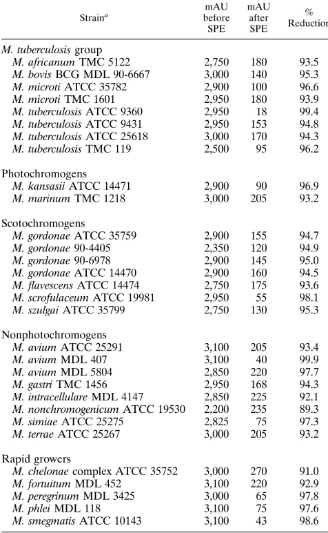
the chromatograms of eluates obtained after SPE of the ex-tracts prepared from the BACTEC 12B culture. In each in-stance (data not shown) the numbers and relative proportions of the diagnostic mycolic acids were essentially identical. Ad-ditionally, SPE resulted in mycolic acid chromatograms con-taining at least one peak with a peak height of$25 mAU that were more than adequate for reliable identification by HPLC. Table 1 shows the percent reduction in contaminants achieved by comparing the contaminants in BACTEC cultures with and without SPE of the 31 strains of mycobacteria assayed. Con-taminants were reduced by 89.3 to 99.9%.
Use of SPE in routine BACTEC culture examination.
[image:3.612.153.467.68.507.2]Eighty-nine cultures with a growth index of$50 that contained acid-fast bacilli (Ziehl-Neelsen stain) were reincubated after addition of OADC as above and then assayed by HPLC using SPE. After subculture to LJ medium, the cultures also were
FIG. 1. Chromatograms of M. tuberculosis ATCC 9360. (a) Growth from LJ medium extracted without SPE; (b) growth from BACTEC 12B medium extracted without SPE; (c) eluate containing mycolic acids after SPE of the extract from the same BACTEC 12B medium as in panel b; (d) effluent containing low-molecular-weight polar contaminants after SPE of the extract from the same BACTEC 12B medium as in panel b; (e) extract without SPE from uninoculated LJ; (f) extract without SPE from uninoculated BACTEC 12B medium. ISTD, internal standard (Ribi R-50).
on May 15, 2020 by guest
http://jcm.asm.org/
identified independently by personnel in the Diagnostic Myco-bacteriology Unit of this laboratory using a standard battery of assay procedures which included staining, morphological, bio-chemical, and growth studies as outlined by Kent and Kubica (9) and DNA probes to confirm M. tuberculosis complex and M. avium complex identifications. Three of the 89 cultures contained only low-molecular-weight mycolic acids that were not characteristic of mycobacteria. These three cultures were later identified as probable Nocardia spp. Of the 86 remaining cultures, two contained no detectable mycolic acids and one contained a mycolic acid pattern resembling the pattern char-acteristic of the M. avium complex but had peak heights that did not exceed 1.6 mAU. Thus, only 3 cultures (of 86) did not produce a sufficient quantity of mycolic acids (QNS) to meet the reporting criteria adopted for this study. Two of these QNS cultures were later identified by conventional methods as members of the M. avium complex, and one was identified as M. xenopi. The remaining 83 of 86 (96.5%) cultures were iden-tified as M. tuberculosis complex: M. bovis (but not M. bovis BCG) (42 cultures), M. bovis BCG (1 culture), M. avium com-plex (28 cultures), M. gordonae (4 cultures), M. terrae comcom-plex (1 culture), M. fortuitum complex (4 cultures), M. mucogeni-cum (1 culture), M. szulgai (1 culture), and M. genavense (1 culture). In each instance, the HPLC identification agreed with the identification independently achieved by both the conven-tional method and the DNA probe method. In the case of M. genavense, identification routinely is based on growth charac-teristics and the HPLC pattern (1, 5). One culture identified as M. tuberculosis complex by HPLC was found to be a mixed culture containing both M. tuberculosis and M. gordonae when analyzed by conventional methods.
DISCUSSION
[image:4.612.156.461.73.228.2]The results show effective use of SPE to improve HPLC identification of mycobacteria in BACTEC cultures. The SPE process removes contaminants from crude p-bromophenylacyl derivatives of mycolic acids obtained directly from the liquid culture medium. By removing compounds not needed for iden-tification of mycobacteria, larger, more concentrated samples may be used to improve detection of mycolic acids, and signif-icant extension of HPLC column life may be expected. Reduc-ing the contaminants permitted routine HPLC assay of myco-lates from cultures grown in liquid media such as BACTEC 12B without shortening column longevity and use of autoscal-ing without limitautoscal-ing data analysis to the later portions of the
FIG. 2. Chromatogram of M. simiae ATCC 25275. (a) Growth from LJ medium extracted without SPE; (b) eluate containing mycolic acids after SPE of the extract from BACTEC 12B medium. ISTD, internal standard (Ribi R-250).
TABLE 1. Reduction in early-eluted, polar compounds by SPE
Straina
mAU before SPE
mAU after SPE
% Reduction
M. tuberculosis group
M. africanum TMC 5122 2,750 180 93.5 M. bovis BCG MDL 90-6667 3,000 140 95.3 M. microti ATCC 35782 2,900 100 96.6 M. microti TMC 1601 2,950 180 93.9 M. tuberculosis ATCC 9360 2,950 18 99.4 M. tuberculosis ATCC 9431 2,950 153 94.8 M. tuberculosis ATCC 25618 3,000 170 94.3 M. tuberculosis TMC 119 2,500 95 96.2
Photochromogens
M. kansasii ATCC 14471 2,900 90 96.9 M. marinum TMC 1218 3,000 205 93.2
Scotochromogens
M. gordonae ATCC 35759 2,900 155 94.7 M. gordonae 90-4405 2,350 120 94.9 M. gordonae 90-6978 2,900 145 95.0 M. gordonae ATCC 14470 2,900 160 94.5 M. flavescens ATCC 14474 2,750 175 93.6 M. scrofulaceum ATCC 19981 2,950 55 98.1 M. szulgai ATCC 35799 2,750 130 95.3
Nonphotochromogens
M. avium ATCC 25291 3,100 205 93.4 M. avium MDL 407 3,100 40 99.9 M. avium MDL 5804 2,850 220 97.7 M. gastri TMC 1456 2,950 168 94.3 M. intracellulare MDL 4147 2,850 225 92.1 M. nonchromogenicum ATCC 19530 2,200 235 89.3 M. simiae ATCC 25275 2,825 75 97.3 M. terrae ATCC 25267 3,000 205 93.2
Rapid growers
M. chelonae complex ATCC 35752 3,000 270 91.0 M. fortuitum MDL 452 3,100 220 92.9 M. peregrinum MDL 3425 3,000 65 97.8 M. phlei MDL 118 3,100 75 97.6 M. smegmatis ATCC 10143 3,100 43 98.6
a
Photochromogens, scotochromogens, nonphotochromogens, and rapid grow-ers are Runyon groups I to IV, respectively.
on May 15, 2020 by guest
http://jcm.asm.org/
[image:4.612.57.295.324.710.2]chromatogram where only mycolic acids are known to be eluted. Availability of the full chromatogram permits visualiza-tion of early-eluted mycolic acid peaks that are sometimes useful in mycobacterial identification and are especially impor-tant in distinguishing nonmycobacterial mycolic acid patterns but are normally not available for analysis when only the later portions of the chromatogram are used. Removing the con-taminants did not alter the relative concentrations of the my-colic acids compared with the same species grown on standard (LJ) medium that were analyzed without SPE. These results also confirm the findings of Cage (3), showing that adding OADC enrichment to BACTEC 12B cultures, followed by reincubation, permitted rapid HPLC identification of myco-bacteria directly from the BACTEC culture with a UV detec-tor. Employing a sensitive fluorescence detector may permit shortened incubation of the BACTEC and other liquid cul-tures, further improving the utility of HPLC as a rapid method for identification of mycobacteria. We also have successfully used SPE with cultures grown on LJ slants, modified Middle-brook 7H10 agar plates, MiddleMiddle-brook 7H9 broth, Dubos Tween broth, and BACTEC 13A cultures with results equiva-lent to those presented here (data not shown). Although we have not used SPE with recently available rapid culture media such as MIGITS (Becton Dickinson) and ESP (Difco), the latter media are similar in composition to Middlebrook 7H9 broth, so SPE should be usable to permit direct HPLC iden-tification from these liquid culture media as well.
REFERENCES
1. Bo¨ttger, E. C., B. Hirschel, and M. B. Coyle.1993. Mycobacterium genavense sp. nov. Int. J. Syst. Bacteriol. 43:841–843.
2. Butler, W. R., K. C. Jost, and J. O. Kilburn. 1991. Identification of myco-bacteria by high-performance liquid chromatography. J. Clin. Microbiol. 29:2468–2472.
3. Cage, G. D. 1994. Direct identification of Mycobacterium species in BACTEC 7H12B medium by use of high-performance liquid chromatography. J. Clin. Microbiol. 32:521–524.
4. Coyle, M. B., L. C. Carlson, C. K. Wallis, R. B. Leonard, V. A. Raisis, J. O. Kilburn, M. Samadpour, and E. C. Bo¨ttger.1992. Laboratory aspects of “Mycobacterium genavense,” a proposed species isolated from AIDS patients. J. Clin. Microbiol. 30:3206–3212.
5. Durst, H. D., M. Milano, E. J. Kitka, Jr., S. A. Connely, and E. Grushka. 1975. Phenylacyl esters of fatty acids via crown ether catalysis for enhanced ultraviolet detection in liquid chromatography. Anal. Chem. 47:1797–1801. 6. Guthertz, L. S., M. E. Griffith, E. G. Ford, J. M. Janda, and T. F. Midura. 1988. Quality control of individual components used in Middlebrook 7H10 medium for mycobacterial susceptibility testing. J. Clin. Microbiol. 26:2338– 2342.
7. Guthertz, L. S., S. D. Lim, Y. Jang, and P. S. Duffey. 1993. Curvilinear-gradient high-performance liquid chromatography for identification of my-cobacteria. J. Clin. Microbiol. 31:1876–1881.
8. Jost, K., Jr., D. F. Dunbar, S. S. Barth, V. L. Headley, and L. B. Elliott. 1995. Identification of Mycobacterium tuberculosis and M. avium complex directly from sputum and BACTEC 12B sputum cultures by high performance liquid chromatography with fluorescence detection and computer-driven pattern recognition models. J. Clin. Microbiol. 33:1270–1277.
9. Kent, P. T., and G. P. Kubica. 1985. Public health mycobacteriology: a guide for the level III laboratory. Publication 86-8230. U.S. Department of Health and Human Services, Washington, D.C.
10. Millipore Corporation. 1992. Waters Sep-Pak cartridge care and use manual. Millipore Corporation, Milford, Mass.
11. Minnikin, D. E., S. M. Minnikin, J. H. Parlett, M. Goodfellow, and M. Magnussen.1984. Mycolic acid patterns of some species of Mycobacterium. Arch. Microbiol. 139:225–231.
12. Schneider, W. 1993. Extend the life of your column, p. 5. In G. Cage (ed.), HPLC users group newsletter, vol. 2. Arizona State Laboratory, Phoenix, Ariz.