Allergen-induced increases in IL-5 receptor
alpha-subunit expression on bone
marrow-derived CD34+ cells from asthmatic subjects. A
novel marker of progenitor cell commitment
towards eosinophilic differentiation.
R Sehmi, … , P M O'Byrne, J A Denburg
J Clin Invest.
1997;
100(10)
:2466-2475.
https://doi.org/10.1172/JCI119789
.
We have proposed previously that hemopoietic myeloid progenitors contribute to the
ongoing recruitment of proinflammatory cells, namely eosinophils, to sites of allergen
challenge in allergic diseases such as asthma. In this study, we investigated the
involvement of bone marrow-derived progenitors in the development of allergen-induced
pulmonary inflammation in mild asthmatic subjects. By flow cytometry, we enumerated the
level of expression of CD34, a hemopoietic progenitor cell marker, on bone marrow
aspirates taken before and 24 h after allergen challenge. In addition, the coexpression of the
alpha-subunits of IL-3 receptor (IL-3R) and IL-5 receptor (IL-5R) on CD34+ cells was
investigated. After allergen-challenge, although no significant change in total BM CD34+
cell numbers was observed, a significant increase in the proportion of CD34+ cells
expressing IL-5R alpha, but not IL-3R alpha, was detected in the 24-h post-allergen,
compared with the pre-allergen bone marrow. This was associated with a significant blood
and sputum eosinophilia and increased methacholine airway responsiveness, 24 h
post-allergen. Using simultaneous in situ hybridization and immunocytochemistry, we
colocalized the expression of messenger RNA for membrane-bound IL-5R alpha to CD34+
cells. In summary, our data suggest that increased expression of IL-5R alpha on CD34+
cells favors eosinophilopoiesis and may thus contribute to the subsequent development of
blood and tissue eosinophilia, a hallmark of allergic inflammation.
Research Article
Find the latest version:
J. Clin. Invest.
© The American Society for Clinical Investigation, Inc. 0021-9738/97/11/2466/10 $2.00
Volume 100, Number 10, November 1997, 2466–2475 http://www.jci.org
Allergen-induced Increases in IL-5 Receptor
a
-subunit Expression on Bone
Marrow–derived CD34
1Cells from Asthmatic Subjects
A Novel Marker of Progenitor Cell Commitment towards Eosinophilic Differentiation
Roma Sehmi,* Lorna J. Wood,* Rick Watson,* Ronan Foley,* Qutayba Hamid,‡ Paul M. O’Byrne,* and Judah A. Denburg* *Asthma Research Group, Division of Clinical Immunology and Allergy, Department of Medicine, McMaster University, Hamilton, Ontario L8N 3Z5, Canada; and ‡Department of Medicine & Pathology, Meakins-Christie Laboratories, McGill University, Montreal,
Canada 3HX 2P2
Abstract
We have proposed previously that hemopoietic myeloid progenitors contribute to the ongoing recruitment of proin-flammatory cells, namely eosinophils, to sites of allergen challenge in allergic diseases such as asthma. In this study, we investigated the involvement of bone marrow–derived progenitors in the development of allergen-induced pulmo-nary inflammation in mild asthmatic subjects. By flow cy-tometry, we enumerated the level of expression of CD34, a hemopoietic progenitor cell marker, on bone marrow aspi-rates taken before and 24 h after allergen challenge. In ad-dition, the coexpression of the a-subunits of IL-3 receptor (IL-3R) and IL-5 receptor (IL-5R) on CD341 cells was in-vestigated. After allergen-challenge, although no significant change in total BM CD341 cell numbers was observed, a significant increase in the proportion of CD341 cells ex-pressing IL-5Ra, but not IL-3Ra, was detected in the 24-h post-allergen, compared with the pre-allergen bone marrow. This was associated with a significant blood and sputum eosinophilia and increased methacholine airway responsive-ness, 24 h post-allergen. Using simultaneous in situ hybrid-ization and immunocytochemistry, we colocalized the ex-pression of messenger RNA for membrane-bound IL-5Ra to CD341 cells. In summary, our data suggest that increased expression of IL-5Ra on CD341 cells favors eosinophilo-poiesis and may thus contribute to the subsequent develop-ment of blood and tissue eosinophilia, a hallmark of allergic inflammation. (J. Clin. Invest. 1997. 100:2466–2475.) Key
words: CD34 • IL-5 receptor • hemopoiesis • asthma
Introduction
Asthma is a complex disorder characterized by reversible air-flow obstruction, airway hyperresponsiveness, and infiltration of the airways by activated inflammatory cells. Increasing evi-dence suggests that the clinical severity of asthma parallels the degree of eosinophilic inflammation and, as such, highlights a
role for eosinophils in the pathology of asthma (1–4). How-ever, the mechanism(s) underlying the development and per-sistence of tissue eosinophilia in allergic inflammation remains unresolved.
We have investigated the hypothesis that activation of spe-cific hemopoietic pathways in the bone marrow may contrib-ute to the allergic diathesis through increased production and traffic of lineage-committed inflammatory progenitor cells such as those for eosinophils. This is supported by findings (in semi-solid liquid cultures) that circulating progenitors (colony-form-ing units, CFU)1 for eosinophils and basophils (Eo/Baso–CFU)
are constitutively increased in atopic individuals(5)and that these cell numbers change in parallel with acute exacerbations and steroid-controlled resolution of clinical asthma(6). Selec-tive and relevant fluctuations in the numbers of blood Eo/ Baso–CFU in allergic rhinitics during seasonal exposure to al-lergen (7, 8) and in atopic asthmatics after alal-lergen inhalation (9) highlight a link between disease severity and progenitor cell numbers. More direct evidence for the involvement of the bone marrow in allergen-driven airway responses is demon-strated in a canine model of airway hyperresponsiveness, where numbers of bone marrow–derived granulocyte–mac-rophage CFU are increased significantly 24 h after allergen inhalation challenge and are abolished by pretreatment with inhaled corticosteroids (10). These studies suggest that a feed-back mechanism exists between the lungs and bone marrow that triggers increased production of bone marrow–derived in-flammatory cell progenitors during allergic inin-flammatory reac-tions. However, the potential involvement of the bone marrow in the genesis of allergic asthma in humans has not been di-rectly investigated to date.
CD34 is an O-sialylated glycoprotein (105–120 kD), whose expression within the hemopoietic system is restricted to prim-itive progenitor cells of all lineages (11, 12). By flow cytometry, we have shown recently that increased numbers of CD341 cells are present in the blood and bone marrow of atopics compared with nonatopic control subjects (13). In addition we showed that in atopics, blood progenitors are skewed towards an creased responsiveness to IL-5 as demonstrated by the in-creased numbers of Eo/Baso–CFU detected in methylcellulose cultures with IL-5 (13). Therefore, we hypothesized that IL-5R1 progenitors would be detectable in the blood and bone marrow of allergic asthmatic subjects and that the level of ex-Address correspondence to Dr. Judah A. Denburg, Department of
Medicine, Room 3V46, Health Sciences Centre, McMaster Univer-sity, 1200 Main St. West, Hamilton, Ontario, L8N 3Z5, Canada. Phone: 905-521-2100 ext. 6714; FAX: 905-521-4971.
Received for publication 5 June 1997 and accepted in revised form
16 September 1997. 1. Abbreviations used in this paper: CFU, colony forming units; DR,
pression of IL-5R would be increased preferentially in situa-tions where eosinophilic inflammation is induced. In order to investigate this hypothesis, we enumerated the level of coex-pression of CD34 and receptors for eosinophilopoietic cyto-kines such as IL-5 and IL-3, on bone marrow–derived progeni-tors from a group of stable atopic asthmatics, taken both before and 24 h after allergen inhalation challenge (a time point associated with increases in circulating and airway eosin-ophils).
Methods
Materials.Materials were obtained as follows: Percoll from Pharma-cia Biotech AB (Uppsala, Sweden); McCoys 5A, Iscove’s modified Dulbecco’s medium, and FCS from GIBCO BRL (Gaithersburg, MD); methylcellulose, BSA grade V, heparin, sodium azide, and paraformaldehyde from Sigma-Aldrich Canada Ltd. (Oakville, On-tario, Canada); May Grunwald Giemsa (MGG) stain from BDH (Mississauga, Canada).
Antibodies. Phycoerythrin (PE)-conjugated IgG1 CD34 antibody (HPCA-2), FITC-conjugated IgG1 CD45 antibody (anti-HLE1), PE-conjugated isotype control antibody (i.e., anti–IgG1-PE specific for keyhole limpet hemocyanin), and streptavidin-conjugated peridinin chlorophyll protein (PerCp) were purchased from Becton Dickinson, Canada (Mississauga, Ontario, Canada). Nonneutralizing monoclo-nal antibodies directed against the a-subunit of IL-3R (IL3Ra; 7G2) and IL-5R (IL5Ra; A16) were kind gifts from Dr. A. Lopez (Institute of Medical and Veterinary Science, Adelaide, Australia) and Roche Laboratories (Ghent, Belgium), respectively. Cytokine receptor anti-bodies and the isotype-matched controls were biotinylated using a long-arm biotin procedure (14) in which biotin was coupled to azide free protein via a hydroxysuccinimide ester after incubation with
N-hydroxysuccinobiotin (Sigma-Aldrich Canada Ltd.); excess biotin was then removed by dialyses against borate buffered solution, pH 8.6.
Subjects. In an attempt to establish if the differential airway sponses to allergen are reflected by different progenitor cell re-sponses in the bone marrow, two groups of asthmatic subjects were examined based on their airway responses to allergen: (a) subjects (n 5 6) who only developed an isolated early asthmatic response with no definite late asthmatic response and no change in airway respon-siveness to methacholine after allergen challenge and, (b) subjects (n 5
7) who developed both an early- and a late-asthmatic response after allergen inhalation together with increased airway responsiveness to methacholine (Table I). All subjects were atopic as determined by skin prick test positivity and were studied at a time when their asthma was mild, stable and treated by inhaled b2-agonist only. All subjects were nonsmokers and had a baseline forced expiratory volume in one second (FEV1) , 70% of the predicted normal on all study days (15) and none had had a respiratory tract infection for at least 4 wk before entering the study. The study was approved by the Ethics Committee of the McMaster University Health Sciences Centre, and each subject gave written informed consent.
[image:3.612.62.555.432.705.2]Study design. Subjects attended the laboratory on three separate occasions. Visit 1: 1 wk before allergen challenge when documenta-tion included a full medical history, skin prick test sensitivity to aller-gen extracts, spirometry, methacholine inhalation test, and induction of sputum to assess baseline airway inflammation. Visit 2: subjects underwent the allergen challenge procedure. Before allergen chal-lenge, a bone marrow aspirate was collected and spirometry measure-ments were taken for 7 h after allergen inhalation in order to follow the allergen-induced bronchoconstrictor response. Blood samples were taken before and 5 h after allergen challenge to enumerate al-lergen-induced changes in the white blood cell count. Visit 3: blood, sputum, and bone marrow aspirates were collected 24 h after allergen challenge. Spirometry measurements and methacholine inhalation
Table I. Subject Characteristics and Allergen-induced Airway Responses
Age Gender Allergen
PC20 Methacholine
EAR LAR Pre-allergen Post-allergen
yr Inhaled dilution Max percent fall in FEV1 mg/ml
Isolated Early Responders
1 23 F HDM (1:32) 25.0 5.4 2.48 8.00
2 22 M Ragweed (1:8) 30.5 28.5 1.00 2.45
3 19 F HDM (1:64) 31.5 11.1 0.27 0.13
4 23 M HDM (1:4) 17.9 7.7 2.99 4.00
5 22 F HDM (1:128) 25.0 11.8 0.13 0.50
6 30 M HDM (1:8) 20.5 4.5 3.48 1.86
Mean 25.1 8.2 1.73 2.82
SEM 2.2 1.2 0.59 1.18
Dual-Responders and Delayed Hypperresponsiveness
1 24 M HDM (1:512) 25.0 40.8 0.17 —
2 22 F HDM (1:4096) 21.2 21.2 1.80 0.29
3 26 F HDM (1:64) 23.5 16.9 2.44 1.65
4 22 M Grass (1:2048) 21.6 22.4 3.38 0.73
5 22 M Cat (1:128) 21.1 15.5 3.31 1.00
6 31 M HDM (1:256) 40.7 18.6 0.62 0.31
7 22 M HDM (1:256) 29.4 23.5 1.30 0.39
Mean 26.1 22.7* 1.86 0.73‡
SEM 2.7 3.2 0.47 0.20
EAR, early asthmatic response; LAR, late asthmatic response; PC20, provocation concentration of methacholine causing a 20% fall in FEV1; HDM,
house dust mite. *P , 0.001 for between group comparisons of maximal % fall in FEV1 during the LAR; ‡P, 0.001 for within group comparison of
challenge were also performed to assess the development of airways hyperresponsiveness.
Methacholine and allergen inhalation challenge. Methacholine in-halation was performed as described by Cockcroft et al. (16). Spirom-etry was measured with a Collins water sealed spirometer and kymo-graph. The test was terminated when a fall in FEV1 of 20% of the baseline value occurred, and the provocative concentration of metha-choline causing a 20% fall in FEV1 (PC20) was calculated. Allergen challenge was performed as described by O’Byrne et al. (17). The early bronchoconstrictor response was taken to be the maximal per-cent fall in FEV1 within 2 h after allergen inhalation and the late asth-matic response was taken to be the maximal percent fall in FEV1 be-tween 3 and 7 h after allergen inhalation.
Sputum and blood differential counts. Sputum was induced by saline inhalation and processed according to the method of Popov et al. (18). Cytospins of cell plugs collected from the sputum sample and processed using 0.1% dithiothreitol (Sputolysin; Calbiochem Corp., San Diego, CA) and Dulbecco’s PBS (GIBCO BRL) were stained with Diff-Quik (American Scientific Products, McGaw Park, IL). Dif-ferential counts are expressed as the mean of duplicate slides (500 cells counted per slide). Venous blood was collected into ethylenedi-aminetetracetic acid (EDTA)-treated tubes. Total cell counts were performed using a Neubauer hemocytometer and differential cell counts were made from blood smears stained by Diff-Quik. Differen-tial cell counts were obtained from the mean of two slides (300 cells counted per slide) and cell populations were expressed as the abso-lute counts (109 cells per liter).
Cells and myeloid cell lines: controls for cytokine receptor stain-ing.Both HL60 clone 15 cells and KG1 myeloid leukemic cell lines were obtained from the American Type Culture Collection (ATCC, Rockville, MD) and were cultured in RPMI 1640 medium (GIBCO BRL). The B9 cell line, an IL-6–dependent mouse B cell hybridoma, was obtained from Dr. Lucien Aarden (Red Cross Transfusion Ser-vices, Amsterdam, The Netherlands) and cultured in RPMI 10 me-dium plus recombinant human IL-6 (19). Peripheral blood-derived neutrophils and eosinophils were isolated by positive and negative se-lection, respectively, using a CD16 dependent magnetic cell separa-tion technique (20) and monocytes were isolated by collecting the ad-herent cell population after adherence of low density mononuclear cells to plastic, as described below.
Preparation of bone marrow cells and immunofluorescence stain-ing. Heparinized (1,000 U/ml) samples of bone marrow (2–3 ml) were aspirated from the iliac crest and low density mononuclear cells (MNC) were isolated by sedimentation on Percoll density gradients (specific gravity 1.077) as described previously (9, 21). Monocytes were depleted from the MNC fraction by incubation in plastic flasks for 2 h at 378C. Samples of 1 3 106 nonadherent mononuclear cells (NAMNC) in a final volume of 100 ml of ice-cold PBS containing 0.1% NaN3 and 0.5% BSA (PAB) were stained with saturating amounts of biotin-conjugated anti–IL-3Ra, anti–IL-5Ra, or IgG1 iso-type control antibody (determined in preliminary studies) for 30 min at 48C. The cells were then washed and stained with streptavidin-con-jugated PerCp, together with saturating concentrations of anti-CD45 FITC and anti-CD34 PE or IgG1 isotype control in a final volume of 100 ml of PAB for 30 min at 48C. The cells were then washed with 3 ml of PBS plus 0.1% azide, fixed in 500 ml of PBS plus 1% paraformalde-hyde, and refrigerated until ready for analysis.
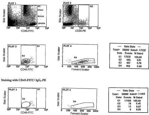
Flow cytometry and gating strategy. Cells were analyzed using a FACScan flow cytometer equipped with an argon ion laser (Becton Dickinson Instrument Systems, BDIS, Mississauga, Canada). Five data parameters were acquired and stored in listmode files: linear forward light scatter (FSC), linear side-angle light scatter (SSC), log FITC, log PE, and log PerCp fluorescence; each measurement contained 50,000 events. Compensation settings were established using CalBrite beads (BDIS) and confirmed using NAMNC stained with anti–CD34-PE, anti–CD45-FITC, or anti–IL-5Ra–PerCp. Off-line analysis was per-formed using the PC lysis software as supplied by BDIS.
We used a multi-parameter sequential gating strategy that, we
have previously shown, accurately enumerates CD341 progenitor cell numbers in various biological samples (22, 23). The rationale for se-quential gating was to gradually eliminate contaminating cells that nonspecifically take up anti-CD34 (24). Briefly, a primary gate using CD45 staining versus SSC (region R1) was set up to quantitate total leukocytes and distinguish contaminating events such as platelet ag-gregates and other debris which can nonspecifically take up anti-CD34 (Fig. 1 A, PLOT 1). Primitive cells characteristically express CD45 at low to intermediate levels (25) and therefore CD451 events generate a stable denominator in the calculation of the absolute CD341 value. Sequential gates were then set up: CD34 staining in re-gion R1 versus SSC (rere-gion R2) (Fig. 1 A, PLOT 2), CD45 versus SSC of the CD341-gated events in R2 (region R3: to identify blast cells) (Fig. 1 A, PLOT 3), and FSC versus SSC to confirm the lym-phoblastoid characteristics of the gated CD341 cells in region R3 (i.e., low to medium SSC and FSC; region R4) (Fig. 1 A, PLOT 4). With-out changing any of the gates, analyses of the same cell sample stained with CD45-FITC and PE linked isotype control antibody were performed (Fig. 1 B, PLOT 3 and PLOT 4). Enumeration data were derived from the gate statistics: events in gate G4 (5 events in R1 to R4) after staining with CD45-FITC/CD34-PE minus events in G4 stained with CD45-FITC/PE–linked control antibody were used to calculate the absolute number of true CD341 blast cells in the test sample.
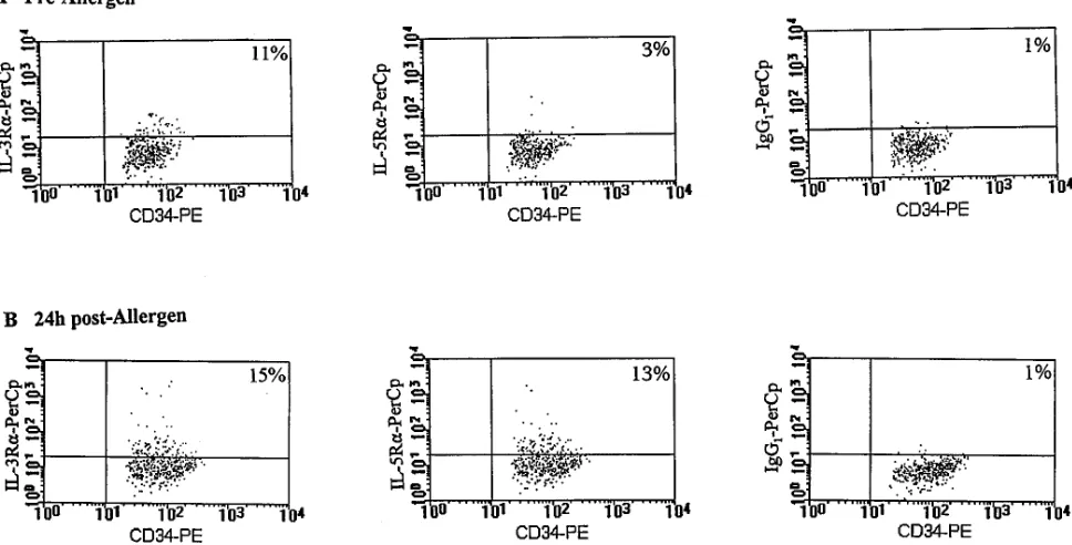
In three-color analysis, events in region R4 were back scattered onto a dot plot of CD34-PE versus staining by PerCp linked cytokine receptor mAbs or control antibody (Fig. 2), and data were collected as percent positive cells at the 99% confidence limit (i.e., relative to a marker set to include only 1% of cells stained with control antibody). The data presented are the mean of duplicate assessments. The intra-assay variability was always less than 5%.
Simultaneous in situ hybridization and immunohistochemistry.
To confirm the association of the membrane bound form of IL-5Ra
messenger RNA to CD341 progenitor cells, simultaneous in situ hy-bridization and immunocytochemistry was performed (26). Messen-ger RNA for membrane bound IL-5Ra was detected by autoradiog-raphy and CD34 immunoreactivity was detected by an alkaline phosphatase antialkaline phosphatase technique (APAAP). A popu-lation of CD341 cells was enriched from cord blood by positive selec-tion using a magnetic cell separaselec-tion technique, as described previ-ously (13). These cells were cytospun on poly-L-lysine–coated slides, fixed in 4% paraformaldehyde in PBS for 30 min, and washed in 15% sucrose in PBS. Preparations were hybridized with 35S- labeled mem-brane-bound IL-5Ra antisense riboprobe and simultaneously immu-nostained with a mouse anti–human mAb against CD34 (QBEND 10; Becton Dickinson, San Jose, CA) (26).
Statistical analysis. The data are presented as absolute numbers of CD341 progenitor cells (Fig. 3) and as arithmetic mean6SEM (Fig. 4, Tables I and II), except PC20 values (Table II) that were loga-rithmically transformed and expressed as geometric means and stan-dard error of geometric means (percent SEM). For statistical analyses of within group comparisons between pre- and post-allergen chal-lenge time points, a paired Student’s t test (two-tailed) was performed (Table I, Figs. 3 and 4). Student’s non-paired t tests (two-tailed) were performed for all between group comparisons (Table I) and changes in blood differentials after allergen were assessed for each group us-ing repeated measures analysis of variance (rmANOVA) (Table II). Significance was accepted at the 95% confidence level.
Results
Allergen-induced bronchoconstrictor responses and airway hy-perresponsiveness. Subjects (n 5 6) in whom the maximal per-cent fall in FEV1 during the late asthmatic response was , 15%
were labeled isolated early responders (IER; mean percent fall in FEV1, 8.261.2%). Subjects in whom that late maximal
(DR; mean percent fall in FEV1, 22.763.2%). The dual
re-sponders, but not the isolated early rere-sponders, developed a significant increase in methacholine airway responsiveness 24 h after allergen challenge (P , 0.001, Table I).
Airway and blood eosinophilia. A significant increase in sputum eosinophilia was detected in DR asthmatics but not in the IER group, when pre-allergen levels were compared with 24 h post-allergen values (Table II). Similarly, a significant in-crease in blood eosinophil levels was observed between 5 and 24 h post-allergen in DR asthmatics (P , 0.05), although this was preceded by a significant reduction in eosinophil numbers 5 h post-allergen compared with baseline values (P , 0.05, Ta-ble II). In contrast, in IER, no significant change in the num-bers of blood eosinophils was observed at either 5 or 24 h post-allergen compared with pre-post-allergen values.
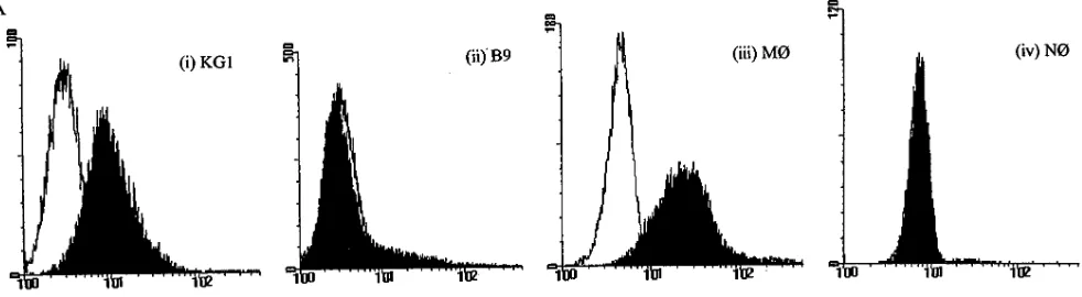
IL-3Ra– and IL-5Ra–subunit expression in various cell types. To verify the specific binding capacity of the cytokine receptor antibodies used in this study, we tested the staining of anti–IL-3Ra (7G2) and anti–IL-5Ra (A16) on various leuke-mic cell lines and mature peripheral blood leukocytes.
Com-pared with the isotype matched control antibody, the signifi-cant expression of the IL-3Ra–subunit was detected on KG1 cells [Fig. 3 A (i)] and peripheral blood monocytes [Fig. 3 A (iii)] (27). As expected, no expression of IL-3Ra was detected on B9 cells, an IL-6–dependent mouse B cell hybridoma cell line [Fig. 3 A (ii)] or on mature neutrophils [Fig. 3 A (iv)] (27). The expression of IL-5Ra was detected on HL60 clone 15 cells, known to overexpress receptors for IL-5 (8), and on pe-ripheral blood eosinophils (Fig. 3 B). No specific expression of IL-5Ra was detected on either neutrophils [Fig. 3 B (iv)] or monocytes (data not shown) (28).
Allergen-induced changes in the phenotype of bone marrow progenitors. By multiparameter flow cytometric analyses, al-lergen-induced changes in the absolute numbers of bone mar-row–derived CD341 progenitor cells and the absolute numbers of CD341 cells expressing the a-subunit of IL-3R and IL-5R were investigated. In DR asthmatics, the total number of bone marrow–derived CD341 cells increased from 3,715 cells/0.25 3 106 white blood cells (WBC) (percent SEM 830) before
[image:5.612.67.547.75.444.2]aller-gen to 5,623 cells/0.25 3 106 WBC (percent SEM 2,627) 24 h
after allergen challenge, although these changes were not sig-nificant (P 5 0.06) (Fig. 4). In IER, no differences in CD341 cell numbers was detected when pre-allergen levels (2,291 cells/ 0.25 3 106 WBC, percent SEM 1,009) were compared
with 24 h post-allergen levels (2,570 cells/0.25 3 106 WBC,
per-cent SEM 782, P 5 0.70) (Fig. 4). No significant differences in either the baseline values of bone marrow CD341 cells or the magnitude of increase of CD341 cells after allergen challenge were detected between the two groups of asthmatic subjects.
In DR asthmatics, the absolute number of bone marrow– derived CD341 cells expressing IL-5Ra increased 30-fold from 26 cells/0.25 3 106 WBC (percent SEM 3.3) before allergen to
724 cells/ 0.25 3 106 WBC (percent SEM 1.6) 24 h after
aller-gen (P 5 0.04) (Fig. 4). In contrast, there was a negligible in-crease in the numbers of CD341IL-5Ra1 cells in IER when pre-allergen levels (6 cells/0.25 3 106 WBC, percent SEM 2.7)
were compared with post-allergen levels (11 cells/0.25 3 106
WBC, percent SEM 2.7, P 5 0.55) (Fig. 4). Although there was a ninefold increase in the absolute number of bone marrow CD341IL-3Ra1 cells in DR asthmatics when pre-allergen lev-els were compared with 24 h post-allergen levlev-els, these changes were not significant (37 cells/0.25 3 106 WBC versus 324 cells/
0.25 3 106 WBC, P 5 0.051) (Fig. 4). In addition, no significant
increase in the absolute numbers of bone marrow CD341 IL-3Ra1 cells was detected in IER 24 h post-allergen (Fig. 4).
It is conceivable that the observed increases in absolute numbers of cytokine receptor positive progenitor cells 24 h post-allergen may be entirely due to increases in total CD341 cell numbers (Fig. 4). In order to exclude this effect and to
de-termine whether a distinct phenotypic change of a fixed pro-genitor cell pool had occurred after allergen challenge, we expressed the data as a percent of total CD341 cell numbers (Fig. 5). In DR, but not IER asthmatics, a significant increase in the percent of CD341 cells expressing IL-5Ra was detected when pre-allergen bone marrow samples were compared with 24 h post-allergen aspirates (DR: 6.663.1 versus 1562.7% of CD341 cells, P 5 0.045, and IER: 1.961.08 versus 2.26 1.2% of CD341 cells, P 5 0.84) (Fig. 5). In contrast, in both groups of asthmatic subjects no significant increase in the pro-portion of CD341 cells expressing IL-3Ra, after allergen chal-lenge, was observed (DR: 8.563.4 versus 1564.5% of CD341 cells, P 5 0.22, and IER: 2.761.2 versus 9.263.5% of CD341 cells, P 5 0.19) (Fig. 5). In comparisons between the two groups of asthmatics, no significant differences were observed in the baseline values of percent CD341 cells expressing either cytokine receptors (Fig. 5). However, allergen-induced in-creases in the proportion of CD341 cells expressing IL-5Ra1 cells were significantly greater in dual responders compared with isolated early responder asthmatics (P 5 0.038).
[image:6.612.63.547.67.313.2]In situ hybridization and immunohistochemistry. Colocal-ization experiments using in situ hybridColocal-ization and immunocy-tochemistry were performed in order to confirm that CD341 progenitor cells can express IL-5Ra mRNA. Because of the small sample sizes of bone marrow aspirates, these experiments were performed on an enriched population of unstimulated CD341 cells isolated from cord blood (purity determined by flow cytometry, . 65%). Of all the CD341 cells, 50% were IL-5Ra mRNA positive. A representative example is shown in Fig. 6.
Discussion
The novel observation from this study is that the proportion of bone marrow–derived CD341 cells expressing IL-5Ra is in-creased preferentially in DR asthmatics who characteristically develop allergen-induced late-bronchoconstrictor responses, methacholine airways hyperresponsiveness, and a significant sputum eosinophilia 24 h after allergen inhalation (Fig. 5, Ta-bles I and 2) (2, 29). In contrast, this distinct change in
[image:7.612.63.554.61.196.2]cyto-kine receptor expression on bone marrow progenitor cells was not seen in asthmatics who did not develop airway hyperre-sponsiveness or a marked eosinophil infiltration after allergen inhalation (Fig. 5). Although the data presented herein do not prove a direct association between activation of the bone mar-row and development of airway pathology as a result of in-creased inflammatory cell production, they are consistent with the view that a feedback mechanism exists between tissues in-volved in allergic inflammation and distal sites such as the
Figure 3. Expression of IL-3Ra and IL-5Ra on various cell types. Cells were incubated with saturating amounts of the biotinylated antireceptor antibodies, washed and incubated with streptavidin-linked PerCp (shaded area). For negative control (blank area), cells were stained with the equivalent amount of the isotype matched biotinylated antibody (mouse IgG1) in place of the antireceptor antibody.
Table II. Inflammatory Cell Count in Sputum and Blood Samples from Asthmatic Subjects
Early single responders Dual responders
Pre-allergen 5 h Post-allergen 24 h Post-allergen Pre-allergen 5 h Post-allergen 24 h Post-allergen
Sputum
Eosinophils (percent) 6.762.0 ND 16.469.9 3.360.9 ND 32.3611.7*
Neutrophils (percent) 15.764.3 ND 28.4610.2 26.0610.5 ND 26.9611.8
Macrophages (percent) 72.264.1 ND 48.0613.5 66.3610.1 ND 36.168.6‡
Blood
Eosinophils (3109/liter) 0.3560.04 0.3660.20 0.4660.50 0.4460.12 0.2360.05* 0.3260.06§
Data for sputum cell counts are presented as the percentage of 500 white cells counted per slide. Cells were stained with Diff-Quik. Data represent the mean6SEM of n5 6 early single responders and n5 7 dual responders. *P, 0.05; ‡P, 0.001 for comparisons of pre-allergen versus post-allergen
[image:7.612.70.554.237.389.2]bone marrow. We have shown that a distinct phenotypic switch occurs within the bone marrow progenitor cell popula-tion and we suggest that in the presence of eosinophil growth factors such as IL-5, increased expression of IL-5Ra on CD341 cells may favor eosinophilopoiesis and thus contribute to the subsequent development of blood and tissue eosinophilia, a hallmark of allergic inflammatory diseases such as asthma.
[image:8.612.62.525.56.642.2]Of the cytokines which can support eosinophilopoiesis (IL-5, IL-3, and GM-CSF), IL-5 is unique in its ability to specifically promote the terminal differentiation and maturation of eosin-ophil/basophil lineage–committed progenitors in liquid and semi-solid cultures (30, 31). In mice that overexpress the IL-5 transgene, IL-5 has been shown to be the predominant regula-tor of eosinophilia (32). Furthermore, a pivotal role for IL-5 in chronic allergic inflammation has been confirmed by the capacity of neutralizing anti–IL-5 mAb to inhibit antigen- or virus-induced airway hyperresponsiveness and eosinophil infil-tration in the airways of mice, guinea pigs, and primates (33– 39). In contrast, IL-3 is a pluripotential hemopoietic factor; mice that overexpress either IL-3 or GM-CSF, show only mod-est eosinophilia, but succumb early owing to massive tissue in-filtration and destruction by myeloid cells, especially neutro-phils and macrophages (32). Our evidence suggests that both GM-CSF and IL-3 commit pluripotential CD341 CD332 pro-genitors to an eosinophil lineage and that IL-5 brings about the terminal differentiation of the less primitive, myeloid lin-eage-committed progenitor cells (i.e., CD342CD331 cells) de-rived from CD341 CD332 precursors (40). In this study, how-ever, we have demonstrated for the first time the expression of IL-5Ra on CD341 cells indicating the existence of specific binding sites for IL-5 on more primitive progenitor cells. This
Figure 4. Flow cytometric enumera-tion of the absolute number of bone marrow derived CD341 progenitor cells expressing the a-subunit of re-ceptors for IL-3 and IL-5. Samples from (n 5 6) isolated early- and (n 5
7) dual-responder asthmatics were taken pre- and 24 h post-allergen challenge. After allergen challenge, a significant increase in the numbers of CD341 IL-5Ra1 cells were de-tected in bone marrow samples taken from DR asthmatics. Hori-zontal bars represent the geometric mean of each data set.
is supported by results from in situ hybridization which dem-onstrate the colocalization of mRNA for membrane bound IL-5Ra to cells immunostained with anti-CD34. Therefore, we propose that the CD341 IL-5Ra1 phenotype may be represen-tative of the earliest eosinophil/basophil lineage–committed progenitor. However, until additional cloning experiments have been performed to assess the progeny of progenitors of this specific phenotype, this proposal cannot be confirmed.
Molecular cloning of cytokine receptors have revealed that IL-3R, IL-5R, and GM-CSFR are uniquely composed of het-erodimeric structures consisting of a distinct a-subunit that binds the cognate cytokine with low affinity and a common, shared, b-subunit which, although failing to bind the ligand it-self, forms high affinity cytokine binding sites in association with the a-subunit (41, 42). Deletion mutation experiments of IL-5R have now revealed that, like the b-subunit, the cytoplas-mic domain of the a-subunit is also essential for signal trans-duction, in particular mediating growth signals through IL-5R (43, 44). Since the a-subunit functions as a cytokine-specific binding site, it has been proposed that this subunit may trans-duce cytokine-specific growth signals while the common b-chain provides the molecular basis for functional redundancy of IL-3, IL-5, and GM-CSF. Therefore, the preferential increase in the proportion of bone marrow–derived CD341 cells expressing membrane bound IL-5Ra-subunit on bone marrow progenitor cells may increase the ability of the cells to respond more readily to IL-5, and thus differentiate terminally into mature eosinophils and basophils. In support of our findings, Wood et al. have shown that bone marrow aspirates taken from DR 24 h post-allergen are more responsive to IL-5 in vitro, as deter-mined by the significantly greater numbers of Eo/Baso–CFU detected in methylcellulose cultures with suboptimal doses of IL-5 compared with bone marrow cells from IER (45).
Since increases in expression of cytokine receptors on CD341 cells were detected within 24 h after allergen challenge in DR asthmatics, these changes may have occurred as a con-sequence of cell division and proliferation (Fig. 4). However, when expressed as a percentage of total CD341 cells, the in-crease in IL-5Ra, but not in IL-3Ra, expression on CD341 cells was independent of changes in the number of progenitors (Fig. 5). This indicates that after allergen challenge in DR asth-matics, a fixed pool of bone marrow CD341 progenitors un-dergo a distinct phenotypic change resulting in increases in IL-5Ra surface expression. In contrast, the increased produc-tion of small amounts of primitive progenitor cells may ac-count for the near significant increases in absolute numbers of CD341IL-3Ra cells observed in DR 24 h post-allergen (Fig. 4) (27).
Evidence of the generation of a serum hemopoietic factor during airway allergen challenge that can prime the bone mar-row for increased production of granulocyte progenitor cells has been recently demonstrated in a canine model of airways hyperresponsiveness (46). Similarly, increased numbers of Eo/ Baso–CFU were grown from the peripheral blood of atopic in-dividuals when antigen-stimulated lymphomononuclear cell conditioned medium was included in colony assays, suggesting the generation of a hemopoietic signal after in vitro allergen challenge (5). Furthermore, in studies of nematode infection in IL-5 transgenic mice, Strath et al. have provided evidence that the level of blood eosinophilia may not only be controlled by the amount of IL-5 produced but, in addition, by the frequency of eosinophil progenitors in the bone marrow during a chronic
inflammatory response (47). Thus, investigation of the nature of the signal(s) that modulate the expression of IL-5R on CD341 progenitor cells may provide insight into the control of eosinophil differentiation from pluripotential stem cells and, potentially provide a novel therapeutic target for controlling the development of the eosinophilic component of the allergic inflammatory response in asthmatic airways. From in vitro studies, little is currently known regarding the modulation of IL-5R expression on normal progenitor cells. Preincubation of peripheral blood CD341 cells with GM-CSF and IL-3 en-hances their subsequent ability to differentiate into eosinophils in response to IL-5 (48). While this finding implies upregula-tion of IL-5R expression, this has not been formally demon-strated, at least not for progenitor cells. On mature eosino-phils, preincubation with GM-CSF, but not IL-3, will enhance expression of IL-5R (49). Downregulation of the IL-5Ra -sub-unit at the mRNA level in myeloid leukemic cell lines has been shown to be due to factors that either promote eosinophil apop-tosis such as TGFb1 (50), or by pharmacological agents, such
as all-trans retinoic acid (RA), which inhibit eosinophil/baso-phil differentiation from pluripotential progenitor cells, while favoring neutrophil maturation (51, 52). This is further support for the view that the level of expression of IL-5Ra on CD341 cells may be directly related to commitment to the eosino-philopoietic pathway.
In conclusion, the results from this study demonstrate that relevant fluctuations occur in the expression of a specific cy-tokine receptor, IL-5Ra, on bone marrow progenitors in re-sponse to allergen challenge in atopic asthmatics. We propose that this selective increase in expression of IL-5Ra on CD341 progenitors may favor eosinophilopoiesis that may play a role in the generation of increased numbers of eosinophils during an allergic inflammatory response in the airways of asthmatic subjects.
Acknowledgments
This work was supported by grants from the Medical Research Coun-cil of Canada and Astra Draco, Sweden. R. Sehmi is a recipient of a joint research fellowship from the Canadian Lung Association and Medical Research Council of Canada.
References
1. Bousquet, J., P. Chanez, Y.J. Lacoste, G. Barnéon, N. Chavanian, I. Enander, P. Venge, S. Ahlstedt, J. Simony-Lafontaine, P. Godard, and F.B. Michel. 1990. Eosinophilic inflammation in asthma. N. Engl. J. Med. 323:1033– 1039.
2. Beasley, R., W.R. Roche, J.A. Roberts, and S.T. Holgate. 1989. Cellular events in the bronchi in mild asthma and after bronchial provocation. Am. Rev. Respir. Dis. 139:806–817.
3. Azzawi, M., B. Bradley, P.K. Jeffery, A.J. Frew, A.J. Wardlaw, G. Knowles, B. Assoufi, J.V. Collins, S.R. Durham, and A.B. Kay. 1990. Identifica-tion of activated T-lymphocytes and eosinophils in bronchial biopsies in stable atopic asthma. Am. Rev. Respir. Dis. 142:1407–1413.
4. Hamid, Q., M. Azzawi, S. Ying, R. Moqbel, A.J. Wardlaw, C.J. Corrigan, B. Bradley, S.R. Durham, J.V. Collins, P.K. Jeffery, et al. 1991. Expression of mRNA for interleukin-5 in mucosal bronchial biopsies from asthma. J. Clin. In-vest. 87:1541–1546.
5. Denburg, J.A., S. Telizyn, A. Belda, J. Dolovich, and J. Bienenstock. 1985. Increased numbers of circulating basophil progenitors in atopic patients.
J. Allergy Clin. Immunol. 76:466–472.
6. Gibson, P.G., J. Dolovich, A. Girgis-Gabardo, M.M. Morris, M. Ander-son, F.E. Hargreave, and J.A. Denburg. 1990. The inflammatory response in asthma exacerbation: changes in circulating eosinophils, basophils and their progenitors. Clin. Exp. Allergy. 20:661–668.
metachro-matic cells in allergic rhinitis. Am. Rev. Respir. Dis. 136:710–717.
8. Linden, M., C. Svensson, M. Andersson, L. Greiff, E. Andersson, J.A. Denburg, J. Seidegard, and C.G.A. Persson. 1994. Increased numbers of circu-lating leukocyte progenitors in patients with allergic rhinitis during natural al-lergen exposure. Am. J. Respir. Crit. Care Med. 149:A602(Abstr.).
9. Gibson, P.G., P.J. Manning, P.M. O’Byrne, A. Girgis-Gabardo, J. Dolo-vich, J.A. Denburg, and F.E. Hargreave. 1991. Allergen induced asthmatic re-sponses. Relationship between increased airway responsiveness and increases in circulating eosinophils, basophils and their progenitors. Am. Rev. Respir. Dis.
143:331–335.
10. Woolley, M.J., J.A. Denburg, R. Ellis, M. Dahlback, and P.M. O’Byrne. 1994. Allergen induced changes in bone marrow progenitors and airways re-sponsiveness in dogs and the effect of inhaled budesonide on these parameters.
Am. J. Respir. Cell. Mol. Biol. 11:600–606.
11. Civin, C., T. Trischman, M.J. Fackler, I. Bernstein, H. Buhring, L. Cam-pos, M.F. Greaves, M. Kamoun, D. Katz, P. Lansdorp, et al. 1989. Leukocyte typing. In Leukocyte Typing IV. W. Knapp, editor. Oxford University Press, Oxford. 818–825.
12. Sutherland, D.R., and A. Keating. 1992. The CD34 antigen: structure, biology and potential clinical applications. J. Hemother. 1:115–129.
13. Sehmi, R., K. Howie, D.R. Sutherland, W. Schragge, P.M. O’Byrne, and J.A. Denburg. 1996. Increased levels of CD341 hemopoietic progenitor cells in
atopic subjects. Am. J. Respir. Cell Mol. Biol. 15:645–654.
14. Coligan, J.E., A.M. Kruisbeek, D.H. Margulies, E.M. Shevach, and W. Strober. 1991. Antibody detection and preparation. In Current Protocols in Im-munology. J.E. Coligan, A.M. Kruisbeek, D.H. Margulies, E.M. Shevach, and W. Strober, editors. John Wiley & Sons, Inc., New York. 2.1–2.13.
15. Morris, J.F., A. Koski, and L.C. Johnson. 1997. Spirometer standards for healthy non-smoking adults. Am. Rev. Respir. Dis. 103:57–67.
16. Cockcroft, D.W. 1996. Measurement of airway responsiveness to in-haled histamine or methacholine; method of continuous aerosol generation and tidal breathing inhalation. In Airway Responsiveness: Measurement and Inter-pretation. F.E. Hargreave and A.J. Woolcock, editors. Astra Pharmaceutical, Canada, Limited, Mississauga, Canada. 22–28.
17. O’Byrne, P.M., J. Dolovich, and F.E. Hargreave. 1987. Late asthmatic response. Am. Rev. Respir. Dis. 136:740–756.
18. Popov, T.A., R. Gottschalk, R. Kolendowicz, J. Dolovich, J. Powers, and F.E. Hargreave. 1994. The evaluation of cell dispersion method of sputum examination. Clin. Exp. Allergy. 24:778–783.
19. Coligan, J.E., A.M. Kruisbeek, D.H. Margulies, E.M. Shevach, and W. Strober. 1991. Cytokines and their cellular receptors. In Current Protocols in Immunology. J.E. Coligan, A.M. Kruisbeek, D.H. Margulies, E.M. Shevach, and W. Strober, editors. John Wiley & Sons, Inc. New York. 6.1–6.22.
20. Sehmi, R., O. Cromwell, A.J. Wardlaw, R. Moqbel, and A.B. Kay. 1993. Interleukin-8 is a chemoattractant for eosinophils purified from subjects with a blood eosinophilia but not from normal healthy subjects. Clin. Exp. Allergy. 23: 1027–1036.
21. Denburg, J.A., M. Richardson, S. Telizyn, and J. Bienenstock. 1983. Ba-sophil/mast cell precursors in human peripheral blood. Blood. 61:775–780.
22. Sutherland, D.R., A. Keating, R. Nayar, S. Anania, and A.K. Stewart. 1994. Sensitive detection and enumeration of CD341 cells in peripheral and
cord blood by flow cytometry. Exp. Hematol. (Charlottesv.). 22:1003–1010. 23. Sehmi, R., L.J. Wood, R.M. Watson, R. Foley, P.M. O’Byrne, and J.A. Denburg. 1996. Increases in the proportion of bone marrow CD341 cells
ex-pressing the alpha-subunit of IL-5 receptors following allergen challenge in dual responder asthmatics. J. Allergy Clin. Immunol. 99:S272. (Abstr.)
24. Sutherland, D.R., L. Anderson, M. Keeney, R. Nayar, and I. Chin-Yee. 1996. The ISHAGE guidelines for CD341 cell determination by flow
cytom-etry: international society of hematotherapy and graft engineering. J. Hemat-other. 5:213–226.
25. Borowitz, M.J., L. Guenther, K.E. Shults, and G.T. Stelzer. 1993. Immu-nophenotyping of acute leukemia by flow cytometric analysis: use of CD45 and right-angle light scatter to gate on leukemic blasts in three-color analysis. He-matopathol. 100:534–540.
26. Hamid, Q., J. Barkans, Q. Meng, S. Ying, J.S. Abrams, A.B. Kay, and R. Moqbel. 1992. Human eosinophils synthesize and secrete interleukin-6, in vitro.
Blood. 806:1496–1501.
27. Kurata, H., T. Arai, T. Yokota, and K. Arai. 1995. Differential expres-sion of granulocyte-macrophage colony stimulating factor and IL-3 receptor subunits on human CD341 cells and leukemic cell lines. J. Allergy Clin. Immu-nol. 96:1083–1099.
28. Lopez, A.F., M.J. Elliott, J. Woodcock, and M.A. Vadas. 1992. GM-CSF, IL-3 and IL-5: cross-competition on human haemopoietic cells. Immunol. Today. 13:495–500.
29. De Monchy, J.G., H.F. Kaufman, P. Venge, G.H. Koëter, H.M. Jansen, and H.J. Sluiter. 1985. Bronchoalveolar eosinophilia during allergen induced late asthmatic reactions. Am. Rev. Respir. Dis. 426:373–376.
30. Denburg, J.A., S. Telizyn, A. Belda, H. Messner, B. Lim, N. Jamal, S.J. Ackerman, G.J. Gleich, and J. Bienenstock. 1985. Heterogeneity of human pe-ripheral blood eosinophil-type colonies: evidence of a common
basophil-eosin-ophil progenitor. Blood. 66:312–318.
31. Clutterbuck, E.J., E.M.A. Herst, and C.J. Sanderson. 1989. Human in-terleukin-5 (IL-5) regulates the production of eosinophils in human bone mar-row cultures: comparisons and interaction with IL-1, IL-3, IL-6 and GM-CSF.
Blood. 73:1504–1512.
32. Dent, L.A., M. Strath, A.L. Mellor, and C.J. Sanderson. 1990. Eosino-philia in mice expressing interleukin-5. J. Exp. Med. 172:1425–1431.
33. Nagai, H., N. Yamaguchi, N. Inagaki, N. Tsuruoka, Y. Hitoshi, and K. Takatsu. 1993. Effect of anti-IL-5 monoclonal antibody on allergic bronchial eosinophilia and airway hyperresponsiveness in mice. Life Sci. 53:PL243– PL247.
34. Kung, T., D.M. Stelts, J.A. Zurcher, G.K. Adams III, R.W. Egan, W. Kreutner, A.S. Watnick, H. Jones, and R.W. Chapman. 1995. Involvement of IL-5 in a murine model of allergic pulmonary inflammation: prophylactic and therapeutic effect of an IL-5 antibody. Am. J. Respir. Cell Mol. Biol. 13:360–365.
35. Chand, R.L., B.W. Seymour, S. Hudak, J. Jackson, and D. Rennick. 1992. Anti-IL-5 monoclonal antibody inhibits allergic late phase bronchial eosin-ophilia in guinea pigs: a therapeutic approach. Eur. J. Pharmacol. 211:121–123.
36. Gulbenkian, A.R., R.W. Egan, X. Fernandez, H. Jones, W. Kreutner, T. Kung, F. Payvandi, L. Sullivan, J.A. Zurcher, and A.S. Watnick. 1992. Interleu-kin-5 modulates eosinophil accumulation in guinea pig lung. Am. Rev. Respir. Dis. 146:263–265.
37. Van Oosterhout, A.J.M., I. Van Ark, G. Folkerts, H.L. Van der Linde, H.F.J. Savelkoul, K.C.P. Verheyen, and F.P. Nijkamp. 1995. Antibody to inter-leukin-5 inhibits virus-induced airways hyperresponsiveness to histamine in guinea pigs. Am. J. Respir. Crit. Care Med. 151:177–183.
38. Mauser, P.J., A.M. Pitman, X. Fernandez, S.K. Foran, G.K. Adams III, W. Kreutner, R.W. Egan, and R.W. Chapman. 1995. Effects of antibody to in-terleukin-5 in a monkey model of asthma. Am. J. Repir. Crit. Care Med. 152: 467–472.
39. Egan, R.W., D. Athwahl, C. Chou, S. Emtage, C. Jehn, T.T. Kung, P.J. Mauser, N.J. Murgolo, and M.W. Bodmer. 1995. Inhibition of pulmonary eosin-ophilia and hyperreactivity by antibodies to interleukin-5. Int. Arch. Allergy Immunol. 107:321–322.
40. Ema, H., T. Suda, K. Nagayoshi, Y. Miura, C.I. Civin, and H. Nakauchi. 1990. Target cells for granulocyte macrophage colony-stimulating factor, inter-leukin-3, and interleukin-5 in the differentiation pathways of neutrophils and eosinophils. Blood. 76:1956–1961(Abstr.).
41. Murata, Y., S. Takaki, M. Migita, Y. Kikuchi, A. Tominaga, and K. Takatsu. 1992. Molecular cloning and expression of the human interleukin-5 re-ceptor. J. Exp. Med. 175:341–351.
42. Tavernier, J., R. Devos, S. Cornelius, T. Tuypens, J. Van der Heyden, W. Fiers, and G. Plaetnick. 1991. A human high affinity interleukin-5 receptor (IL-5R) is composed of an IL-5 specific a-chain and a b-chain shared with a re-ceptor for GM-CSF. Cell. 66:1175–1184.
43. Takaki, S., Y. Murata, T. Kitamura, A. Miyajima, A. Tominaga, and K. Takatsu. 1993. Reconstitution of the functional receptors for murine and hu-man interleukin 5. J. Exp. Med. 177:1523–1529.
44. Takagi, M., H. Kanazawa, M. Shiiba, and K. Takatsu. 1994. A critical cy-toplasmic domain of the interleukin-5 (IL-5) receptor a chain and its function in IL-5-mediated growth signal transduction. Mol. Cell. Biol. 14:7404–7413.
45. Wood, L.J., M.D. Inman, R.M. Watson, R. Foley, J.A. Denburg, and P.M. O’Byrne. 1997. Inhaled allergen-induced changes in bone marrow eosino-phil/basophil progenitors in mild asthmatic subjects. J. Allergy Clin. Immunol.
99:A1485(Abstr.).
46. Inman, M.D., J.A. Denburg, R. Ellis, M. Dahlback, and P.M. O’Byrne. 1996. Allergen-induced increase in bone marrow progenitors in airway hyperre-sponsive dogs: regulation by a serum hemopoietic factor. Am. J. Respir. Cell. Mol. Biol. 15:305–311.
47. Strath, M., L.A. Dent, and C.J. Sanderson. 1992. Infection of IL-5 trans-genic mice with Mesocestoides corti induces very high levels of IL-5 but de-pressed production of eosinophils. Exp. Hematol. 20:229–234.
48. Shalit, M., S. Sekhsaria, and H.L. Malech. 1995. Modulation of growth and differentiation of eosinophils from human peripheral blood CD341 cells by
IL-5 and other growth factors. Cell. Immunol. 160:50–57.
49. Chihara, J., J. Plumas, V. Gruart, J. Tavernier, L. Prin, A. Capron, and M. Capron. 1990. Characterization of a receptor for interleukin 5 on human eosinophils: variable expression and induction by granulocyte macrophage col-ony stimulating factor. J. Exp. Med. 172:1347–1351.
50. Jacobson, S.E.W., F.W. Ruscetti, A.B. Roberts, and J.R. Keller. 1993. TGF-b is a bidirectional modulator of cytokine receptor expression on murine bone marrow cells. Differential effects of TGF-b1 and TGF-b3. J. Immunol.
151:4534–4544.
51. Paul, C.C., S. Mahrer, M. Tolbert, B.L. Elbert, I. Wong, S.J. Ackerman, and M.A. Baumann. 1995. Changing the differentiation program of hematopoi-etic cells: retinoic acid-induced shift of eosinophil-committed cells to neutro-phils. Blood. 86:3737–3744.