Microreview
Polymerizing the fibre between bacteria and host cells:
the biogenesis of functional amyloid fibres
Elisabeth Ashman Epstein and Matthew R. Chapman*
Department of Molecular, Cellular and Developmental Biology, University of Michigan, 830 North University, Ann Arbor, MI 48109, USA.
Summary
Amyloid fibres are proteinaceous aggregates asso-ciated with several human diseases, including Alzheimer’s, Huntington’s and Creutzfeldt Jakob’s. Disease-associated amyloid formation is the result of proteins that misfold and aggregate intobsheet-rich fibre polymers. Cellular toxicity is readily asso-ciated with amyloidogenesis, although the molecular mechanism of toxicity remains unknown. Recently, a new class of ‘functional’ amyloid fibres was discov-ered that demonstrates that amyloids can be utilized as a productive part of cellular biology. These functional amyloids will provide unique insights into how amyloid formation can be controlled and made less cytotoxic. Bacteria produce some of the best-characterized functional amyloids, including a surface amyloid fibre called curli. Assembled by enteric bacteria, curli fibres mediate attachment to surfaces and host tissues. Some bacterial amyloids, like harpins and microcinE492, have exploited amy-loid toxicity in a directed and functional manner. Here, we review and discuss the functional amyloids assembled by bacteria. Special emphasis will be paid to the biology of functional amyloid synthesis and the connections between bacterial physiology and pathology.
Amyloid: a convergence of diverse proteins along a common folding pathway
The ability of bacteria to interact with their environment is often mediated by the presence of cell-surface organelles
composed of protein polymers. These extracellular protein fibres are implicated in diverse processes like locomotion, attachment to surfaces, natural competence and host–pathogen interactions (Fernandez and Beren-guer, 2000; Jonson et al., 2005). The extracellular envi-ronment can be a harsh locale with little energy available for protein folding, and dynamic physical conditions like hydrophobicity, salt concentrations, pH fluctuations and bombardment with denaturing chemicals. Therefore, assembly of surface structures in the extracellular envi-ronment would be predicted to feature proteins that can self-assemble without assistance from cellular chaper-ones, have low-energy folding requirements and are resistant to denaturation and chemical perturbation.
One protein folding assembly pathway that satisfies these conditions is amyloidogenesis. Amyloid fibril assembly is a hallmark of diverse neurological diseases, such as Alzheimer’s disease, Parkinson’s disease, Hun-tington’s disease and bovine spongiform encephalopathy (mad cow disease). Disease-associated amyloid forma-tion is the result of misfolding and aggregaforma-tion of peptides into structurally conserved fibres (Ross and Poirier, 2004). However, amyloid formation is also integrated as a critical element of cellular physiology, and many organisms direct amyloid fibre formation to accomplish important biological tasks (Fowler et al., 2007). Whether the product of disease-associated protein misfolding or a dedicated assembly pathway, amyloid fibres are characterized by a ‘cross-bstrand’ structure, where thebsheets are oriented anti-parallel to the fibre axis (Sundeet al., 1997; Nelson et al., 2005). This structure gives rise to common physical properties. Amyloid fibres are self-assembling protein polymers that are resistant to chemical and temperature denaturation, and to digestion by proteinases (Nordstedt et al., 1994). The b sheet-rich fibres also have similar tinctorial properties: binding to amyloid-specific dyes like Congo red and thioflavin T is diagnostic of amyloid (Elghetany and Saleem, 1988). Amyloids are abundant in bacterial biofilms, and a recent report suggested that up to 40% of the biomass in activated sludge consists of amyloid-like filaments (Larsenet al., 2007; 2008).
Much of our knowledge concerning the highly adaptive amyloid fold comes from studies of eukaryotic protein-Received 1 February, 2008; revised 13 March, 2008; accepted 13
March, 2008. *For correspondence. E-mail [email protected]; Tel. (+1) 734 764 7592; Fax (+1) 734 647 0884.
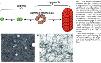
misfolding diseases. Each of these diseases shares a common etiology: the conversion of soluble, non-toxic protein into highly ordered and aggregative fibres. Remarkably, the amyloid-forming proteins of different dis-eases share little to no sequence homology while the fibres themselves converge on a biochemically similar b sheet-rich conformation. Indeed, a common folding pathway characterizes most amyloid fibres where oligo-meric intermediates form that give way to the mature b sheet-rich fibres (Fig. 1). Conformation-specific antibod-ies have been isolated that recognize the transient oligo-meric intermediate, but not soluble monomers or mature fibres (Kayedet al., 2003). Conformationally reactive anti-bodies raised against oligomers from the Alzheimer’s disease-associated amyloid peptide (Ab) also recognize oligomers of non-related amyloid proteins. This suggests that the amyloid folding pathway is conserved, even among diverse amyloidogenic proteins (Kayed et al., 2003; 2007; Larsenet al., 2007; 2008; Wanget al., 2007; Yoshiikeet al., 2007).
Evidence is accumulating that suggests that the most toxic species formed by the amyloid folding pathway is the oligomeric intermediate, not the fibres themselves (Caughey and Lansbury, 2003). Further, the conformation of the oligomers as viewed by electron microscopy seems to be quite similar between different amyloid proteins. Indeed, the similar structure of the pre-amyloid oligomers may underlie their common mechanism of toxicity: spheri-cal pre-amyloid oligomers may compromise the integrity of membranes by acting as ion-conducting channels
(Caughey and Lansbury, 2003). This model of toxicity has led to the speculation that the mature amyloid fibres may serve a detoxifying function in cells, and so amyloid for-mation may offer physiological benefits for cells.
[image:2.595.65.487.53.318.2]Functional amyloids have been identified in mammals, yeast and bacteria (Fowleret al., 2007). In bacteria, these amyloids play diverse physiological roles, from providing the building blocks of the extracellular matrix to mediating toxicity in plants and bacteria – physiological processes that are highly relevant to study in their own right. Func-tional bacterial amyloids and their disease-associated counterparts converge on a folding pathway that converts soluble proteins tobsheet-rich fibres (Fig. 1). Functional amyloid formation involves a transient oligomeric interme-diate species akin to one detected in disease-associated amyloids (Wanget al., 2007), and a pre-amyloid oligomer has been implicated in the functional toxicity of the harpins and microcinE492 (Bieler et al., 2005; Oh et al., 2007). Notably, the properties that are the most exploited, and even beneficial for the organisms that assemble functional amyloids, are precisely those characteristics that are the most detrimental when amyloids form in the context of disease. This review focuses on some of the best-studied bacterial amyloids and the insights on amyloid formation that we can draw from their respective properties. In par-ticular, we examine bacterial amyloids that appear to func-tionally favour either the mature, non-toxic amyloid fibres (e.g. curli) or the toxic oligomeric intermediate (e.g. MccE492), as well as survey several newly discovered functional bacterial amyloid-like proteins.
Fig. 1. The amyloid polymerization pathway proceeds through a common intermediate. A. Hypothetical amyloid folding pathway. Soluble pre-amyloid peptides (green circles) may accumulate and form a toxic oligomeric common intermediate (tan circles) before the appearance of mature fibres (red circles). Arrow length corresponds to the relative favourability of the folding step.
B. Electron micrograph of biologically active pre-amyloid oligomer formed by the harpin HpaG. Purified HpaG oligomers were negatively stained with uranyl acetate before visualization (Ohet al., 2007). Scale bar is 100 nm.
Curli amyloid fibres: biology and pathology
Curli are highly aggregative surface fibres assembled by many Enterobacteriaceae, including Escherichia coli (Bokranz et al., 2005). First observed in 1989 in fibronectin-binding E. coli isolates from bovine faecal samples (Olsenet al., 1989), curli fibres have been shown to mediate interactions between individual bacteria, between bacteria and host tissues, and even to inert surfaces that often resist bacterial colonization, like Teflon and stainless steel (Gophna et al., 2001; Zogaj et al., 2003; Ryuet al., 2004; Bokranzet al., 2005; Pawaret al., 2005; Uhlichet al., 2006). Curli stimulate the host inflam-matory response and contribute to persistence within the host (Bianet al., 2000; Lawleyet al., 2006). Curli are also required for formation of bacterial multi-cellular communi-ties called biofilms (Olsen et al., 1998; Gophna et al., 2001; Johansson et al., 2001; Uhlichet al., 2002). Curli fibres are 4–6 nm-wide, densely tangled masses of extra-cellular structures that entangle cells (Fig. 2). Curli, like other amyloid fibres, are resistant to proteinase digestion and are insoluble when boiled in 1% SDS (Olsenet al., 1989; Collinsonet al., 1991). Curli fibres display a char-acteristicbsheet-rich spectrum when analysed by circular dichroism spectroscopy and bind the amyloid-specific dyes Congo red and thioflavin T (Chapmanet al., 2002).
The curli biogenesis machine
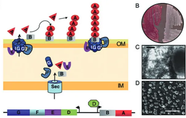
Curli assembly requires the products of at least two divergently transcribed operons, csgBACand csgDEFG (Fig. 2). The csgBAC operon encodes the protein sub-units of curli fibres, CsgA and CsgB (Arnqvistet al., 1992; Collinson et al., 1996; Johansson et al., 2001; Knobl et al., 2001; La Ragioneet al., 2001). A third gene in the operon,csgC, has no described function, nor has a tran-script or protein been detected inE. coli. A recent report on AgfC, encoded by thecsgChomologue inSalmonella spp., suggested that this protein influences the ultra-structural characteristics of curli (Gibsonet al., 2007).
[image:3.595.148.452.54.248.2]CsgA is secreted from cells in a soluble, unfolded state in the absence of CsgB (Hammar et al., 1996). CsgB, which is associated with the bacterial outer membrane, mediates the conversion of soluble CsgA into insoluble and aggregative cell-surface fibres (Hammaret al., 1996; Bian and Normark, 1997; Chapmanet al., 2002; Hammer et al., 2007). Interestingly, CsgA and CsgB need not be expressed from the same cell to display these polymer-ization activities. Curli fibres assemble on the surface of csgA- (CsgB+) cells if grown adjacent to cells secreting CsgA (csgB-) (Hammar et al., 1996; Chapman et al., 2002). Interbacterial complementation suggests a model for curli assembly termed ‘nucleation-precipitation’ and it Fig. 2. A secretion and assembly machine directs amyloid fibre formation inE. coli.
A. Model of curli assembly. Curli biosynthesis requires the products of the divergently transcribedcsgBACandcsgDEFGoperons. CsgD is a transcriptional activator of thecsgBACoperon. CsgB, CsgA, CsgE, CsgF and CsgG have SEC signal sequences that target them across the inner membrane (IM). CsgG, CsgE and CsgF are non-structural proteins that interact at the outer membrane (OM). CsgA and CsgB are secreted across the outer membrane in a CsgG-dependent manner (see text). CsgB interacts with the outer membrane and presents an amyloid-like template to soluble CsgA (red triangles). CsgA adopts the amyloid conformation (red ovals) and becomes anchored to the cell surface, where it can propagate thebsheet-rich amyloid fold onto unpolymerized CsgA monomers. It is unclear if CsgB is anchored directly to the outer membrane or to the assembly complex via a protein–protein interaction (both are shown).
B. Curli fibres bind the amyloid-specific dye Congo red. Curliated bacteria stain red when grown on media containing the diazo dye Congo red (left), butcsg-bacteria remain white (right).
C. Electron micrograph of curliated bacteria, where the bacteria were grown for 40 h at 26°C prior to analysis (Wanget al., 2007). Scale bar is 1mm.
provides an elegant means to control amyloid formation (Fig. 2). By separating the nucleator and polymerization activities into two proteins, the cell can direct amyloid formation by controlling when and where the CsgA and CsgB proteins interact.
ThecsgDEFGoperon encodes non-structural proteins essential for production, stability and secretion of the subunit proteins (Hammaret al., 1995; Lofereret al., 1997; Chapman et al., 2002; Robinson et al., 2006). Efficient secretion and polymerization of CsgA and CsgB requires CsgE, CsgF and CsgG. CsgD is a transcriptional activator of thecsgBACoperon, and is also a critical activator of several other operons necessary for biofilm formation, most notably, the cellulose biosynthesis pathway (Brom-bacheret al., 2003; Gerstel and Romling, 2003; Gerstel et al., 2003). The intergenic region betweencsgDEFGand csgBACis one of the largest inE. coli, and the expression of the curli operons is regulated by a complex blend of factors that fine-tune operon expression according to envi-ronmental conditions. The interplay between different regulatory factors leads to strain-specific responses to any given condition or set of conditions (Romling, 2005; Barn-hart and Chapman, 2006). CsgB, CsgA, CsgE, CsgF and CsgG each have putative Sec translocation sequences that target them for translocation across the cytoplasmic (inner) membrane. CsgG is an outer membrane-localized lipoprotein that is essential for stability and secretion of CsgA and CsgB (Loferer et al., 1997; Robinson et al., 2006). Purified CsgG forms barrel-shaped oligomeric structures (Fig. 2), and its expression is correlated with pore-like properties such as increased sensitivity to eryth-romycin (Robinsonet al., 2006). CsgG must be localized to the outer membrane in order to stabilize CsgA or CsgB, and CsgG variants localized either to the inner membrane or the periplasm are unable to stabilize or secrete the subunits (Robinsonet al., 2006). CsgG interacts with a specific domain at the N-terminus of CsgA; this domain can be used to target non-csg proteins to CsgG (Robinson et al., 2006). CsgG has also been demonstrated to co-immunoprecipitate with CsgE and CsgF at the outer membrane. CsgG is sufficient to mediate CsgA secretion out of the cell, but CsgE and CsgF are required for efficient curli fibre assembly under most conditions (Hammaret al., 1995; Chapman et al., 2002). The molecular details of CsgE and CsgF function are unclear, although some clues point to their activities.csgEmutants do not assemble curli fibres, and the stability of the CsgA and CsgB proteins is greatly reduced relative to wild type (Chapman et al., 2002).csgFmutants are deficient in nucleation and exhibit a delay in assembly of curli fibres (Chapmanet al., 2002; Hammer et al., 2007). Increased amounts of CsgA are secreted when cells lack CsgF, which suggests that CsgF has a negative effect on CsgA secretion (Chapmanet al., 2002; Hammeret al., 2007).
Curli biogenesis is an extremely efficient process, but the mechanism by which assembly is achieved is only beginning to be described. How does the cell prevent internal amyloid assembly (that is, association of CsgA and CsgB prior to their localization to the cell surface)? No accumulation of intracellular intermediates is detected in the absence of CsgG, although the reasons for this remain uncharacterized (Robinsonet al., 2006). In addi-tion, the functions of CsgE and CsgF appear to modulate some undefined aspect of CsgG activity, as secretion and stability of CsgA and CsgB are altered in this strain (Chapmanet al., 2002; Hammeret al., 2007). Fortunately, E. coli is highly amenable to biochemical and genetic analysis, making possible molecular dissection of nucle-ation and polymeriznucle-ation.
Molecular dissection of curli nucleation and polymerization
The amino acid sequences of CsgA and CsgB contain three readily identifiable domains: a Sec secretion signal, an N-terminal sequence that, at least for CsgA, targets the protein to CsgG, and an amyloid core domain that is incorporated into amyloid fibres (Collinson et al., 1991; Robinson et al., 2006; Wang et al., 2007). The amyloid core can be further divided into five repeating units. The repeating units are characterized by a fivefold internal symmetry, where each unit is comprised of 19–24 amino acids containing conserved serine, glutamine and aspar-agine residues (Wang et al., 2007). Interestingly, the CsgA repeating units have different propensities for aggregation, and three of the five units are distinctly amy-loidogenic (Olsenet al., 2002; Wanget al., 2007).
CsgA can be readily purified from the culture superna-tants of a strain overexpressing both CsgA and CsgG; CsgA is an unstructured, soluble monomer immediately following purification (Chapman et al., 2002). Purified CsgA self-assembles into amyloid fibres several hours after purification (Wanget al., 2007). Amyloid polymeriza-tion of CsgA in vitro can be followed in real time by monitoring the fluorescence emitted when the protein is mixed with the amyloid-specific dye thioflavin T (Wang et al., 2007).In vitrokinetic studies on CsgA polymeriza-tion suggest that the elongapolymeriza-tion process can happen in three distinct phases: a lag phase, a fast phase and a stationary phase (Fig. 1). The lag phase of CsgA polymer-ization can be abolished by adding a small amount of pre-formed CsgA fibres, or ‘seeds’ (Wanget al., 2007).
C-terminus mediates interaction with the outer mem-brane; truncation of CsgB results in its secretion away from the cells (Hammeret al., 2007). How the C-terminal domain mediates membrane interaction is undefined. The N-terminal domain of CsgB self-assembles into amyloid fibresin vitroand in some conditions can nucleate CsgA into curli fibresin vivo(Hammer et al., 2007). The CsgB N-terminal domain can abolish the lag phase of CsgA polymerization in vitro, suggesting that CsgB-mediated nucleation of CsgA may occur via presentation of an amyloid-like template to soluble CsgA molecules (Hammeret al., 2007).
The ability of CsgA and CsgB to self-assemble into fibres coupled with the ability of the fibres to seed CsgA amyloid polymerization suggests a template-driven mechanism for the assembly of curli fibres (Hammer et al., 2007; Wanget al., 2007). First, CsgA may adopt the amyloid conformation presented to it by CsgB at the cell surface. Second, amyloid CsgA can itself act as a tem-plate for the subsequent conversion and incorporation of unpolymerized CsgA monomers to the growing tip of the curli fibre (Fig. 2). It is noteworthy that full-length CsgB does not form fibres in vivo, and thus may be an ex-ample of a non-fibre-forming amyloid-like protein. The membrane-associated C-terminus of CsgB may prevent CsgB from polymerizing by anchoring the protein to the membrane, but the exact mechanism underlying this aspect of CsgB behaviour remains to be elucidated. CsgA, on the other hand, seems to be adapted to poly-merize into amyloid fibres and yet to only undergo that process, in vivo, when CsgB is available to nucleate it. Elucidation of the molecular details of the CsgB–CsgA interaction and the determinants of CsgA response to nucleation and self-seeding will provide new insights into amyloid assembly.
Amyloid formation of microcin E492: exploitation of toxic oligomers
Microcin E492 (MccE492) ofKlebsiella pneumoniaeis a toxic, bactericidal protein that assembles into oligomeric pores in the inner membrane of neighbouring bacteria. MccE492, which specifically targets Enterobacter species, is imported across the bacterial outer membrane in a receptor-mediated fashion (Destoumieux-Garzon et al., 2003). MccE492 has been shown to form voltage-independent ion channels in planar lipid bilayers. The molecular weight of MccE492 is 6 kDa, but conductance observed for MccE492 ion channels suggests that an oligomeric species forms this pore (Lagoset al., 1993). The toxicity of the MccE492 peptide changes over the growth cycle of the K. pneumoniae: toxicity is greatest during exponential phase and lowest during stationary phase. No difference in the mass of the peptide,
post-translational modification or protein stability can be detected during these different phases (Bieler et al., 2005).
The change in bactericidal activity of MccE492 when cells transition into stationary phase is proposed to be dependent on amyloidogenesis. MccE492 forms amyloid fibres in vivo that correspond to MccE492 toxicity decrease. The assembly of amyloid fibres can be moni-toredin vitro, and corresponds to a loss of toxicity (Bieler et al., 2005). Therefore, as MccE492 forms toxic oligo-meric channels in membranes, the biologically active MccE492 species is presumably a transient pre-amyloid oligomer. A tempting speculation is that the toxic MccE492 species has structural similarity with the toxic, transient oligomeric species formed by disease-associated amyloids. Interestingly, MccE492 has been shown to trigger apoptosis in some human cell lines, reminiscent of the toxic effect observed for disease-associated pre-amyloid oligomers (Hetzet al., 2002).K. pneumoniae pro-duces a small protein localized to the inner membrane, MceB, which confers resistance to MccE492 toxicity (Lagos et al., 1999). The mechanism of MceB-mediated immunity has not been reported.
Emerging bacterial amyloids: harpins, chaplins and MTP
domain is dispensable for hypersensitive response (Kimet al., 2004).
Streptomyces coelicolor are soil bacteria, akin to fila-mentous fungi, which produce aerial hyphae to dispense spores. The biogenesis of aerial hyphae is a physical challenge that requires a dramatic change in the surface hydrophobicity of the organism: the subterranean hyphae are hydrophilic while the aerial hyphae and spores are hydrophobic (Elliotet al., 2003). This change in hydropho-bicity is a result of the SDS-insoluble ‘surface layer’ on the aerial hyphae and spores. The formation of the surface layer requires a group of proteins called chaplins (Claessen et al., 2003). Chaplins in S. coelicolor are encoded by chpA-Hand are necessary for formation of aerial hyphae (Claessen et al., 2003). The chaplins assemble into a network of amyloid fibres in the surface layer; the fibres self-assemble tobsheet-rich fibresin vitro and bind amyloid-specific dyes. The amyloid properties of chaplins might impart a hydrophobic nature to the surface layer and cause a decrease in the surface tension at the air–water interface. The hyphae then could be freed from the soil and continue to grow up into the air (Claessen et al., 2004). The mechanism by which chaplin amyloids assemble and the individual contributions of the eight chaplin proteins to the amyloid network remain largely undefined. Remarkably, addition of crude chaplin-containing cell extract restores formation of aerial hyphae to mutantS. coelicolorfilaments lacking some of thechp genes, an observation reminiscent of the interbacterial complementation phenotype of curliated bacteria (Claessenet al., 2003). Further, there may be a temporal regulation influencing the expression of the genes encod-ing chaplins (Claessen et al., 2003), suggesting that a co-ordinated amyloid biosynthesis pathway is at work in the assembly of chaplin amyloids.
A recent report describes the presence of thin, highly aggregative protein fibres on the surface of Mycobacte-rium tuberculosis (Alteri et al., 2007). Electron micro-scopic analysis of the M. tuberculosispili (MTP) shows fibres that are strikingly similar to curli fibres. MTP are SDS-insoluble and bind Congo red, suggesting that MTP indeed may be an amyloid fibre. The significance of MTP fibres forM. tuberculosisphysiology is unknown, but more than half of recently examined tuberculosis patients possess antiserum to MTP, suggesting that MTP pro-duction may be part of M. tuberculosis pathogenesis (Alteriet al., 2007).
In conclusion: protein misfolding done right
The discovery of proteinaceous plaques in the brains of deceased dementia patients launched a hundred-year exploration for the factors that mediate the conversion of soluble, non-toxic proteins into aggregative amyloid
fibres. Despite this effort, the exact nature of amyloid toxicity and the initiation of amyloid protein aggregation remains unclear. Bacteria exploit amyloid folding path-ways to accomplish a variety of tasks, from attachment and colonization to toxicity and pathogenesis. Bacteria apparently utilize the toxic intermediate species (in the cases of MccE492 and the harpins) as well as the mature amyloid fibre (as in curli and the chaplins) in positive ways. The study of bacterial amyloid fibres may hold the key to understanding the methods nature has evolved to harness the power of amyloid formation and direct it to beneficial uses.
Acknowledgements
The authors thank Dr Ingyu Hwang for providing the electron micrograph of HpaG oligomers in Fig. 1. We also gratefully acknowledge members of the Chapman lab, especially Xuan Wang and Neal Hammer, for critical evaluation of this manuscript. The authors were supported by the NIH National Research Service Award T32GMO7544 to E.A.E. and by NIH RO1A1073847 to M.R.C.
References
Alteri, C.J., Xicohtencatl-Cortes, J., Hess, S., Caballero-Olin, G., Giron, J.A., and Friedman, R.L. (2007)Mycobacterium tuberculosis produces pili during human infection. Proc Natl Acad Sci USA104:5145–5150.
Arnqvist, A., Olsen, A., Pfeifer, J., Russell, D.G., and Normark, S. (1992) The Crl protein activates cryptic genes for curli formation and fibronectin binding in Escherichia coliHB101.Mol Microbiol6:2443–2452.
Barnhart, M.M., and Chapman, M.R. (2006) Curli biogenesis and function.Annu Rev Microbiol60:131–147.
Bian, Z., and Normark, S. (1997) Nucleator function of CsgB for the assembly of adhesive surface organelles in Escheri-chia coli.EMBO J16:5827–5836.
Bian, Z., Brauner, A., Li, Y., and Normark, S. (2000) Expres-sion of and cytokine activation by Escherichia coli curli fibers in human sepsis.J Infect Dis181:602–612. Bieler, S., Estrada, L., Lagos, R., Baeza, M., Castilla, J., and
Soto, C. (2005) Amyloid formation modulates the biological activity of a bacterial protein. J Biol Chem 280: 26880– 26885.
Bokranz, W., Wang, X., Tschape, H., and Romling, U. (2005) Expression of cellulose and curli fimbriae byEscherichia coliisolated from the gastrointestinal tract.J Med Microbiol 54:1171–1182.
Brombacher, E., Dorel, C., Zehnder, A.J., and Landini, P. (2003) The curli biosynthesis regulator CsgD co-ordinates the expression of both positive and negative determinants for biofilm formation inEscherichia coli.Microbiology149:
2847–2857.
Caughey, B., and Lansbury, P.T. (2003) Protofibrils, pores, fibrils, and neurodegeneration: separating the responsible protein aggregates from the innocent bystanders. Annu Rev Neurosci26:267–298.
Heuser, J., Hammar, M.,et al.(2002) Role ofEscherichia coli curli operons in directing amyloid fiber formation.
Science295:851–855.
Claessen, D., Rink, R., de Jong, W., Siebring, J., de Vreugd, P., Boersma, F.G.,et al.(2003) A novel class of secreted hydrophobic proteins is involved in aerial hyphae formation inStreptomyces coelicolorby forming amyloid-like fibrils.
Genes Dev17:1714–1726.
Claessen, D., Stokroos, I., Deelstra, H.J., Penninga, N.A., Bormann, C., Salas, J.A.,et al.(2004) The formation of the rodlet layer ofstreptomycetesis the result of the interplay between rodlins and chaplins.Mol Microbiol53:433–443. Collinson, S.K., Emody, L., Muller, K.H., Trust, T.J., and Kay, W.W. (1991) Purification and characterization of thin, aggregative fimbriae from Salmonella enteritidis.
J Bacteriol173:4773–4781.
Collinson, S.K., Clouthier, S.C., Doran, J.L., Banser, P.A., and Kay, W.W. (1996) Salmonella enteritidis agfBAC
operon encoding thin, aggregative fimbriae. J Bacteriol 178:662–667.
Destoumieux-Garzon, D., Thomas, X., Santamaria, M., Goulard, C., Barthelemy, M., Boscher, B., et al. (2003) Microcin E492 antibacterial activity: evidence for a TonB-dependent inner membrane permeabilization on Escheri-chia coli.Mol Microbiol49:1031–1041.
Elghetany, M.T., and Saleem, A. (1988) Methods for staining amyloid in tissues: a review.Stain Technol63:201–212. Elliot, M.A., Karoonuthaisiri, N., Huang, J., Bibb, M.J., Cohen,
S.N., Kao, C.M., and Buttner, M.J. (2003) The chaplins: a family of hydrophobic cell-surface proteins involved in aerial mycelium formation in Streptomyces coelicolor.
Genes Dev17:1727–1740.
Fernandez, L.A., and Berenguer, J. (2000) Secretion and assembly of regular surface structures in Gram-negative bacteria.FEMS Microbiol Rev24:21–44.
Fowler, D.M., Koulov, A.V., Balch, W.E., and Kelly, J.W. (2007) Functional amyloid – from bacteria to humans.
Trends Biochem Sci32:217–224.
Gerstel, U., and Romling, U. (2003) The csgD promoter, a control unit for biofilm formation in Salmonella typhimurium.Res Microbiol154:659–667.
Gerstel, U., Park, C., and Romling, U. (2003) Complex regu-lation of csgD promoter activity by global regulatory proteins.Mol Microbiol49:639–654.
Gibson, D.L., White, A.P., Rajotte, C.M., and Kay, W.W. (2007) AgfC and AgfE facilitate extracellular thin aggrega-tive fimbriae synthesis in Salmonella Enteritidis. Micro-biology153:1131–1140.
Gophna, U., Barlev, M., Seijffers, R., Oelschlager, T.A., Hacker, J., and Ron, E.Z. (2001) Curli fibers mediate inter-nalization of Escherichia coli by eukaryotic cells. Infect Immun69:2659–2665.
Hammar, M., Arnqvist, A., Bian, Z., Olsen, A., and Normark, S. (1995) Expression of two csgoperons is required for production of fibronectin- and congo red-binding curli poly-mers inEscherichia coliK-12.Mol Microbiol18:661–670. Hammar, M., Bian, Z., and Normark, S. (1996) Nucleator-dependent intercellular assembly of adhesive curli organelles inEscherichia coli.Proc Natl Acad Sci USA93:
6562–6566.
Hammer, N.D., Schmidt, J.C., and Chapman, M.R. (2007)
The curli nucleator protein, CsgB, contains an amy-loidogenic domain that directs CsgA polymerization.Proc Natl Acad Sci USA104:12494–12499.
Heath, M.C. (2000) Hypersensitive response-related death.
Plant Mol Biol44:321–334.
Hetz, C., Bono, M.R., Barros, L.F., and Lagos, R. (2002) Microcin E492, a channel-forming bacteriocin from Kleb-siella pneumoniae, induces apoptosis in some human cell lines.Proc Natl Acad Sci USA99:2696–2701.
Johansson, C., Nilsson, T., Olsen, A., and Wick, M.J. (2001) The influence of curli, a MHC-I-binding bacterial surface structure, on macrophage–T cell interactions. FEMS Immunol Med Microbiol30:21–29.
Jonson, A.B., Normark, S., and Rhen, M. (2005) Fimbriae, pili, flagella and bacterial virulence.Contrib Microbiol12:
67–89.
Kayed, R., Head, E., Thompson, J.L., McIntire, T.M., Milton, S.C., Cotman, C.W., and Glabe, C.G. (2003) Common structure of soluble amyloid oligomers implies common mechanism of pathogenesis.Science300:486–489. Kayed, R., Head, E., Sarsoza, F., Saing, T., Cotman, C.W.,
Necula, M., et al. (2007) Fibril specific, conformation dependent antibodies recognize a generic epitope common to amyloid fibrils and fibrillar oligomers that is absent in prefibrillar oligomers.Mol Neurodegener2:18. Kim, J.G., Jeon, E., Oh, J., Moon, J.S., and Hwang, I. (2004)
Mutational analysis ofXanthomonas harpin HpaG identi-fies a key functional region that elicits the hypersensitive response in nonhost plants.J Bacteriol186:6239–6247. Knobl, T., Baccaro, M.R., Moreno, A.M., Gomes, T.A., Vieira,
M.A., Ferreira, C.S., and Ferreira, A.J. (2001) Virulence properties ofEscherichia coliisolated from ostriches with respiratory disease.Vet Microbiol83:71–80.
La Ragione, R.M., Coles, K.E., Jorgensen, F., Humphrey, T.J., and Woodward, M.J. (2001) Virulence in the chick model and stress tolerance ofSalmonella entericaserovar Orion var. 15+.Int J Med Microbiol290:707–718. Lagos, R., Wilkens, M., Vergara, C., Cecchi, X., and
Monas-terio, O. (1993) Microcin E492 forms ion channels in phos-pholipid bilayer membrane.FEBS Lett321:145–148. Lagos, R., Villanueva, J.E., and Monasterio, O. (1999)
Iden-tification and properties of the genes encoding microcin E492 and its immunity protein.J Bacteriol181:212–217. Larsen, P., Nielsen, J.L., Dueholm, M.S., Wetzel, R., Otzen,
D., and Nielsen, P.H. (2007) Amyloid adhesins are abun-dant in natural biofilms.Environ Microbiol9:3077–3090. Larsen, P., Nielsen, J.L., Otzen, D., and Nielsen, P.H. (2008)
Amyloid-like adhesins in floc-forming and filamentous bac-teria in activated sludge.Appl Environ Microbiol74: 1517– 1526.
Lawley, T.D., Chan, K., Thompson, L.J., Kim, C.C., Govoni, G.R., and Monack, D.M. (2006) Genome-wide screen for
salmonellagenes required for long-term systemic infection of the mouse.PLoS Pathog2:e11.
Lee, J., Klusener, B., Tsiamis, G., Stevens, C., Neyt, C., Tampakaki, A.P.,et al.(2001) HrpZ (Psph) from the plant pathogenPseudomonas syringaepv. phaseolicola binds to lipid bilayers and forms an ion-conducting pore in vitro.
Proc Natl Acad Sci USA98:289–294.
during assembly of fibronectin-binding curli is limited by the intracellular concentration of the novel lipoprotein CsgG.
Mol Microbiol26:11–23.
Nelson, R., Sawaya, M.R., Balbirnie, M., Madsen, A.O., Riekel, C., Grothe, R., and Eisenberg, D. (2005) Structure of the cross-beta spine of amyloid-like fibrils.Nature435:
773–778.
Nordstedt, C., Naslund, J., Tjernberg, L.O., Karlstrom, A.R., Thyberg, J., and Terenius, L. (1994) The Alzheimer A beta peptide develops protease resistance in association with its polymerization into fibrils.J Biol Chem269:30773–30776. Oh, J., Kim, J.G., Jeon, E., Yoo, C.H., Moon, J.S., Rhee, S., and Hwang, I. (2007) Amyloidogenesis of type III-dependent harpins from plant pathogenic bacteria.J Biol Chem282:13601–13609.
Olsen, A., Jonsson, A., and Normark, S. (1989) Fibronectin binding mediated by a novel class of surface organelles on
Escherichia coli.Nature338:652–655.
Olsen, A., Wick, M.J., Morgelin, M., and Bjorck, L. (1998) Curli, fibrous surface proteins ofEscherichia coli, interact with major histocompatibility complex class I molecules.
Infect Immun66:944–949.
Olsen, A., Herwald, H., Wikstrom, M., Persson, K., Mattsson, E., and Bjorck, L. (2002) Identification of two protein-binding and functional regions of curli, a surface organelle and virulence determinant ofEscherichia coli.J Biol Chem 277:34568–34572.
Pawar, D.M., Rossman, M.L., and Chen, J. (2005) Role of curli fimbriae in mediating the cells of enterohaemorrhagic
Escherichia colito attach to abiotic surfaces.J Appl Micro-biol99:418–425.
Robinson, L.S., Ashman, E.M., Hultgren, S.J., and Chapman, M.R. (2006) Secretion of curli fibre subunits is mediated by the outer membrane-localized CsgG protein.Mol Microbiol 59:870–881.
Romling, U. (2005) Characterization of the rdar morphotype, a multicellular behaviour inEnterobacteriaceae.Cell Mol Life Sci62:1234–1246.
Ross, C.A., and Poirier, M.A. (2004) Protein aggregation and neurodegenerative disease. Nat Med 10 (Suppl.): S10– S17.
Ryu, J.H., Kim, H., Frank, J.F., and Beuchat, L.R. (2004) Attachment and biofilm formation on stainless steel by
Escherichia coliO157: H7 as affected by curli production.
Lett Appl Microbiol39:359–362.
Sunde, M., Serpell, L.C., Bartlam, M., Fraser, P.E., Pepys, M.B., and Blake, C.C. (1997) Common core structure of amyloid fibrils by synchrotron X-ray diffraction.J Mol Biol 273:729–739.
Uhlich, G.A., Cooke, P.H., and Solomon, E.B. (2006) Analy-ses of the red-dry-rough phenotype of anEscherichia coli
O157: H7 strain and its role in biofilm formation and resis-tance to antibacterial agents.Appl Environ Microbiol72:
2564–2572.
Uhlich, G.A., Keen, J.E., and Elder, R.O. (2002) Variations in thecsgDpromoter ofEscherichia coliO157: H7 associated with increased virulence in mice and increased invasion of HEp-2 cells.Infect Immun70:395–399.
Wang, X., Smith, D.R., Jones, J.W., and Chapman, M.R. (2007) In vitro polymerization of a functional
Escherichia coliamyloid protein.J Biol Chem282:3713– 3719.
Yoshiike, Y., Kayed, R., Milton, S.C., Takashima, A., and Glabe, C.G. (2007) Pore-forming proteins share structural and functional homology with amyloid oligomers. Neuro-molecular Med9:270–275.