Open Access
R E S E A R C H A R T I C L E
Bio
Med
Central
© 2010 Windolf et al; licensee BioMed Central Ltd. This is an Open Access article distributed under the terms of the Creative CommonsAttribution License (http://creativecommons.org/licenses/by/2.0), which permits unrestricted use, distribution, and reproduction in any medium, provided the original work is properly cited.Research article
Biomechanical investigation of an alternative
concept to angular stable plating using
conventional fixation hardware
Markus Windolf*
1, Kajetan Klos
1, Dirk Wähnert
1, Bas van der Pol
1, Roman Radtke
1, Karsten Schwieger
1and
Roland P Jakob
2Abstract
Background: Angle-stable locking plates have improved the surgical management of fractures. However, locking implants are costly and removal can be difficult. The aim of this in vitro study was to evaluate the biomechanical performance of a newly proposed crossed-screw concept ("Fence") utilizing conventional (non-locked) implants in comparison to conventional LC-DCP (limited contact dynamic compression plate) and LCP (locking compression plate) stabilization, in a human cadaveric diaphyseal gap model.
Methods: In eight pairs of human cadaveric femora, one femur per pair was randomly assigned to receive a Fence construct with either elevated or non-elevated plate, while the contralateral femur received either an LCP or LC-DCP instrumentation. Fracture gap motion and fatigue performance under cyclic loading was evaluated successively in axial compression and in torsion. Results were statistically compared in a pairwise setting.
Results: The elevated Fence constructs allowed significantly higher gap motion compared to the LCP
instrumentations (axial compression: p ≤ 0.011, torsion p ≤ 0.015) but revealed similar performance under cyclic loading (p = 0.43). The Fence instrumentation with established bone-plate contact revealed larger fracture gap motion under axial compression compared to the conventional LC-DCP osteosynthesis (p ≤ 0.017). However, all contact Fence specimens survived the cyclic test, whereas all LC-DCP constructs failed early during torsion testing (p < 0.001). All failures occurred due to breakage of the screw heads.
Conclusions: Even though accentuated fracture gap motion became obvious, the "Fence" technique is considered an alternative to cost-intensive locking-head devices. The concept can be of interest in cases were angle-stable implants are unavailable and can lead to new strategies in implant design.
Background
The devices known as angular stable internal fixators have enhanced the armamentarium for surgical fracture treatment [1-3]. The mechanical principle of these implants is the locking of the screw head into the plate, resulting in a load transfer via plate and screws [1-3]. This increases the stability of the construct and eliminates the risk of loss of reduction due to screw toggling. Further-more, the periosteal blood supply of the bone under the device is preserved, since there is no need for contact or
compression between plate and bone. Biomechanical studies [4-6] have shown the advantages of angle-stable plate fixation over conventional plating. However, several unique complications have been noted, such as difficulty with implant removal and implant cut out in osteoporotic bone [7]. Furthermore, locking implants increase the cost of surgery, which is why many surgeons are restricted in the use of angle-stable fixation hardware. Developing countries and countries with a small budget health care system rarely use these techniques [8,9].
The objective of this study was to compare the biome-chanical performance of a newly proposed crossed screw technique ("Fence") utilizing a conventional LC-DCP (limited contact dynamic compression plate) to LCP
* Correspondence: [email protected]
1 AO Research Institute Davos, AO Foundation, Clavadelerstrasse 8, 7270 Davos,
Switzerland
(locking compression plate) and standard LC-DCP stabi-lization in a human cadaveric diaphyseal gap model. Frac-ture gap motion and fatigue properties under cyclic loading were evaluated under axial compression and tor-sion.
The null hypothesis was that the construct created with conventional screws in a crossed configuration would yield biomechanical results comparable to those achieved with the other instrumentations.
Methods
Specimens and study-groups
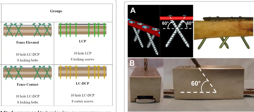
Eight pairs of fresh frozen (-20°C) human cadaveric fem-ora (7 male, 1 female donors; mean donor age 74 years; range 64 - 83 years) were obtained from the department of Pathology, Kantonsspital Basel, Switzerland, where they had been harvested post mortem with appropriate consent of the relatives. Use of the specimens for the pur-pose of the present study was approved by the ethical commission of Kantonsspital Basel. Soft tissue was removed before instrumentation and mechanical testing. Bone mineral density (BMD) was measured by means of CT-scanning (XtremeCT, SCANCO Medical AG, Bassersdorf, Switzerland) in the cortical bone of the femur diaphysis. The specimens were pairwise assigned to four study-groups according to Figure 1. Within each pair of femora, one femur was randomly assigned to receive a Fence (elevated or contact) construct, while the contralateral femur was assigned to receive an LCP or a conventional LC-DCP instrumentation. Two test-series were established. Series 1: Pairwise comparison between the elevated Fence construct and the LCP instrumenta-tion. Series 2: pairwise comparison between the contact
Fence construct and the conventional LC-DCP instru-mentation.
Instrumentations
For the Fence constructs, LC-DCP plates (10-hole, 4.5-mm broad LC-DCP) and conventional 4.9-4.5-mm self-tap-ping non-locking head locking bolts were used. Locking bolts were preferred over cortex screws, because of an increased core diameter and altered loading environ-ment. In contrast to conventional compression osteosyn-thesis, where screws are loaded in tension, shear and bending is expected here. The bolts were routed in a criss-cross pattern resembling a fence, inserted with 60° angulation to the longitudinal shaft axis in pairwise con-verging bicortical arrangement (Figure 2A). The Fence technique is based on the geometrical principle that the plate can not be displaced along non-parallel screw axes and is therefore constrained. To avoid contact between neighboured screws at the crossing point, a custom-made drill guide was used (Figure 2B). For the elevated Fence constructs (Series 1), the plates were offset from the bone surface by 5-mm-thick plastic spacers (Figure 3B); for the contact Fence constructs (Series 2), the plates were placed directly on the cortex.
[image:2.595.60.309.505.695.2]For the LCP (Series 1) and the conventional LC-DCP constructs (Series 2), standard plating techniques were used (Figure 3A). The LCP plates (10-hole, 4.5/5.0-mm broad LCP) were attached with 4.9-mm self-tapping head locking screws inserted through the threaded portion of the combination hole provided in the plate. Head locking screws were tightened using a torque limiter. 5-mm-thick spacers were used to offset the plates from the cortex (Figure 3B). The LC-DCP plates were placed directly on
[image:2.595.87.532.507.703.2]Figure 1 Study-groups and test-series. Pairwise comparisons were carried out investigating the elevated Fence construct versus the LCP instrumentation (Series 1) and the contact Fence technique versus the LC-DCP instrumentation (Series 2).
the cortex and attached using 4.5-mm self-tapping bicor-tical cortex screws, since 4.9-mm screws of this type are not available. All conventional screws and bolts were tightened by hand following the clinical practise.
All implants were obtained from the same manufac-turer (Synthes GmbH, Bettlach, Switzerland). Plate mate-rial was stainless steel; all screws and bolts were made of Titanium. All plates were positioned in the centre of the femoral shaft. A 10-mm transverse osteotomy was cre-ated with an oscillating saw below plate holes 5 and 6 to simulate an unstable diaphyseal fracture (Figure 3B). All instrumentations were performed by the same experi-enced surgeon. The investigated constructs and test-series are visualized in Figure 1. For details of the hard-ware see Figure 4.
Mechanical testing
The bones were cut proximally and distally at a distance of 60 mm from the ends of each plate, and potted in Polymethylmethacrylate (PMMA, Beracryl, W. Troller Kunststoffe AG, Jegenstorf, Switzerland). At either end of the plate, a 5-mm distance was ensured between the plate and the potting material (Figure 3B).
Fatigue performances, construct stiffness and fracture gap motion were investigated in a biomechanical experi-ment consisting of a cyclic axial compression test fol-lowed by cyclic torsion until failure of the construct. The test was carried out on a servo-hydraulic test system (858 Mini Bionix® II, MTS Systems Corporation, Eden Prairie, USA) in axial-torsional configuration, equipped with a 25-kN/250-Nm load cell. For the axial compression test the load was proximally introduced via a metal sphere centred on the axis of the femur. A second sphere was located at the distal end of the specimen. The spheres were chosen to replicate the function of the hip and knee joints. The distance between the plate and the centre of the sphere in mediolateral direction was kept constant within each bone pair, so as to ensure a constant lever arm. Sinusoidal axial compression was performed between 100 and 1000 N at 1 Hz for 5000 cycles (Figure 5A). In case no fatal failure occurred, the axial test was continued in cyclic torsion. The proximal sphere was replaced by a double-cardanic joint for the transfer of torque. The distal part was rigidly affixed to the base-plate. Sinusoidal loading was carried out between +20 and -20 Nm at 1 Hz for another 5000 cycles or until struct failure (Figure 5B). The axial load was kept con-stant at 0 N throughout the torsion test.
Data acquisition and analysis
[image:3.595.59.290.98.199.2]Displacement, load, angle, and torque were recorded, at a rate of 50 Hz, from the transducers of the test system. Additionally, an optical 3D motion tracking system with five ProReflex MCU digital cameras (Qualisys Motion Capture System; Qualisys AB, Gothenburg, Sweden) was used to identify relative motion in the fracture gap. Reflective marker-sets were attached to the proximal and distal femur shaft fragments (Figure 5). The fracture gap angle under axial loading and the torsional deformation were calculated for all specimens throughout the test (Figure 6). Initial construct stiffness in axial direction was determined at the beginning of the test as change in frac-ture-gap angulation per unit load. Torsional stiffness was
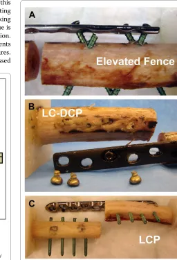
[image:3.595.59.288.540.702.2]Figure 3 Fracture model. A 10-mm mid-diaphyseal gap was created to simulate a severely comminuted fracture. (A) LC-DCP instrumenta-tion. (B) Fence construct with 5 mm elevated plate.
Figure 4 Implants. Implants used in the study. Left: 10-hole LCP plate and locking screws. Right: 10-hole LC-DCP plate with conventional and "Fence" screw configuration.
[image:3.595.302.540.592.704.2]defined as torsional deformation per unit torque at the beginning of the torsion test. Additionally, the range of motion in the fracture gap was defined as amplitude of the gap angle/torsional angle within one load cycle and was evaluated at 1, 2000 and 4000 test cycles for the axial as well as for the torsion test, if applicable. Fatigue perfor-mance of the constructs was quantified by the number of load cycles until an angular deformation larger then 15° was reached or when an obvious failure of the osteosyn-thesis occurred. A threshold of 15° was chosen from pilot experiments using artificial bones.
For comparisons within test-series 1 and 2 (elevated Fence versus LCP; contact Fence versus LC-DCP), paired t-tests were employed on cycles to failure and range of gap motion at 1, 2000 and 4000 cycles. Furthermore, a Repeated Measures ANOVA (analysis of variance) was used to compare between gap motion at 1, 2000 and 4000 cycles within each group. A statistical software package (SPSS 18.0, SPSS Inc., Chicago, USA) was used. Level of significance was set to α = 0.05.
Results
Cortical bone density was 625 ± 204 mgHA/cm3 (mean ± SD) for the elevated Fence specimens, 612 ± 202 mgHA/ cm3 for the LCP samples, 608 ± 70 mgHA/cm3 for the contact Fence group and 585 ± 129 mgHA/cm3 for the
LC-DCP specimens. The donor's mean age was 76 years (range 70 - 83 years, 7 male and 1 female).
At the beginning of the axial test (cycle 1), the highest gap motion was observed for the elevated Fence speci-mens (3.52 ± 0.16°, mean ± SD) compared to 2.41 ± 0.18° for the LCP constructs. This difference in gap motion remained statistically significant for all time points (all p ≤ 0.011, Figure 7). The gap motion of the contact Fence group was 2.66 ± 0.51° compared to 1.51 ± 0.33° for the LC-DCP constructs which was the highest observed rigidity in the course of testing. This difference in gap motion was also found statistically significant for all time points (all p ≤ 0.017, Figure 7). When comparing the motion in the fracture gap between time points (1, 2000, 4000 cycles) within each group, no statistical differences were observed (all p ≥ 0.097).
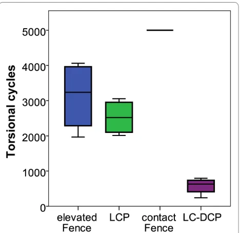
Regarding torsional gap motion at cycle 1, the elevated Fence constructs revealed again the highest values of the experiment (20.5 ± 2.1°). In comparison, the LCP speci-mens showed a significantly lower gap motion in torsion of 14.3 ± 0.6° (all p ≤ 0.015, Figure 8). The lowest torsional gap motion at cycle 1 was observed for the contact Fence group (11.9 ± 2.5°) compared to 15.9 ± 3.3° for the LC-DCP specimens. This difference was, however, not signif-icant (p = 0.273, Figure 8). Corresponding values for con-struct stiffness are shown in Table 1.
All specimens survived the cyclic test in axial compres-sion. During cyclic torsion, failures occurred due to breakage of the screws at the screw heads (Figure 9 A-C). The elevated Fence and LCP constructs showed similar numbers of load cycles to failure (p = 0.43): 3'125 ± 1'008 for the elevated Fence group and 2'526 ± 505 for the LCP specimens. The LC-DCP constructs failed earliest (574 ± 239). In contrast, all contact Fence instrumentations
[image:4.595.56.292.93.344.2]sur-Figure 6 Data evaluation. Schematic sketch of axial and torsional de-formations as determined from optical motion tracking. Left: definition of the fracture gap angulation as measure for axial deformation. Right: torsional deformation angle.
[image:4.595.304.538.516.679.2]vived the cyclic torsion test. This difference was statisti-cally significant (p < 0.001, Figure 10).
Discussion
We investigated the concept of a lower-cost plating tech-nique that would confer the same benefits as those offered by locking plates. The hallmark feature of this technique was the criss-cross pattern of screw routing ("Fence"), using conventional (non-locking-head) locking bolts. An additional advantage of the Fence technique is the variable direction of angulation and screw insertion. Thus, it might be possible to fix additional fragments especially when treating multi-fragmentary fractures. Furthermore, periprosthetic fractures may be addressed
using the Fence technique passing the stem of the pros-thesis anterior or posterior and at the same time achiev-ing angular stability. The technique with its advantages and opportunities might, however, be more demanding to apply compared to e.g. an LCP instrumentation. A cer-tain experience and skill level of the surgeon is required to avoid complications like screw collisions during implant placement. For further commercialization of the technique an easy-to-use drill template might be an option for ease of the procedure.
[image:5.595.58.277.113.206.2]In a first step, we compared the Fence technique with established bone contact to conventional, non-locked plating. The conventional constructs were most rigid under axial loading, but failed earliest during cyclic tor-sional testing, while none of the contact Fence specimens failed. This suggests that the contact Fence technique car-ries potential to enhance the construct's fatigue proper-ties under cyclic loading conditions compared to
Table 1: Construct stiffness
Axial stiffness [N/°]
Torsional stiffness [Nm/°]
elevated Fence 92 ± 25 2.1 ± 0.6
LCP 171 ± 19 2.9 ± 0.1
contact Fence 148 ± 29 3.5 ± 0.4
LC-DCP 299 ± 118 3.0 ± 1.0
[image:5.595.275.531.326.701.2]Axial and torsional stiffness for all study groups as obtained form a static loading ramp at the beginning of the cyclic axial and torsion tests. Values represent mean ± SD.
Figure 8 Torsional range of gap motion. Boxplots of the derived motion in the fracture gap during the cyclic torsion test for all study groups. The evaluation was carried out from the motion tracking data at 1 and 2000 cycles if applicable. The LC-DCP specimens already failed before the second evaluation step. Comparisons at a later time-point are not feasible.
[image:5.595.63.304.429.675.2]conventional plating. However, it has to be taken into account that different screw types with slightly different core diameters were used (cortex screw vs. locking bolt). An influence of this factor can not be excluded. Several authors have investigated the biomechanical properties of locking plates versus conventional plates; findings have been mixed [5,10-14]. Even though not tested in a direct comparison, we found that the LC-DCP constructs failed markedly earlier than did the LCP instrumentations, which would agree with the findings by Lill et al. [15] that flexible constructs are better able to withstand cyclic loading.
In a second test-series, we evaluated a non-contact Fence instrumentation and compared it to an LCP fixa-tion. Both osteosyntheses reflected comparable fatigue properties. However, the Fence technique showed signifi-cantly higher fracture gap motion under axial and tor-sional loading. There is insecurity about the optimal amount of micro-motion in the fracture gap for enhanced bone healing. Hypothetically, a less rigid construct could be advantageous by potentially stimulating callus forma-tion. On the other hand, extensive motion could lead to delayed unions or could cause pseudarthrosis. Although the senior author treated 12 patients successfully with this technique in his trauma centre, further studies-and, in particular, clinical trials-will be required for a defini-tive assessment of the utility of the technique described in this paper. However, such work would appear to be jus-tified in light of the results of the present study.
Other aspects requiring further investigation might be the screw angulation and the distance of an elevated con-struct from the cortex of the bone. Ahmad et al. [10] compared LCPs applied at different distances (flush to bone; 2 mm, and 5 mm off the bone), with a DCP control, and found comparable biomechanical behaviour and sim-ilar results in the DCP and the LCP constructs in which the plate was applied at or less than 2 mm from the bone. LCP constructs 5 mm off the cortex showed increased plastic deformation and lower failure loads. Similarly, Fulkerson et al. [5], investigating locked-screw con-structs, found that increasing the bone-plate distance sig-nificantly decreased construct stability. We believe that in our study 5-mm elevation of the plates produced a lever-arm effect at the unsupported free part of the screws which considerably affected the mechanical behaviour of the elevated constructs. Regarding angulation of the Fence pattern, a standardized screw angle of 60° was cho-sen. The potential effect of this angle on the construct stability was not subject to our investigation. With increasing screw angle the entry points of adjacent screws would approach each other at the near cortex, which could induce a potential weak point. We concluded that fatigue performance and rigidity of the Fence construct may be further optimized by adjusting the bone-plate dis-tance and the screw pattern angulation. Another draw-back of the method might be the interdependency within screw pairs. Given only one screw pair is used, failure of one screw would lead to simultaneous loss of stability of the second screw and hence, to failure of the construct.
Our experiment was subject to the limitations common to biomechanical studies. The in vivo loading environ-ment could only be mimicked in a restricted way. We decided to test successively in axial compression and tor-sion considered as most relevant loading patterns. The sample size was small due to limited availability of bone specimens. We, therefore, decided to carry out only pair-wise comparisons without considering the relations between unmatched study-groups. However, conclu-sions drawn from our findings, based on a low sample size still need to be viewed critically.
Conclusions
[image:6.595.58.292.94.321.2]This study introduces a plating technique with crossed screw configuration ("Fence") as a potential alternative to cost-intensive locking-head devices. The fatigue perfor-mance was found comparable to angular stable plating, whereas the "Fence" construct allowed larger motion in the fracture gap. A potential influence on bone healing can not be evaluated here. The technique can be of inter-est in cases were angle-stable implants are unavailable or may lead to new strategies for implant development.
Competing interests
The authors declare that they have no competing interests.
Authors' contributions
MW planned the study, carried out the statistics and drafted the manuscript. KK and DW evaluated the data and drafted the manuscript. BV and RR devel-oped the setup and carried out the experiments, KS supported in study plan-ning and helped in the paper draft. RPJ developed the idea of the study and performed the instrumentations. All authors read and approved the final man-uscript.
Acknowledgements
We thank B. Gueorguiev for his valuable support and expertise in optical motion tracking and data evaluation.
Author Details
1AO Research Institute Davos, AO Foundation, Clavadelerstrasse 8, 7270 Davos,
Switzerland and 2Orthopädie und Traumatologie HFR, Kantonsspital Freiburg,
1708 Freiburg, Switzerland
References
1. Perren SM: Backgrounds of the technology of internal fixators. Injury 2003, 34(Suppl 2):B1-B3.
2. Egol KA, Kubiak EN, Fulkerson E, Kummer FJ, Koval KJ: Biomechanics of locked plates and screws. J Orthop Trauma 2004, 18:488-493.
3. Wagner M: General principles for the clinical use of the LCP. Injury 2003, 34(Suppl 2):B31-B42.
4. Fitzpatrick DC, Doornink J, Madey SM, Bottlang M: Relative stability of conventional and locked plating fixation in a model of the osteoporotic femoral diaphysis. Clin Biomech (Bristol, Avon) 2009, 24:203-209.
5. Fulkerson E, Egol KA, Kubiak EN, Liporace F, Kummer FJ, Koval KJ: Fixation of diaphyseal fractures with a segmental defect: a biomechanical comparison of locked and conventional plating techniques. J Trauma 2006, 60:830-835.
6. Miller DL, Goswami T: A review of locking compression plate biomechanics and their advantages as internal fixators in fracture healing. Clin Biomech (Bristol, Avon) 2007, 22:1049-1062.
7. Haidukewych GJ, Ricci W: Locked plating in orthopaedic trauma: a clinical update. J Am Acad Orthop Surg 2008, 16:347-355. 8. Dewo P, Sharma PK, Tas HF van der, Houwen EB van der, Timmer M,
Magetsari R, Busscher HJ, van Horn JR, Verkerke GJ: Surface properties of Indonesian-made narrow dynamic compression plates. Med J Malaysia 2008, 63(Suppl A):21-22.
9. Angelini AJ, Livani B, Flierl MA, Morgan SJ, Belangero WD: Less invasive percutaneous wave plating of simple femur shaft fractures: A prospective series. Injury 2010.
10. Ahmad M, Nanda R, Bajwa AS, Candal-Couto J, Green S, Hui AC: Biomechanical testing of the locking compression plate: when does the distance between bone and implant significantly reduce construct stability? Injury 2007, 38:358-364.
11. DeTora M, Kraus K: Mechanical testing of 3.5 mm locking and non-locking bone plates. Vet Comp Orthop Traumatol 2008, 21:318-322. 12. Gardner MJ, Griffith MH, Demetrakopoulos D, Brophy RH, Grose A, Helfet
DL, Lorich DG: Hybrid locked plating of osteoporotic fractures of the humerus. J Bone Joint Surg Am 2006, 88:1962-1967.
13. O'Toole RV, Andersen RC, Vesnovsky O, Alexander M, Topoleski LD, Nascone JW, Sciadini MF, Turen C, Eglseder WA Jr: Are locking screws advantageous with plate fixation of humeral shaft fractures? A biomechanical analysis of synthetic and cadaveric bone. J Orthop Trauma 2008, 22:709-715.
14. Uhl JM, Seguin B, Kapatkin AS, Schulz KS, Garcia TC, Stover SM: Mechanical comparison of 3.5 mm broad dynamic compression plate, broad limited-contact dynamic compression plate, and narrow locking compression plate systems using interfragmentary gap models. Vet Surg 2008, 37:663-673.
15. Lill H, Hepp P, Korner J, Kassi JP, Verheyden AP, Josten C, Duda GN: Proximal humeral fractures: how stiff should an implant be? A
comparative mechanical study with new implants in human specimens. Arch Orthop Trauma Surg 2003, 123:74-81.
Pre-publication history
The pre-publication history for this paper can be accessed here: http://www.biomedcentral.com/1471-2474/11/95/prepub
doi: 10.1186/1471-2474-11-95
Cite this article as: Windolf et al., Biomechanical investigation of an alterna-tive concept to angular stable plating using conventional fixation hardware
BMC Musculoskeletal Disorders 2010, 11:95
Received: 22 January 2010 Accepted: 21 May 2010 Published: 21 May 2010
This article is available from: http://www.biomedcentral.com/1471-2474/11/95 © 2010 Windolf et al; licensee BioMed Central Ltd.
This is an Open Access article distributed under the terms of the Creative Commons Attribution License (http://creativecommons.org/licenses/by/2.0), which permits unrestricted use, distribution, and reproduction in any medium, provided the original work is properly cited.