LEABHARLANN CHOLAISTE NA TRIONOIDE, BAILE ATHA CLIATH TRINITY COLLEGE LIBRARY DUBLIN OUscoil Atha Cliath The University of Dublin
Terms and Conditions of Use of Digitised Theses from Trinity College Library Dublin
Copyright statement
All material supplied by Trinity College Library is protected by copyright (under the Copyright and Related Rights Act, 2000 as amended) and other relevant Intellectual Property Rights. By accessing and using a Digitised Thesis from Trinity College Library you acknowledge that all Intellectual Property Rights in any Works supplied are the sole and exclusive property of the copyright and/or other I PR holder. Specific copyright holders may not be explicitly identified. Use of materials from other sources within a thesis should not be construed as a claim over them.
A non-exclusive, non-transferable licence is hereby granted to those using or reproducing, in whole or in part, the material for valid purposes, providing the copyright owners are acknowledged using the normal conventions. Where specific permission to use material is required, this is identified and such permission must be sought from the copyright holder or agency cited.
Liability statement
By using a Digitised Thesis, I accept that Trinity College Dublin bears no legal responsibility for the accuracy, legality or comprehensiveness of materials contained within the thesis, and that Trinity College Dublin accepts no liability for indirect, consequential, or incidental, damages or losses arising from use of the thesis for whatever reason. Information located in a thesis may be subject to specific use constraints, details of which may not be explicitly described. It is the responsibility of potential and actual users to be aware of such constraints and to abide by them. By making use of material from a digitised thesis, you accept these copyright and disclaimer provisions. Where it is brought to the attention of Trinity College Library that there may be a breach of copyright or other restraint, it is the policy to withdraw or take down access to a thesis while the issue is being resolved.
Access Agreement
By using a Digitised Thesis from Trinity College Library you are bound by the following Terms & Conditions. Please read them carefully.
Tr in it y c o L L E G ^
2 7 MAY 2008
Systemic Inflammation after Cardiac
Surgery
By
Edel Duggan, MB, BCh, BAO, FCARCSI
A Thesis Submitted in Fulfillment of the Requirements for the
Degree of Doctor of Medicine (M.D)
Faculty of Medicine
Trinity College Dublin
This work was carried out under the supervision o f
Dr. Thomas Ryan and Dr Ross Me Manus,
Declaration
I wish to declare that this thesis has not been submitted for a degree at this or any
other University and it is entirely m y own work. I agree that the Library may lend or
Summary of Contents
Summary o f Contents 3
Detailed Table o f Contents 4
List o f Tables 7
List o f Figures 8
Abbreviations 9
Acknowledgem ents 14
Dedication 15
Summary o f Thesis 16
1. Introduction 18
2. Study Design and M ethodology 52
3. Tum our Necrosis Factor and Interleukin 10 Gene Expression in Peripheral Blood M ononuclear Cells after Cardiac Surgery: Experimental Series I 73 4. Coagulopathy following Cardiac Surgery :Influence o f Plasminogen Activator
Inhibitor Polymorphism: Experimental Series I 94
5. Discussion 110
6. Appendices 129
7. Publications and Presentations 132
DETAILED TABLE OF CONTENTS
1.1 Introduction...19
1.2 Systemic Inflammatory Response Syndrom e...20
1.2.1 Definition and Diagnostic C riteria...20
1.2.2 Predisposing factors to systemic inflam m ation... 22
1.2.2.1 The Systemic Inflam m ato)y response to Cardiac Surgery... 22
1.2.2.2 Influence o f cardiopulmonary bypass on systemic inflam m ation. 22 1.2.2.3 Effects o f Anaesthestic technique on systemic inflam m ation... 23
1.3 Lactic A c id o sis... 25
1.3.1 M etabolism o f L actate...25
1.3.2 Lactic acidosis and systemic inflam m ation...26
1.3.3 Causes o f Lactic acidosis after cardiac surgery... 26
1.3.4 Relationship between lactate production and cytokine release... 27
1.4 Genetic polym orphism s... 29
1.4.1 Importance o f Genetic Polymorphisms on Disease Susceptibility...29
1.4.2 Single Nucleotide Polymorphisms...30
1.4.3 Importance o f Haplotype an aly sis...30
1.5 Role o f Cytokines in systemic inflamm ation... 33
1.5.1 Cytokine production after cardiopulmonary bypass...34
1.5.2 Tumour Necrosis F acto r... 35
1.5.2.1 Genetics o f Tumour Necrosis Factor A lp h a...37
1.5.3 Interleukin 10... 39
1.5.3.1 Genetics o f Interleukin 10... 40
1.5.4 Cardiac surgery and cytokine related genetic polym orphism s... 41
1.6 Fibrinolytyic sy ste m ...42
1.6.1 Regulation o f the fibrinolytic system ...43
1.6.2 Plasminogen Activator inhibitor-1... 43
1.6.2.1 Genetics o f Plasminogen Activator Inhibitor-1...45
1.6.3 Plasminogen Activator Inhibitor and Systemic Inflam m ation... 46
1.6.4 Influence o f Cardiac Surgery on P A I-1 levels... 47
1.6.5 Functional Relevance o f 4G/5G PAI-1 polym orphism ... 48
1.7 Hypothesis and A im s...50
1.7.1 A i m # l ... 50
1.7.2 Aim # 2 ... 50
1.8 Sum m ary... 51
2.1 S ubj ect Recruitm ent... 53
2.2 Cardiac Surgery and A naesthesia... 53
2.3 R andom isation...54
2.4 Physiological V ariables... 54
2.5 Blood sampling protocol... 55
2.5.1 RNA E xtraction...55
2.6 RNA processing...56
2.6.1 Reverse T ranscriptase... 56
2.6.2 Real time P C R ...57
2.6.3 Relative Quantification o f gene expression...58
2.6.3.1 Validation E xperim ent... 58
2.6.3.2 The Comparative M ethod o f Relative Quantification...60
2.7 DNA processing... 65
2.7.1 Restriction fragment length polymorphisms (RFLP’s ) ... 65
2.7.1.1 PCR ofT N pp/LT A Biallelicpolym orphic site...65
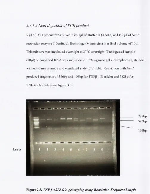
2.7.1.2 N co l digestion o f PCR p ro d u ct...66
2.7.2 Taqman™ SNP Allelic discrim ination... 67
2.7.2.1 ILIO Single nucleotide polym orphism s... 67
2.7.2.2 Plasminogen Activator Inhibitor-1 4G/5G S N P... 68
2.7.2.3 Am plifluor Technology - remaining polym orphism s...70
2.8 Outcome m easures... 70
2.9 Statistical analysis... 71
2.9.1 Haplotype A nalysis... 72
3.1 A bstract...74
3.2 Introduction... 76
3.3 Aims and H ypotheses... 77
3.4 M eth o d s...78
3.4.1 Patient population... 78
3.4.2 M easurem ents...78
3.4.3 Statistical A nalysis... 79
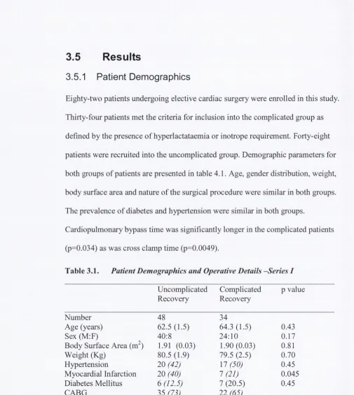
3.5 R esults... 80
3.5.1 Patient D em ographics... 80
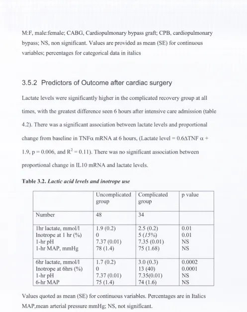
3.5.2 Predictors o f Outcome after cardiac su rg ery ... 81
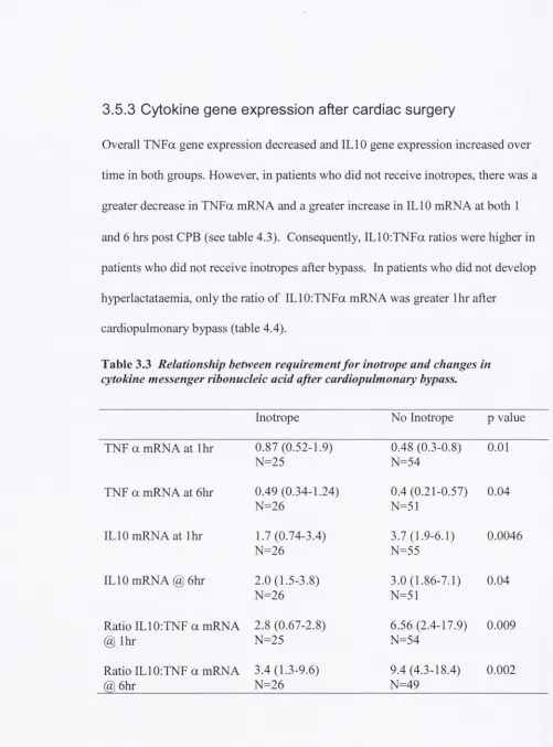
3.5.3 Cytokine gene expression after cardiac surg ery... 82
3.5.4 Cytokine gene polymorphisms associated with outcom e...88
3.6 D iscussion... 91
4.1 A bstract...95
4.2 Introduction... 96
4.3 Aims and H ypotheses... 98
4.4 M eth o d s...99
4.4.1 Patient population... 99
4.4.2 M easurem ents...99
4.4.3 Statistical A nalysis... 100
4.5 Results... 101
4.5.1 Patient D em ographics... 101
4.5.2 PAI-1 Gene Expression after Cardiac Surgery...102
4.5.3 PAI-1 4G/5G polymorphism and O utcom e...104
4.5.4 PAI-1 4G/5G polymorphism and PAI gene expression... 106
4.5.5 PAI-1 gene expression related to cytokine gene expression... 107
5.1 General D iscussion...I l l
5.2 Assumptions and L im itations...120
5.3 Clinical Implications o f our fin din gs... 124
5.4 Direction for future research...126
List of Tables
Table 1.1 :
Table 4.1. : Table 4.2 : Table 4.3 :
Table 4.4 :
Table 4.5 :
Table 4.6 :
Table 4.7 :
Table 4.8 :
Table 5.1 :
Table 5.2 ;
Table 5.3 :
Table 5.4 :
Expanded list o f signs for diagnosis o f systemic inflammation/sepsis.
Patient Demographics and Operative Details: Experimental Series I Lactic acid levels and inotrope use
Relationship between requirement for inotrope and changes in cytokine m essenger ribonucleic acid after cardiopulmonary bypass. Relationship between hyperlactataem ia and changes in cytokine mRNA after cardiopulm onary bypass.
M ultivariate logistic regression analysis o f cardiopulmonary bypass and IL 10: TN F a mRNA ratio with lactate, inotrope and composite endpoints
IL 10 allele frequencies in 82 cardiac surgical patients
HAPLOTYPE ANALYSIS FOR TNF for 82 patients - Uncomplicated (U) vs. Complicated (C)
HAPLOTYPE ANALYSIS for IL 10 for 82 patients Uncomplicated (U) vs. Complicated (C)
Patient Demographics and Operative Details: Experimental Series II
N-fold change in PAI-1 mRNA after cardiac surgery compared with baseline values in patients w ith com plicated and
uncom plicated recovery
4G/5G genotype frequencies in relation to PAI-1 mRNA 1 hour after cardiopulm onary bypass.
List of Figures
Figure 2 .1 : PCR Efficiency curve
Figure 2.2 : RT- PCR amplification curve for PAI-1 mRNA.
Figure 2.3 : TNF 13 +252 G/A genotyping using Restriction Fragment Length Polymorphisms with Ncol.
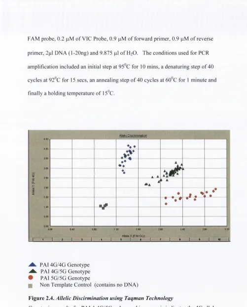
Figure 2.4 : Allelic Discirmination using Taqman Technology
Figure 3 .1 : N-fold change in TN Fa mRNA after cardiac surgery in patients with uncomplicated versus complicated postoperative recovery. Figure 3.2 : N-fold change in ILIO mRNA after cardiac surgery in patients with
uncomplicated versus complicated postoperative recovery. Figure 3.3 : Ratio ILIO: TNFa mRNA after cardiac surgery.
[image:10.516.4.501.26.776.2]Abbreviations
A Adenine
ACCP American College o f Chest Physician
ACT Activated Clotting Time
ANOVA Analysis o f Variance
APTT Activated Partial Throm boplastin Time
AT A Adenine Thym ine Adenine
ATP Adenosine triphosphate
Bp Base pair
C Cytosine
°C Degrees Celsius
CABG Coronary Artery Bypass Graft
cDNA Complementary Deoxyribonucleic acid
Cl Confidence Interval
CPB Cardiopulmonary Bypass
Cr Creatinine
Ct Threshold cycle
ACt Difference in threshold cycle between the target and the reference.
dNTP Deoxyribonucleotide triphosphate
EDTA Ethlyenediamine Tetraacetic Acid
ELISA Enzyme-Linked Immunosorbent Assay
Fig. Figure
Fi02 Fractional inspired O2 concentration
G Guanine
GAPDH Glyceraldehyde-3 -phosphate dehydrogenase
gm gram
HCL Hydrogen chloride
HCO
3 Bicarbonate ionH2O Water
hrs hours
ICU Intensive Care Unit
IkP Inhibitor kappa beta
IL Interleukin
INR International Normalised Ratio
KCL Potassium Chloride
kDa Kilodalton
KG Kilogram
L Litre (s)
LPS Lipopolysaccharide
M A P M ean Arterial Pressure
M G B M inor G roove B inding
M in. M inutes
m g M iligram m es
M gC l M agn esiu m Chloride
ml M ililitres
m M M ilim o les
m m H g M ilim etres o f M ercury
m M L V M urine M o lo n ey L eukaem ia Virus M O D S M ultiple Organ D ysftinction Syndrom e m R N A m essen ger R ibonucleic acid
M O F M ultiple Organ Failure
N Num ber
N A D H N icotin am id e A d en ine D in u cleotid e N FkP N u clear Factor Kappa B eta
n g nanogram
nm nanom etres
N S N o t significant
O2 O xygen
p Probability
PAI-1 P lasm in ogen A ctivator Inhibitor-1
Oxygen Concentration
PATS Patient Administration and Tracking System PBMC Peripheral Blood M ononuclear Cells
PCR Polymerase Chain Reaction
PDH Pyruvate Dehydrogenase
pH potential o f Hydrogen ion
pit platelets
pmol picomoles
R Correlation coefficient (Pearson’s)
RFLP Restriction Fragment Length Polymorphism
rpm Revolutions per minute
rRNA ribosomal Ribonucleic acid
SBP Systolic Blood Pressure
SCCM Society o f Critical Care Medicine
SD Standard Deviation
SE Standard error
s Seconds
SEM Standard Error o f the Mean
SIRS Systemic Inflammatory Response Syndrome
SNP Single Nucleotide Polymorphism
T Thymine
T N F a Tum our Necrosis Factor Alpha
t-PA Tissue Plasminogen Activator.
U Units
Microlitre
|im Micrometres, microns
^mol Micromole
u-PA Urokinase Plasminogen Activator
VLDL Very Low Density Lipoprotein
Vs Versus
Acknowledgements
Dedication
Summary of Thesis
Chapter 1
1.1
Introduction
M odulation o f the inflammatory cascade during sepsis has been considered as a possible means to improve survival in sepsis and prevent septic shock. However, outcome in patients with sepsis is also dependant on the presence o f co existing disease and the interaction between infection and inflammation. The situation is fiarther com plicated by patient selection and presentation, with patients who acquire infection and develop systemic inflammation almost exclusively represented among the critically ill, whereas those who experience a lesser degree o f systemic
inflammation in response to infection remain unobtrusive and thereby difficult to identify. Thus the role o f genetic factors in the generation o f systemic inflammation is difficult to study in septic pafients as the initial insult has already occurred and is complicated by an infective component. The ability to measure the activation o f the systemic inflammatory response exists in a cardiac surgical setting. Systemic inflammation occurs after surgery in cardiac surgical patients in a sterile
1.2 Systemic Inflammatory Response Syndrome
1.2.1
Definition and Diagnostic Criteria
In 1991 the American College o f Chest Physicians (A CC?) and the Society of Critical Care M edicine (SCCM) convened a “Consensus Conference”, in an attempt to provide a conceptual and practical frame-work to define systemic inflammation. The 1992 statement from the ACCP/SCCM Consensus Conference introduced into the term “systemic inflamm atory response syndrome”(SIRS) (1992). The term provided a reference for systemic activation o f the immune response, regardless o f cause. SIRS was considered to be present when patients had more than one o f the following clinical findings:
Body temperature higher than 38^ C or lower than 36° C Heart rate higher than 90/min
Respiratory rate > 20/min or PaC02 < 32mmHg W hite blood cells > 12,000cells/|il or < 4,000/|jl
The systemic inflamm atory response can be triggered by a variety o f infectious and noninfectious conditions. Signs o f systemic inflammation can and do occur in the absence o f infection among patients with bums, pancreatitis, and after
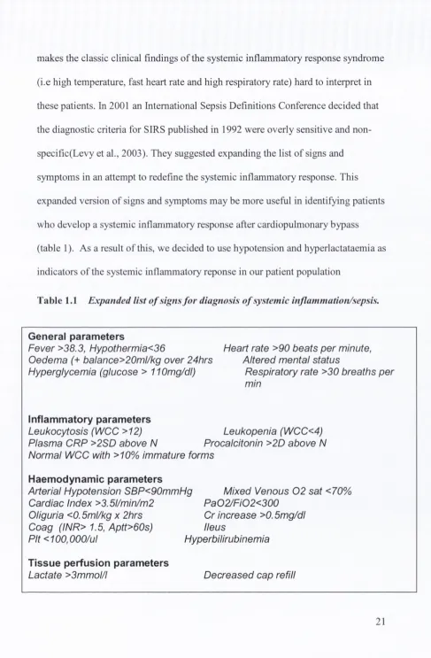
makes the classic clinical findings o f the systemic inflammatory response syndrome (i.e high temperature, fast heart rate and high respiratory rate) hard to interpret in these patients. In 2001 an International Sepsis Definitions Conference decided that the diagnostic criteria for SIRS published in 1992 were overly sensitive and non- specific(Levy et al., 2003). They suggested expanding the list o f signs and symptoms in an attempt to redefine the systemic inflamm atory response. This expanded version o f signs and symptoms may be more usefiil in identifying patients who develop a systemic inflammatory response after cardiopulmonary bypass (table 1). As a result o f this, we decided to use hypotension and hyperlactataemia as indicators o f the systemic inflammatory reponse in our patient population
Table 1.1 Expanded list o f signs f o r diagnosis o f system ic inflammation/sepsis.
General parameters
Fever >38.3, Hypothermia<36 Heart rate >90 beats per minute, Oedema (+ balance>20ml/kg over 24hrs Altered mental status
Hyperglycemia (glucose > 110mg/dl) Respiratory rate >30 breaths per mm
Inflammatory parameters Leukocytosis (WCC >12)
Plasma CRP >2SD above N Pn Normal WCC with >10% immature forms
Leukopenia (WCC<4) Procalcitonin >2D above N
Haemodynamic parameters
Arterial Hypotension SBP<90mmHg Cardiac Index >3.5l/min/m2
Oliguria <0.5ml/kg x 2hrs
Mixed Venous 0 2 sat <70% Pa02/Fi02<300
Cr increase >0.5mg/dl Ileus
Coag (INR> 1.5, Aptt>60s)
Pit <100,000/ul Hyperbilirubinemia
Tissue perfusion parameters
[image:23.516.19.506.39.782.2]1.2.2
Predisposing factors to systemic inflammation
1.2.2.1 The Systemic Inflammatory response to Cardiac Surgery
Cardiac surgery provokes a vigorous inflamm atory response, which has important clinical implications. At least 12.6% o f cardiac surgical patients develop systemic inflammation after cardiac surgery (Taneja et al., 2001). This pathophysiologic condition reflects a hyperdynamic circulation resulting in a high cardiac output state with reduced systemic vascular resistance, requiring treatment with fluid
replacement and agents that help increase the blood pressure (Hall et al., 1997). Subsequently, lactic acidosis can occur with the risk o f bleeding, respiratory distress, kidney dysfunction and ultimately multi-organ failure (Cremer et al.,
1996). Although systemic inflammation after cardiac surgery is usually brief, coagulopathy with bleeding necessitating operation, and prolonged mechanical ventilation are documented sequelae. In addition, where organ dysfunction is severe, or in those with limited functional reserve, systemic inflammation after cardiac surgery can result in increased m orbidity and mortality. Factors
contributing to the inflammatory response after cardiac surgery are currently not well understood.
1.2.2.2 Influence o f cardiopulmonary bypass on systemic inflammation
However, since its introduction it has become clear that cardiopulmonary bypass (CPB) is associated with an undesirable systemic inflamm atory response. Many factors during CPB induce a complex inflammatory response e.g exposure o f blood to nonphysiologic surfaces, surgical trauma, reperfiision to the organs after CPB, and release o f endotoxin. Once initiated, the systemic inflamm atory response is maintained by several factors. This consists o f a broad range o f host responses that include the production o f protein and lipid mediators, expression o f cell surface receptors and adhesion molecules, induction o f enzymes and production o f acute phase proteins (Hall et al., 1997). Activation o f pro-coagulation proteins as well as fibrinolytic activators and inhibitors on the endothelial surface is also a feature and this represents an important link between the inflammatory, coagulation and fibrinolytic systems (Paparella et al., 2004). This complex chain o f events has strong similarities with sepsis.
1.2.2.3 Effects o f Anaesthestic technique on system ic inflammation
1.3 Lactic Acidosis
Hyperlactataemia (high blood lactate levels) was included in the list o f signs for diagnosing the systemic inflammatory response syndrome at the International
Sepsis Definition Conference in 2001. Lactic acid was first isolated from sour milk by Scheele in 1780 (Fall and Szerlip, 2005). In 1918, Cannon made an important observation that metabolic acidosis was associated with decreased blood flow and shock. Shortly thereafter, Clausen introduced an assay for lactate, and subsequently it was demonstrated that the accumulation o f lactic acid accounted for the metabolic acidosis described in diabetic patients without ketoacidosis (Fall and Szerlip, 2005). Lactic acid exists as two stereoisomers, L-lactic acid and D-lactic acid. In healthy humans, serum lactate is considered entirely L-lactate, as this is the isomer exclusively produced in the body (Uribarri et al., 1998).
1.3.1
Metabolism of Lactate
To understand the clinical syndrome o f lactic acidosis requires knowledge o f normal lactate metabolism. Lactate is a metabolic end product o f anaerobic
glycolysis and is produced by the reduction o f pyruvate. U nder hypoxic conditions
glucose is glycolytically converted to pyruvate and lactate is used as substrate to produce ATP by the enzyme lactate dehydrogenase with NADH as a co-factor.
1998, Fall and Szerlip, 2005). Once produced, lactate can be either slow ly oxidized in cells or rapidly converted back to glucose in the liver.
1.3.2
Lactic acidosis and systemic inflammation
The most common cause o f lactic acidosis in the intensive care setting is the systemic inflamm atory response syndrome (Stacpoole et al., 1994). Because these patients are frequently haem odynamically unstable, it has been assumed that the increase in lactate production is the result o f inadequate oxygen delivery and poor tissue perfusion (Hurtado et al., 1992). However, more recently this belief had been challenged. These studies have shown that increased pyruvate production, decreased PDH activity, regional differences in lactate production, and release o f lactate from lung tissue are all possible mechanisms o f lactic acidosis in SIRS (Gore et al., 1996, Brown et al., 1996, Vary et al., 1998). In addition, lactate levels have been shown to correlate with the development o f M ulti-Organ Dysfunction Syndrome (MODS) and prolonged hyperlactataem ia has been associated with increased m ortality (McNelis e ta l., 2001).
1.3.3
Causes of Lactic acidosis after cardiac surgery
blood flow to the internal organs) during surgery. However Haisjackl, measuring splanchnic blood flow with indocyanine green and using gastric tonom etry for mucosal pH measurement, found no evidence o f reduced splanchnic blood flow (Haisjackl et al., 1998). Indeed post CPB splanchnic blood flow and lactic acid levels were both increased compared to pre CPB levels suggesting that splanchnic lactic acid production after cardiac surgery is related to systemic inflammation and not hypoperfusion (reduced blood flow).
Decreased lactate clearance has also been demonstrated with SIRS in animal models. Chrusch et al found a combination o f increased splanchnic production and decreased hepatic clearance o f lactate in a model o f canine sepsis (Chrusch et al., 2000). Severin et al demonstrated decreased hepatic lactate clearance even in haemodynamically stable septic rats (Severin et al., 2002). Therefore, lactic acidosis in patients with SIRS not only develops in haem odynamically unstable patients but also can occur in the setting o f adequate perfusion and oxygenation. This leads us to believe that lactic acidosis is not a result o f hypoperfusion and hypoxia but rather that other factors contribute to the increase in lactate production.
1.3.4
Relationship between lactate production and cytokine
release.
1.4 Genetic polymorphisms
Since W atson and Crick introduced in 1953 the m olecular structure for
deoxyribonucleic acid, molecular biology has revolutionized medicine by increasing our understanding o f the pathophysiological mechanisms o f diseases. Genetic polym orphisms account for traits and varied susceptibility to complex diseases. A genetic polymorphism is an allelic variant that occurs in >1% o f the population, is stable in frequency, and cannot be accounted for by new mutations.
1.4.1
Importance of Genetic Polymorphisms on Disease
Susceptibility
Susceptibility to many diseases appear to have a genetic component. A study by Sorensen et al in 1988 looked at the genetic influences on the risk to die from various diseases (Sorensen et al., 1988). The authors followed 960 families that included adopted children. The risk o f dying from specific groups o f diseases for adoptees with a biologic or adoptive parent, who died o f the same cause before, was compared. The death o f a biological parent before the age o f 50 resulted in relative risks o f death in the adoptees o f 1.71 for all causes, 1.19 for cancer, 4.52 for cardiovascular and cerebrovascular causes, and 5.81 for infections. Therefore, genetic factors appear to play an important role in the susceptibility to
1.4.2
Single Nucleotide Polymorphisms
Single nucleotide polym orphisms (SNPs) are DNA sequence variations that occur when a single nucleotide (A, adenine; T, thymine; C, cytosine; G, guanine) in the genome sequence is altered. For a variation to be considered a SNP, it m ust occur in at least 1% o f the population and SN P’s occur approximately one in every 300 base pairs in the human genome (2003). SN P’s constitute a major com ponent to human genetic variation (Brookes, 1999). Changes to protein structure m ay be defined as conservative or non-conservative and may or may not alter the structure and or function o f the gene product. SN P’s in the promoter or other regulatory regions o f a gene may change the binding affinity o f nucleic acid binding proteins (an obvious example o f which are transcription factors) thus altering the rate o f transcription and/or translation leading ultimately to changes in protein levels. Besides influencing the rate o f transcription directly, SNP’s in coding regions can influence gene expression in other ways; for example by altering the stability or otherwise the availability o f transcribed mRNA. Finally it should be noted that distal elements (such as enhancers and silencers) may influence gene expression from considerable distances either upstream or downstream o f a gene.
1.4.3
Importance of Haplotype analysis
pronounced LD, called haplotype blocks, within which a limited subset o f all possible haplotypes are found at appreciable frequencies for a given population (Gabriel et al., 2002). W ithin these blocks, a relatively small number o f SNPs can mark common haplotypes and capture some o f the genetic diversity in a sample (Patil et al., 2001, Daly et al., 2001). Haplotype analysis can increase the pow er to detect disease associations. It allows for the possibility o f an ungenotyped
fiinctional variant to be in linkage disequilibrium with the genotyped polymorphism. M any common diseases in humans such as systemic
1.5 Role of Cytokines in systemic inflammation
Cytokines are early mediators o f the systemic inflamm atory reaction. They serve to
initiate the systemic inflammatory response and determine the magnitude and nature
o f the immune response. Cytokine synthesis and release are generally a b rief
regulated, self limiting event. The release o f cytokines is triggered by many
different stimuli including endotoxin exposure, tissue damage and oxidative stress
(Larmann and Theilmeier, 2004). Cytokines appear to have a local effect and a
systemic effect (due to the consequences o f high cytokine levels circulating in the
blood). The local effect involves recruitment o f phagocytic cells, essential for the
elimination o f noxious stimuli, while the systemic effects (if prolonged or
excessive) can have long lasting deleterious consequences. Excess production or
activity o f cytokines can lead to tissue injury, respiratory disease, shock with
decreased perfusion (blood flow) to organs leading to multi-organ failure such as
renal, cardiac and respiratory failure. Clinical manifestations o f systemic cytokine
release include fever, reduced level o f consciousness, haemodynamic instability,
high lactate levels and myocardial depression (Hennein et al., 1994, Dinarello,
2000). These features can also be found in the early postoperative course after CPB.
In general, cytokines are not stored as preformed molecules, and their synthesis is
limited to newly transcribed mRNA. M onocytes-macrophages produce T N F a,
IL l, IL6 and IL8 in response to an activating stimuli (Hagiwara et al., 1995). These
peptides act on other cells and blood elements such as polym orphonuclear cells,
cytokines induce the production and release o f other mediators. TN F a appears to be a key mediator in the systemic inflamm atory response syndrome (Giroir, 1993). T N F a, ILl and IL6 stimulate the synthesis o f acute phase proteins by the liver. They also prime the lym phocyte response. The important biological effects o f TNFa and IL l can be modulated by I L l0 (Moore et al., 2001). IL 10 is an anti
inflammatory cytokine produced by macrophages/monocytes and subsets o f T cells which lowers the production o f TNFa and IL l by macrophages and monocytes (Fiorentino et al., 1991). In addition, ILIO appears to enhance clotting activity (Pajkrt et al., 1997). Other anti-inflammatory cytokines which specifically inhibit TNFa and ILl have been detected in the blood such as IL l receptor antagonist, and soluble receptors for TNFa and IL l. The balance o f pro-inflammatory and anti inflammatory responses m ay be important in determining the extent o f the inflammatory response and clinical outcome.
1.5.1
Cytokine production after cardiopulmonary bypass.
Furthermore, measured plasm a cytokine protein may not be a good index o f
cytokine activity after surgery, as many o f these cytokines bind to cell surfaces receptors with such high affinity that unbound protein levels can not be measured (Munoz et al., 1991, Brix-Christensen et al., 2003, Butler et al., 1993, M isoph and Babin-Ebell, 1997). It is notable that the ligand receptor dissociation constant for
both TNFa and ILIO are several orders o f magnitude greater than the dissociation constant for other common biologic receptor ligand interactions, such as the
interaction between antigen and immunoglobulin (Grell et al., 1998). An alternative approach to quantifying the cytokine components o f inflammation by measuring cytokine protein levels would be useful. It is possible to accurately quantify
inflammatory cell mRNA levels using a real-tim e polymerase chain reaction (PCR) assay, and this might represent an important index o f cytokine mediated
inflammation. Furthermore, a recent study by Fu et al discovered a positive correlation between mRNA expression and protein expression (Fu et al., 2007).
Therefore, mRNA not only reflects the DNA genetic code but may also give us some insight into what is occurring at the protein level.
1.5.2
Tumour Necrosis Factor
Tum our Necrosis Factor was named over 20 years ago, on the basis o f its ability to
kill tum our cells in vitro and cause haemorrhagic necrosis o f transplantable tumours
preprotein o f 233 amino acids, from which the N-terminal 76 amino acids are later
removed (Wang et al., 1985). TNFa occurs in two forms, as a cell membrane
protein (26 kd) which can be cleaved into a soluble form (17 kd) by a specific TNFa
converting enzyme (TACE)(Black et al., 1997). The primary sources o f TNFa
synthesis include monocytes/macrophages and T cells. Although the half life o f
TNFa is less than 20 minutes, this brief appearance is sufficient to evoke marked
metabolic and haemodynamic changes and activate mediators distally in the
cytokine cascade (Tonnesen et al., 1996). TNFa is known to be a pleiotropic
cytokine with many complex effects which can be viewed as both beneficial and
harmful in the context o f human disease (Bazzoni and Beutler, 1996). On the one
hand TNFa is essential for a proper immune response to be mounted; on the other
hand, it is clear that TNFa plays a key role in orchestrating the inflammatory
response which contributes to numberous inflammatory diseases. With particular
reference to this study, TNFa is a central endogenous mediator of endotoxic shock
which is a manifestation o f an acute systemic inflammatory response.
TNFa initiates signalling reponses by binding to at least two receptors; TNF
receptor l(p55) and TNF receptor 2 (p75) which are expressed on most nucleated
cells (Engelmann et al., 1990). However, although both receptors are expressed on
cells, one is typically dominant. In vivo studies have suggested that p55 receptor
binding is critical primarily for TN Fa’s proinflammatory properties (Van Zee et al.,
1994). These show that administration o f p55 TNFa receptor agonists to healthy
changes simulating a systemic inflammatory response. In another study, antibodies that prevented TNFa binding to the p55 receptor and not the p75, protected mice from lethal endotoxic shock but blocked development o f a protective response against infection with Listeria monocytogenes (Sheehan et al., 1995).
Furthermore, TNFa stimulates mononuclear phagocytes and other cell types to secrete chemokines that contribute to leukocyte recruitment and activation of neutrophils and other mononuclear phagocytes (Dinarello, 2000). TNFa, an endogenous pyrogen, stimulates prostaglandin and nitric oxide synthesis, and can induce a fever directly or through the secondary induction o f ILl and IL6. In addition, these three cytokines act on hepatocytes to increase synthesis o f acute phase proteins like C-reactive protein, a\ antitrypsin, and haptoglobin.
1.5.2.1 Genetics o f Tumour Necrosis F actor Alpha
The gene encoding TNFa is located within the major histocompatibility complex (MHC) on chromosome 6p21.3. Multiple bi-allelic polymorphisms have been identified in the human TNFa promoter region. These may influence TNFa production and hence inflammatory responses. A TNFa polymorphism at position -
308 involves replacement o f guanine with adenine. Mira et al. studied six TNFa
death or severe neurologic disease and that this effect was independent o f HLA variation (McGuire et al., 1999). Previous studies have associated the TNF2 (- 308A) allele with increased TNFa production (Wilson et al., 1997). Nadel studied children with meningococcal disease and determined that carriage o f the TNF2 allele was associated with a significantly increased risk o f severe disease and death (Nadel et al., 1996). A bi-allelic jVco/restriction fragment length polymorphism has also been described within the first intron o f the TNFfi gene (which is situated contiguous to TNFa) at position +252 with an adenine to guanine substitution (Wilson et al., 1992). The TNFB2 (A allele) has also been associated with higher TNFa levels in patients with sepsis (Stuber et al., 1996). However, Stuber inadvertently links a polymorphism associated with lesser TNFp production with
syndrome or sepsis. In addition, it must be remembered that although TNFa is an
important mediator, other cytokine related polymorphisms may play a role in
determining susceptibility to SIRS.
1.5.3
Interleukin 10
Interleukin 10 is an anti-inflammatory cytokine expressed and secreted by a variety
o f cell types, including T cells, monocytes/macrophages, dendritic cells and
epithelial cells usually after an activation stimulus such as cardiopulmonar>' bypass
(Moore et al., 2001). It circulates as a homodimer consisting o f two tightly packed
160 amino acid proteins. After engaging its high-affm ity 110-kd cellular receptor,
ILIO inhibits monocyte/macrophage-derived T N F a, IL ip , IL8 and nitric oxide
production (Moore et al., 2001). It also inhibits the generation o f cytokines by
neutrophils and natural killer cells (Opal and DePalo, 2000). Prior exposure to ILIO
inhibits nuclear factor kappa B (NFkB) translocation in response to
lipopolysaccharide stimulation. In addition, ILIO attenuates surface expression of
TNF receptors and promotes the shedding o f TNF receptors into the systemic
circulation (M oore et al., 2001, Opal and DePalo, 2000). Therefore ILIO appears to
inhibit non-specific inflammatory responses. In fact, inflammation is a major
stimulus for the production o f ILIO since previous studies have shown that both ILl
and T N F a can stimulate ILIO production directly indicating the existence o f a
1.5.3.1 Genetics o f Interleukin 10
T he ILIO gene is located on chrom osom e 1 (lq 3 1 -3 2 ). ILIO secretion in response to LPS show s a large interindividual variation w hich has a genetic com ponent o f over 70% (W estendorp et al., 1997) P olym orphism s w ithin the ILIO gene m ay plausibly account for different levels o f ILIO production. T h e S N P ’s at position -1 0 8 2 (A/G), -819 (T/C) and -5 9 2 (A /C ) are in strong linkage disequilibrium (Fife et al., 2006).
These variants have been reported to influence the transcription rate o f ILIO and therefore the production o f this cytokine. T urner associated reduced ILIO secretion w ith the presence or absence o f an ‘A ’ at position 1082 o f the hum an ILIO prom oter region (T urner et al., 1997). T he frequency o f the low producing 1082 A /A
genotype w as show n to be increased in patients w ith acute m yocardial infarction in a study by Lio (Lio et al., 2004). In addition, B alding show ed that the frequency o f the ILIO 1082 A allele w as h igher in patients w ith severe m eningococcal disease (2003). H ow ever, other studies have found conflicting results. To assess the genetic influence on ILIO production, W estendorp determ ined the capacity to produce ILIO in fam ilies o f patients w ho had m eningococcal disease. Fam ilies with high ILIO production to endotoxin stim ulus had a 20 fold increased risk o f fatal
outcom e from m eningococcal disease (W estendorp et al., 1997). G allagher associated high ILIO production and the 1082 G allele w ith increased m ortality in
patients w ith SIRS due to pneum onia (W estendorp et al., 1997, G allagher et al.,
The ILIO promotor SNP’s -1082A, -819T, and -5 92A (ATA haplotype) have been
associated low ILIO production. A study by Opdal associated the ATA haplotype
with sudden unexpected infant death due to infection (Opdal et al., 2003).
Conversely, frequency o f the ATA haplotype is higher in patients who are
asymptomatic carriers of hepatitis C than in patients with chronic progressive liver
disease (Mangia et al., 2004). Overall, the evidence supporting a role for ILIO
polymorphisms in determining outcome in inflammatory disease states is
controversial and requires further investigation.
1.5.4
Cardiac surgery and cytokine related genetic
polymorphisms
Genetic polymorphisms in genes coding for cytokines have been associated with
poor outcome after cardiac surgery. Tomasdottir et al related a TNFp -252
polymorphism to increased TNFa levels which was associated with prolonged ICU
stay and cardiopulmonary dysfunction after cardiac surgery (Tomasdottir et al.,
2003). Polymorphisms in the promoter o f IL6 promotor (-572 G-C and -174 G-C)
have been associated with higher postoperative plasma IL6 levels and prolonged
hospitalization after cardiac surgery with CPB. A study by Podgoreanu et al found
that the -572 G IL6 polymorphism was an independent predictor of postoperative
myocardial infarction. (Podgoreanu et al., 2006). Gaudino provided evidence that
homozygous patients having higher IL6 levels and a greater incidence of
postoperative renal and pulm onary dysfunction after cardiac surgery.
1.6 Fibrinolytyic system
The plasminogen activator (PA)/plasmin system, also known as the fibrinolytic
system serves as one o f the endogenous defense mechanisms for the prevention of
intravascular throm bosis (Vaughan, 2005). Fibrin deposition occurs on activation of
the coagulation cascade. This results in the ultimate conversion o f prothrom bin into
thrombin, which then catalyzes conversion o f soluble fibrinogen to fibrin. Fibrin
removal occurs on activation o f the fibrinolytic system. This system comprises an
inactive proenzyme (plasminogen) that is converted to the active enzyme (plasmin)
that degrades fibrin into soluble fibrin-degradation products. Two immunologically
distinct types o f physiological plasminogen activators have been identified in
humans: tissue plasminogen activator (tPA) and urokinase-type plasminogen
activator (u-PA) (Lijnen and Collen, 1997). t-PA mediated plasm inogen activation
is mainly involved in the dissolution o f fibrin in the circulation. u-PA binds to a
specific cellular receptor resulting in enhanced activation o f cell-bound
plasminogen. Plasminogen activation is regulated by the presence o f very specif c
and rapid acting, plasminogen activator inhibitors that are also present in plasma
properly regulated, they perform their physiologic functions remarkably well.
However, when unbalanced, the consequences can lead to an increased tendency to
bleed or conversely an increased tendency for thrombotic events.
1.6.1
Regulation of the fibrinolytic system
Regulation o f the fibrinolytic system occurs via 1) inhibition o f plasmin by a z
-antiplasmin, 2) thrombin-activatable fibrinolysis inhibitor which attenuates plasmin
generation and 3) PAI-1, the primary inhibitor o f both plasminogen activators
(Lijnen and Collen, 1995). Together these inhibitors constitute a powerful, negative
regulatory system for controlling the formation and activity o f plasmin.
Plasminogen activator inhibitor (PAI-1) inhibits plasminogen activators (u-PA and
t-PA) by forming inert, covalent complexes. The rapid inhibition o f both t-PA and
u-PA involves a high affinity region o f PAI-1. There appears to be no endogenous
mechanism for recycling PA-PAI-1 complexes, which are cleared through the low-
density lipoprotein-related (LRP) receptor and the VLDL receptor (Orth et al.,
1992).
1.6.2
Plasminogen Activator inhibitor-1
PAI-1 was first identified in human endothelial cells and subsequently shown to be
produced by vascular smooth muscle cells, mesangial cells,
monocytes/macrophages and by stromal cells from adipose tissue (Orth et al.,
stored within cells but is rapidly and constitutively secreted after synthesis (Lijnen,
2005). Synthesis and secretion o f PAI-1 can be modulated by various agonists such
as endotoxin and cytokines (Sawdey et al., 1989). PAl-1, a member o f the serpin
family, is a single-chain glycoprotein o f about 45 kDa consisting o f 379-381 amino
acids (Lijnen, 2005). PA l-1 reacts very rapidly with t-PA and u-PA. In humans,
increased levels o f PAI activity resulting in a decreased fibrinolytic capacity have
been reported in several thrombotic disease states, including thromboembolism,
coronary artery disease and acute myocardial infarction (Hamsten et al., 1987,
Ridker et al., 1992). Excessive fibrinolysis due to decreased PAl-1 levels has been
reported in a few cases. A complete deficiency o f PAI-1 has been associated with
episodes o f major haemorrhage, all in response to trauma or surgery (Schleef et al.,
1989, M inowa et al., 1999). The effect o f PAl-1 gene disruption on haemostasis,
thrombosis and thrombolysis has been investigated in mice. Spontaneous bleeding
or delayed bleeding was not observed after partial amputation o f the tail or the
caecum in PAI-1 deficient mice (Carmeliet et al., 1993). This is in contrast to the
delayed rebleeding observed after traum a or surgery in patients with reduced or
absent PAI-1 levels. This difference may be due to the 5-fold lower basal plasma
levels o f active PAl-1 in wild-type mice than in man. PAI-1 has also been shown to
inhibit vascular wound healing via regulation o f cell adhesion and migration
(Lijnen, 2005). Elevated plasm a PAI-1 levels have been associated with the
progression o f atherosclerosis, by inhibiting the clearance o f fibrin incorporated into
1.6.2.1 Genetics o f Plasminogen A ctivator Inhibitor-1
PAI-1 expression has been observed in various cell types including mononuclear
cells, and multiple regulatory factors have been identified that play a role in PAl-1
transcription. The human PAI-1 gene is mapped on chromosome 7q21.3-q22 and
several polymorphisms within the PAI-1 gene have been described (Strandberg et
al., 1988, Westendorp et al., 1999). While the genetic architectures o f PAI-1 have
not been fully elucidated, there is accumulating evidence suggesting that
interindividual variation in plasma enzymes such as PAI-1 is significantly
influenced by polymorphisms. A common functional polymorphism exists in the
PA I-I promoter. A single base pair insertion (5G)/deletion (4G) polymorphism
675bp upstream from the start o f transcription is fiinctionally important in
regulating the PAl-1. Artifical constructs containing the P A I-l prom oter have
shown that the 4 0 allele produces six times more RNA than the 5 0 allele in
response to interleukin IB (Dawson et al., 1993). Individuals homozygous for the
4 0 allele have higher basal and inducible concentrations o f PAI-1 than those with
one or two copies o f the 5 0 allele (Dawson et al., 1993, Hermans et al., 1999).
Transcription studies o f this promoter region revealed that both alleles bind a
transcription activator (at position -683 to -6 76 ), whereas the 5 0 allele also binds a
repressor protein to an overlapping binding site (at position -6 7 2 to -67 6). The
extent to which this polymorphism influences PAI-1 protein levels has also been
investigated. A study o f 464 German twins estimated the heritability o f PAI-1
approximately 56% o f the PAI-1 heritability is explained by interindividual
variation in the PAI-1 4G/5G polymorphism. In addition, Asselbergs found that the 4G/5G polym orphism is a significant predictor o f plasm a PAI-1 levels (Asselbergs et al., 2006).
1.6.3
Plasminogen Activator Inhibitor and Systemic
Inflammation
The systemic inflammatory response is a complex clinical syndrome that involves the activation o f many cells and cascade reactions. Activation o f the coagulation and fibrinolytic systems are an integral part o f the inflammatory response and is an important pathogenic factor in sepsis associated organ injury (Idell, 2001, Johnson et al., 1998). Endotoxin infusions in healthy individuals induces up-regulation o f both tissue plasminogen activator and plasm inogen activator inhibitor (PAI-1) (Suffredini et al., 1989). This is not surprising as PAI-1 is an acute phase protein, and is increased by inflammatory stimuli such as interleukin 1 13 and tumour
necrosis factor (Dawson et al., 1993, Ryan et al., 1996). The inflammatory process leads to the production o f a great number o f inflamm atory mediators. These
mediators induce tissue factor- mediated throm bin production and activation of the coagulation system (Binder et al., 2002). Ultimately this can result in microvascular
fibrin deposition, platelet depletion and disseminated intravascular coagulopathy leading to multiorgan failure (Hermans and Hazelzet, 2005, Kidokoro et al., 1996).
which is downregulated by a sustained increase in plasm a PAI-1 levels (Biemond et al., 1995, Suffredini et al., 1989). The increase in PAI-1 effectively eliminates plasma t-PA leaving undissolved fibrin deposits which is commonly seen in sepsis and multiorgan failure (Levi et al., 2003). PAI-1 levels correlate closely with the severity o f disseminated intravascular coagulation and disease. Substantially increased PAI-1 levels are observed during acute lung injury, experimental sepsis, endotoxemia in volunteers, and meninogococcal sepsis (Schultz et al., 2004, Hermans et al., 1999).
1.6.4
Influence of Cardiac Surgery on PAI-1 levels
after cardiac bypass with either an increase or no change in active PAI-1 (Chandler
and Velan, 2003). Inhibition o f gene expression may result in undetectable protein
levels which can be difficult to quantify with the current assays available. The
difference in measurement technique m ay account for discordance between the
results o f these studies.
1.6.5
Functional Relevance of 4G/5G PAI-1 polymorphism
Concentrations o f PAI-1 in plasm a are very high in children with meninogococcal
sepsis, with the highest concentrations being found in severe and fatal disease
(Komelisse et al., 1996). Carriage o f the 4G deletion polym orphism in the PAI-1
gene has been associated with increased mortality from meningococcal sepsis
(Hermans et al., 1999). In a similar study, Westendorp analysed 50 patients who
survived meningococcal infection and 131 control subjects from the same
geographic region, as well as 183 first-degree relatives o f patients with
meningococcal infection for the 4G/5G polymorphism (W estendorp et al., 1999 .
Because Westendorp had no information on genotypes o f patients who died, he
included first-degree relatives o f patients with meningococcal infection to avoid
under-representation o f the patients who did not survive. The 5G/5G genotype was
more common among relatives o f patients with meningitis (31% vs 11%, p=0.001).
However, patients whose relatives were carriers o f the 4G/4G genotype had a 6 'old
1.7 Hypothesis and Aims
1.7.1
Aim#1
The hypothesis that interindividual variability in cytokine gene expression after
cardiac surgery is associated with clinically important events, can be investigated by
measuring change in cytokine mRNA before and after cardiac surgery using real
time PCR. With this approach, the interrelation o f pro-inflammatory and anti
inflammatory cytokine gene expression in the pathophysiology o f systemic
inflammation could be examined in vivo. Hence, the aim o f the study was to
investigate the relation between cytokine gene expression in peripheral blood
mononuclear cells, genotype and clinical events after cardiac surger>'.
1.7.2
Aim #2
The purpose o f this study was to examine the temporal pattern o f changes in PAI-1
messenger RNA (mRNA) levels in peripheral blood mononuclear cells in patients
undergoing cardiac surgery, to determine w hether PAI 4G/5G gene polymorphism
influences PAI-1 mRNA expression after cardiac surgery, and to obtain preliminary
information on the relation between PAI-1 mRNA levels and clinical outcomes. We
hypothesized that PAI-1 gene expression and indices o f systemic inflammation in
cardiac surgical patients might be linked. Furthermore the pattern o f change in PAI-
1 gene expression after cardiac surgery might be modulated by carriage o f
1.8 Summary
Systemic inflammation may occur after cardiopulmonary bypass, when it frequently
presents as arterial vasodilation and lactic acidosis. Inflam mation is cytokine
mediated with tum our necrosis factor a recognized as a critically important
molecule involved in the initiation, propagation and regulation o f the systemic
inflammataory response while interleukin 10 is a potent anti-inflammatory cytokine
that down-regulates the proinflammatory response. The balance between the
proinflammatory and anti-inflammatory response may significantly influence
outcome after cardiac surgery. In addition, the inflamm atory cascade is linked to the
fibrinolytic system with both TNFa and ILl influencing the release o f P A I- I.
Indeed, susceptibility to thrombosis has been confirmed in the setting o f
meningococcal septicemia, trauma, and deep vein thrombosis. Additionally, there
are clinical data associating deficiencies in PAI-1 production with an increased
likelihood o f haemorrhagic events. Analyzing SNPs and haplotypes may help
Chapter 2
2.1 Subject Recruitment
Following approval o f the study protocol by the institutional ethics committee, a
written informed consent was obtained for each patient before inclusion in to the
study. Eighty-two patients scheduled for routine cardiac surgery were recruited
over a 2 year period. Exclusion criteria included: renal impairment as defined by a
creatinine concentration o f > 150nmol/L, history o f liver disease, coagulation
disorders as defined by International Normalised Ratio (INR) > 1.5, haematological
malignancies, coronary artery bypass graft (CABG) surgery without
cardiopulmonary bypass (CPB), emergency surgery, and use o f corticosteroids or
anticoagulant drugs before surgery.
2.2 Cardiac Surgery and Anaesthesia
Perioperative clinical care included anaesthesia w ith propofol, fentanyl,
pancuronium, and isoflurane. Non-pulsatile CPB was performed using a
membrane oxygenator (Cobe Inc, Denver, CO, USA) with flow maintained between
2.0 and 2.4 L/min/m^. Suction systems were controlled. Body temperature was kept
between 32 and 35^^C during CPB. Heparin 3mg/kg was given to obtain an ACT o f
> 480 seconds prior to CPB and reversed after CPB with protamine (1:1 ratio of
mechanical ventilation was continued until the patient was ready for extubation.
Extubation was determined by a consultant anaesthetist.
2.3 Randomisation
Post operatively patients were divided into two groups;
a) the uncomplicated group and
b) the complicated group i.e those that developed a systemic inflammatory
response within 24hrs.
The complicated group fulfilled the following criteria:
1) Hypotension including those requiring the infusion o f a potent
vasoconstrictor or an intra-aortic balloon pump to maintain mean blood
pressure > 70mmHg, or
2) Hyperlactataemia as defined by lactate levels > 4.0mmol/L within 24hrs post
surgery.
2.4 Physiological Variables
Demographics such as age, gender, weight, preoperative chronic disease state and
operative details were recorded on a patient information database (PATS system).
Agilent Technology) including hourly haemodynamics, inotropic requirement,
requirement for mechanical assist devices such as intra-aortic balloon counter
pulsation, duration o f mechanical ventilation and indices o f oxygenation,
coagulation parameters and blood loss in the first 24 hours after surgery, and
duration o f intensive stay.
2.5 Blood sampling protocol
2.5.1
RNA Extraction
Blood samples for RNA analysis were obtained at three time points: a) prior to
cardiac surgery, b) 1 hour post cardiopulmonary pump, and c) 6 hours post
cardiopulmonary pump. A 24hr sample was taken on a subgroup o f patients who
remained in ICU after 24hrs. Blood was collected in EDTA containing tubes and
processed immediately. PBM C’s (peripheral blood mononuclear cells) were
obtained by density gradient centrifugation on Lymphoprep (Nycomed, Oslo,
Norway) using a standard protocol (Woollard et al., 2002, Bugalho et al., 2001).
Briefly, this involved diluting 10ml blood samples in H anks’ buffered saline (1:1
dilution) (Life Technologies Inc.), and layering it on 5 ml o f Lymphoprep. Each
sample was spun at 1,200 rpm for 30 minutes, and the m ononuclear cell interphase
was collected into a fresh tube and washed with H anks’ medium for 10 minutes at
l,200rpm . The resultant cell pellet was reconstituted with Hanks Medium at 4°C
rpm thus producing a cell pellet. The cell pellets were stored in a lysis buffer (RLT
buffer, Rneasy mini Kit, Qiagen, Sussex, England) at -SO'^C. Total RNA was
extracted using RNeasy mini kit (Qiagen, Sussex, England). The RNA was treated
with 10 units o f RNase-free DNase for 15min during the extraction procedure in
order to avoid amplification of contaminating genomic DNA. Concentration and
purity o f RNA was determined by measuring the absorbance at 260nm and 280nm
on a spectrophotometer (Eppendorf, Biophotometer). Ribosomal RNA (28S rRNA
and 18S rRNA) was visualized on a 1.2% agarose/formaldehyde gel following
electrophoresis to demonstrate the intergrity o f the RNA.
2.5.2
DNA extraction
Blood was collected in ethlyenediamine-tetraacetic acid (EDTA) tubes pre-
operatively. DNA was extracted using the QIAamp DNA Blood Midi Kit (Qiagen,
Sussex, England) according to manufacturers instructions, and DNA was stored at -
20^C until amplification. Concentration o f DNA was determined by measuring the
absorbance at 260nm and the purity was assessed by calculating the ratio of
absorbances at 260nm and 280nm on a spectrophotometer (Eppendorf,
Biophotometer).
2.6
RNA processing
2.6.1
Reverse Transcriptase
Total RNA was reversed transcribed to produce complementary DNA (cDNA)
transcription w as perform ed at 37° C for a duration o f 60 m in in a final volum e o f
30)al containing; 500ng sam ple RN A , 1.25|il (250U ) m M L V reverse transcriptase
(Prom ega,) w ith 2\i\ random hexam ers (pd (N)6), 0.2m M d N T P ’s, 0 .6|il R N A sin
1U/|^1, lOmM dithiothreitol, 4 .5|il 11% D M SO and 6fil 5 X RT buffer (250m M
Tris-H C L, pH 8.3, 375 m M K Cl, 15mM M g C y . T he cD N A w as analysed
im m ediately or stored at -2 0 °C for later analysis.
2.6.2
Real time PCR
The extent o f cytokine gene expression after cardiopulm onary bypass w as
determ ined using quantitative real tim e RT-PC R. M essenger R N A for interleukin
10 (ILIO), plasm inogen activator inhibitor 1 (PA I-1) and tum our necrosis Factor
alpha (TN F a ) w ere m easured. C om plim entary D N A (cD N A ) w as used as a
tem plate for the polym erase chain reaction. All sam ples and controls w ere assayed
in triplicate in 25|^1 reactions. Q uantification o f m R N A using group-specific prim er
pairs and PAM probes (A ssay b y D em and, A pplied B isosystem s Perkin Elm er,
Foster City, CA ) for the detection o f IL 10, PAI-1 and T N F a w as carried out on an
ABI PR ISM G eneA m p 7000 Sequence D etection System (A pplied B iosystem s
Perkin Elm er, Foster City, CA ) w ith real-tim e detection and quantification taking
advantage o f the fluorescence T aqM an technology.
The reaction conditions were: T aqm an U niversal M asterm ix (12.5|al), tem plate
cD N A (50ng), A ssay on dem and (1.25|al). T he balance o f the 25|al reaction w as
following contidions: 50° C for 2minutes, 95° C for 1 Ominutes and then 40 cycles o f
95° C for 15s and 60° C for 1 minute. Normalisation o f cDNA templates was
achieved by glyceraldehyde-3-phosphate dehydrogenase (GAPDH) quantification.
All probes used in the Taqman reaction were designed to have nonfluorescent
quenchers and m inor groove-binding (MGB) modifications. N egative template
controls and no amplification controls were included in each Taqm an run. The
comparative C j method o f quantification was used comparing the timed and
baseline samples (ABI Prism 7700 Sequence Detection System U ser Bulletin 2, PE
Applied Biosystems).
2.6.3
Relative Quantification of gene expression
Relative quantification can be performed using the standard curve method or the
comparative method using ABI prism 7000. The comparative Ct method is similar
to the standard curve method, except it uses arithmetic formulas to achieve the same
result for relative quantification (Livak and Schmittgen, 2001). The comparative
method can be used provided a validation experiment is performed.
2.6.3.1 Validation Experiment
Before using the AACt method for quantification, a validation experiment was
performed to demonstrate that the efficencies o f the target and reference are
approximately equal. A sensitive method for assessing if two amplications have the
ACt
10
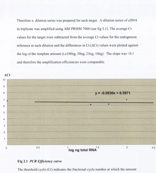
Therefore a dilution series was prepared for each target. A dilution series o f cDNA
in triplicate was amplified using ABI PRISM 7000 (see fig 3.1). The average Ct
values for the target were subtracted from the average Ct values for the endogenous
reference at each dilution and the differences in Ct (ACt) values were plotted against
the log o f the template amount (i.elOOng, 50ng, 25ng, lOng). The slope was <0.1
and therefore the amplification efficiencies were comparable.
7
6
5
4
3
2
1
y = -0.0936X + 6.5971
0.5 1 1.5 2 2.5
[image:61.518.6.512.50.610.2]log ng total RNA
Fig 2.1 PCR Efficiency curve
The threshold cycle (Ct) indicates the fractional cycle number at which the amount
o f the amplified target reaches a fixed threshold. The difference in Ct values
(between the reference and the target) were plotted against the log o f the RNA
amount. Slope = 0.0936
2 .6 3 .2 The Comparative Method o f Relative Quantification
The com parative (AACt ) m ethod o f relative quantification w as used to determ ine
the am ount o f m R N A at each tim e point relative to the baseline sam ple. A
correlation betw een PCR efficiency o f the endogenous reference (G A P D H )
com pared to targets (IL-10, T N F a , PA I) allow ed the use o f the AACt m ethod for
quantification (see above). The com parative m ethod uses an arithrim etic form ula
for relative quantification. The relative quantification o f the target, norm alized to
the endogenous reference (G A PD H ) and relative to a baseline sam ple is given by:
Relative quantification = ,
W here AACt is defined as the difference o f m ean ACt (tim ed sam ple) and the Tiean
AACt (baseline sam ple)
A A Ct = A Ct (timed sample) — A A Ct (baseline sample)
and the A C t is d e fin e d as th e d iffe r e n c e in m ean Ct( il-iq.tnfo, PAoand m ean
Ct(Gapdh) as e n d o g e n o u s co n tro l,
CT(IL-IO,TNFa, PAI)- Ct (GAPDH).
Derivation o f the formula:
T he equation that describes the exponential am plification o f PC R is:
Where:
Xn Number o f target molecules at cycle n
Xo — Initial number o f target molecules
E x — Efficiency o f target amplification
n Number o f cycles
The threshold cycle (Ct) indicates the fractional cycle number at which the amount
o f amplified target reaches a fixed threshold. Thus,
Xt= Xo X (1 = K x
Where:
Xt = Threshold num ber o f target molecules
C t, x Threshold cycle for target amplification
= Constant 1.8.1.1.1.1.1.1.1 Kx
A similar equation for the endogenous reference reaction is:
R
t= R
oX
+
" = K „
Ry = Threshold number of reference molecules
Ro Threshold number o f reference molecules
Er Efficiency o f reference amplification
C t, r Threshold cycle for reference amplification
Kr
Constant
Dividing X j by R j gives the following expression:
XT = XO
X(1 + EX)CT, X = KX
_________________________
= K
RT = RO
X(1 + ER)CT, R = KR
The exact values o f XT and RT depend on a number o f factors, including:
• Reporter dye used in the probe
• Sequence context effects on the fluorescence properties of the probe
• Efficiency o f probe cleavage
• Purity o f probe
• Setting of the fluorescence threshold.
Therefore, the constant K does not have to be equal to one.
Assuming efficiencies of the target and the reference are the same:
E x = Er=E,
XO X (1 + E)CT, X - CT, R = K
Or
XN
X(1 + E)ACT =
K
Where:
ACt = C'j' ’ ’ X - Cy ’ ’ the difference in threshold cycles for targetR
and reference
Rearranging gives the follow ing expression:
Xn= K x (1+E)
The final step is to divide the Xn for any sample q by the for the calibrator (cb):
Xn,, = K x ( l + E ) ) ’ ' ' ‘^ ^ ‘'
-AACt
( 1 + E ) )
XN,cb K x ( l + E ) )-A C x,cb
Where:
A A Ct ACT,q - ACtj,cb
Therefore, the amount o f target, normalized to an endogenous reference and relative
to a calibrator is given by:
2 'A A C T
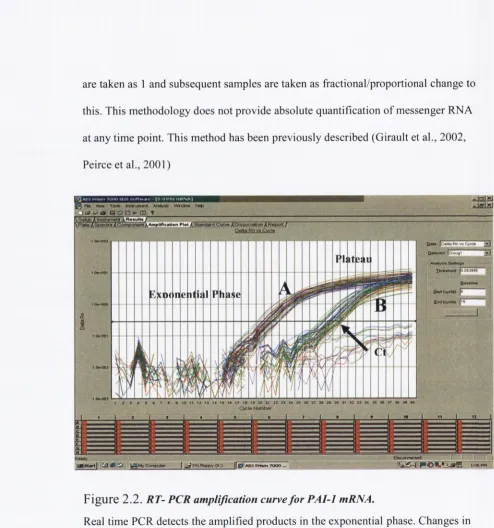
In summary, gene expression is measured as increased fluorescence corresponding
to amplification o f the target mRNA. The cycle in which fluorescence exceeds the
background signal is termed the threshold cycle (Ct) (see fig 3.2). The Ct w ill
always occur during the exponential phase o f amplification. The higher the starting
quantity o f the target mRNA, the earlier a significant increase in fluorescence, and
the smaller the Ct value obtained. The Ct values o f the timed samples (i.e Ihr, 6hr,
and 24hr) were compared to the preoperative baseline sample to determine the