1
TLR-dependent T cell activation in autoimmunity
Kingston H.G. Mills
Immunology Research Centre and School of Biochemistry and Immunology, Trinity College Dublin, Ireland
Abstract
Autoimmune disease can develop as a result of a breakdown in immunological tolerance, leading to the activation of self-reactive T cells. There is an established link between infection and human autoimmune diseases. Furthermore, experimental autoimmune diseases can be induced by immunization with autoantigens that are administered together with complete Freund’s adjuvant containing killed Mycobacteria tuberculosis, which in some cases can be replaced with individual pathogen-associated molecular patterns (PAMPs). Exogenous PAMPs and endogenous danger signals from necrotic cells bind to pathogen recognition receptors, including Toll-like receptors, and activate signaling pathways in innate immune cells and on T cells, leading to inflammatory cytokine production and T cell activation, and this is now considered to be a major factor in the development of autoimmunity.
Introduction
2
mechanism is thought to involve a breakdown of immunological tolerance, mainly due to defective regulatory T (TReg) cell function, which leads to the uncontrolled generation of
autoantigen-specific antibodies and pathogenic T cells that mediate tissue inflammation3.
Inflammatory T cells are induced by pathogen- or commensal- derived molecules, as well as by endogenous stress-induced self molecules. These microbe-associated molecular patterns (MAMPs) and damage-associated molecular patterns (DAMPs) [G] induce inflammatory T cells either indirectly through the induction of pro-inflammatory cytokine production by innate immune cells, or directly through their binding to PRRs on T cells. Evidence for this is provided by the described association between infections and autoimmune diseases (such as multiple sclerosis and rheumatoid arthritis), which develop following activation of self-reactive B and T cells, or between infections and chronic inflammatory diseases (such as Crohn’s disease) which result from uncontrolled innate and adaptive immune responses to normal constituents of the gut microflora.
There are several families of PPRs: Toll-like receptors (TLRs) sense conserved molecules from bacteria, virus and parasites; the cytosolic nucleotide binding oligomerization domain (NOD)-like receptors (NLRs) sense bacterial products; c-type lectin receptors (CLRs) bind β-glucans; and RIG-1-like receptors (RLRs) bind nucleic acids4-5
. There have been a number of exciting recent discoveries that have linked the activation of PRRs with autoimmune diseases, especially around the capacity of exogenous and endogenous ligands to promote innate inflammatory responses that drive the induction of autoreactive T cells, in particular IL-17-producing TH17 cells and γδ T cells6-11. Coupled with the discoveries around the pathogenic role
of these IL-17-producing T cells in autoimmunity, this has significantly advanced our understanding of the disease process in many autoimmune disorders. TLRs are a key family of PRRs involved in driving autoimmune inflammation, therefore, inhibitors of TLR binding or signaling are now considered to have great potential as therapeutics for autoimmune and other inflammatory diseases.
3
PAMPs shape innate and adaptive immune responses PRRs are expressed by a variety of cell types, especially cells of the innate immune system, such as dendritic cells (DC) and macrophages, where they sense danger or damage signals. The PRR ligands include pathogen-associated molecular patterns (PAMPs) [G] — conserved microbial structures, such as lipopolysaccharide (LPS), flagellin, viral and bacterial nucleic acids — but also endogenous danger signals from dead and dying cells, called DAMPs, such amyloid-β and saturated fatty acids (BOX 1). The primary function of PRRs is to mediate innate immune responses to pathogens and tumors, but they also have a role in sterile inflammation [G] and have been linked with autoimmune and chronic inflammatory diseases1,4.
Innate immune cells, especially DCs, direct adaptive immunity, not only by acting as antigen presenting cells (APCs) for naïve T cells, but also by providing the cytokine stimulus (the third signal) for T cell activation1-2. Binding of agonists to TLRs promotes DC maturation and production of T cell-promoting cytokines1, using distinct signaling pathways (FIG. 1). Activation of NF-κB results in production of the pro-inflammatory cytokines, including tumour necrosis factor (TNF), interleukin-6 (IL-6), pro-IL-1β and pro-IL-184-5. Activation of the interferon regulatory factor (IRF) pathways mediate IL-12p35 and type I interferon (IFN) production, whereas active extracellular signal-regulated kinase (ERK) and p38 MAP kinases are required for IL-23p19 and IL-10 production respectively5,12-13 (FIG. 1). TLR agonists have potent immunomodulatory activity and can promote adaptive immune responses to co-administered antigens and consequently have been exploited as adjuvants in infectious disease and cancer vaccines and as tumor immunotherapeutics14-15. Conversely, because of their ability to activate innate immunity and inflammation, molecules that antagonize TLR ligand binding or inhibit TLR signaling pathways are being developed as therapies against autoimmune and other inflammatory diseases5.
Targeting innate cytokines that promote pathogenic T cell responses has become a major focus in drug development against autoimmune diseases (FIG. 2). Whereas much of the initial focus was on IL-12, which is the key driver of pathogenic TH1 cells, the discovery of TH17 cells
and their role in autoimmunity (BOX 2) has shifted the emphasis to the innate regulatory cytokines IL-6, IL-23 and IL-1, which promote the development of TH17 cells. It is now
recognized that IL-6 and TGF promote differentiation of murine TH17 cells from naïve CD4+ T
4
the NLRP3 inflammasome [G] in TLR-primed DCs8) promote expansion and IL-17 production by memory TH17 cells19-20 (FIG 3). Moreover, it has been suggested that IL-23 can also induce
differentiation of TH17 cells21-22.
As well as having an indirect role in promoting IL-17 production by T cells, there is increasing evidence that PRR-triggered innate immune cells can act as a direct source of IL-17. T cells6,23, natural killer T (NKT) cells24 and lymphoid tissue inducer-like (LTi) [G] cells25 have all been shown to be important sources of IL-17 and associated cytokines. We have demonstrated that IL-1 or IL-1 can synergize with IL-23 to promote IL-17 secretion from T cells without T cell receptor engagement6,19. T cells secrete IL-17, IL-21 and IL-22 and act in an amplification loop to expand TH17 cells6. A high frequency of IL-17-producing T cells are
found in the CNS of mice with experimental autoimmune encephalitis (EAE), where they function with TH17 cells to mediate autoimmune inflammation6,26.
Although much of the focus on TLRs has center around their role in sensing and responding to infection by the innate immune system, it is now clear that TLRs agonists also have a crucial role in adaptive immunity. Indeed activation of DC and other innate immune cells through PRRs appears to be a critical step in the induction of T cell responses to pathogens, but also in the early inflammatory responses that lead to the development of autoreactive B and T cells that mediate many autoimmune diseases.
The link between PAMPs and autoimmunity
Expression of PRRs has been found in tissues of patients with organ-specific autoimmunity. Furthermore a polymorphism in TLR2 is associated with type 1 diabetes27 and polymorphisms in TLR4 and TLR9 have been described in patients with Crohn’s disease28. Mutations in NOD2 have been strongly associated with Crohn’s disease, and the synergy between NOD2 and TLR9 activation of inflammatory cytokine production by peripheral blood mononuclear cells is lost in cells from patients with the NOD2 mutation29. Moreover, the presence of PAMPs in tissues following infection has been associated with autoimmunity. These observations provide indirect evidence that PAMPs may promote autoimmune and chronic inflammatory diseases.
5
attributed to either molecular mimicry between pathogen-derived antigens and self antigens or non-specific activation of innate immunity leading to a breakdown in immunological tolerance and the development of self antigen-specific T cell- and antibody-responses.
Transgenic mice expressing a T cell receptor (TCR) specific for a myelin-derived peptide develop spontaneous EAE when housed in conventional, but not specific pathogen free conditions30. This suggests that infection may precipitate autoimmunity in mice. Several studies in multiple sclerosis patients have suggested that disease may be triggered or exacerbated by infections with Chlamydia pneumonia, human herpes virus 6 or Epstein-Barr virus (EBV). The strongest evidence comes from studies on EBV, as viral latent protein has been found in the brain of patients with multiple sclerosis31. In patients with relapsing remitting multiple sclerosis, there is an association between upper respiratory tract infections and relapse32. It has also been reported that systemic infections may trigger relapse in multiple sclerosis by bystander enhancement of myelin-specific T cell responses33.
Parvovirus B19 is considered to be one triggering factor for rheumatoid arthritis; the virus was detected by PCR in synovial biopsies of 75% of patients with rheumatoid arthritis compared with 17% of patients with osteoarthritis and other arthritides34. Furthermore, EBV DNA and RNA were detected in 34% of patients with rheumatoid arthritis compared with 10% of healthy individuals35.
Commensal microorganisms may also be involved in the development of autoimmune diseases, as suggested by the finding that some species of gut microbiota promote the pathogenesis of inflammatory bowel disease, especially Crohn’s disease36. Commensal microbes, in particular segmented filamentous bacteria have been shown to promote the induction of TH17
cells9 and as well contributing to intestinal inflammation, these TH17 cells can also drive
autoimmune arthritis37. It has also been reported that commensal bacteria-derived ATP promotes IL-6, TGFβ and IL-23 production by DC, leading to local activation of naturally occurring and pathogenic TH17 cells38. However, this was independent of TLR signaling. Conversely, studies
in a T cell transfer model of colitis showed that commensal bacteria promote TH1 and TH17
responses by MYD88-dependant activation of innate immune responses39. Although IL-6 was implicated, a role for commensal-induced IL-1 in promoting TH17 responses has not been ruled
6
PAMPS may exacerbate autoimmunity. In addition to initiating autoimmune and chronic inflammatory diseases, PAMPs may exacerbate these diseases. Infection with Streptococcus pneumonia 7 days after induction of EAE exacerbates autoimmunity in wildtype but not Tlr2 -/-mice40. This suggests that pathogens may exacerbate autoimmune diseases via activation of TLRs. Furthermore, PAMPs are present in diseased tissues of patients with autoimmunity. For example, peptidoglycans, which include NLR and TLR2 ligands, has been found in synovial tissue macrophages and DCs isolated from patients with rheumatoid arthritis41, in the bowel wall of patients with Crohn’s disease42, in the brain of primates with EAE43 and in macrophages and DCs in the brain of patients with multiple sclerosis44. Interestingly, the expression of some TLRs (such as TLR2 and TLR3) is enhanced in astrocytes and oligodendrocytes from multiple sclerosis patients, suggesting that increased TLR signaling may occur during ongoing autoimmunity45. This in turn may exacerbate autoimmunity. Indeed, treatment of cancer patients with the TLR7/8 agonist imiquimod exacerbates psoriasis46.
Some infections suppress autoimmunity. In addition to driving autoimmunity, certain infections have also been associated with suppression of autoimmune diseases. Epidemiological data has shown that incidence of multiple sclerosis and type 1 diabetes is increasing in developed countries, and this correlates with a decrease in the number of infections47. Conversely, a higher incidence of infection with helminth parasites in rural parts of developing countries is associated with a lower incidence of allergy and autoimmune diseases48. The so-called ‘hygiene hypothesis’ suggests that anti-inflammatory cytokines or TReg cells induced by such infections exert
bystander suppression on the immune responses that mediate allergy and autoimmunity48-49. This hypothesis is supported by mouse studies showing that helminth infections can suppress autoimmunity through bystander suppression of autoantigen-specific TH17 cells, through
induction of anti-inflammatory cytokines and TReg cells50. It has also been shown that infection
of germ-free mice with a single human commensal Bacteroides fragilis protected against EAE through TReg cell induction51. So, although much of the evidence points to a role for pathogens
and commensal bacteria, especially those that induce potent TH17 responses, in promoting
7 Role of DAMPs in autoimmuneity
While pathogens and their products have been implicated in precipitating autoimmune diseases in humans and can induce experimental autoimmunity in animal models, there is also evidence that DAMPs or alarmins [G] released from dead and dying cells can stimulate innate immune responses leading to autoimmunity (FIG. 4). Polly Matzinger first suggested that, as well as responding to PAMPs, APCs can also respond to danger or alarm from endogenous molecules produced by distressed tissues, called DAMPs52. The term alarmin was coined by Joost Openheim53 to describe a group of mediators released by necrotic cells in response to infection or injury that interact with PRRs and activate innate immune cells, including DC and thereby enhance T cell responses. It has been suggested that alarmins are members of a larger family of DAMPs that also includes PAMPs54.
Human mobility group box protein 1 (HMGB1) is considered to be a DAMP capable of activating innate immune responses. Although there are reports that it may bind to TLR4 and other TLRs, many of the early studies on HMGB1 and other putative DAMPs were compromised by contamination with PAMPs, especially LPS. However, it has recently been reported that HMGB1 can stimulate innate immune responses by acting as a universal sentinel for nucleic acids55. HMGB1 binds to most or all nucleic acids facilitating their interaction with nucleic acid-sensing TLRs (TLR3, TLR7 and TLR9) and cytosolic nucleic acid binding sensors (RIG-1, MDA5, DAI, AIM2) on innate immune cells. Interestingly, HMGBI concentrations are elevated in the blood of patients with SLE, compared with healthy controls and there was a positive correlation between HMGB1 concentrations and disease activity56. This provides indirect evidence that this endogenous DAMP may be involved in the pathogenesis of human autoimmune diseases.
8
IL-1α promotes IL-17 production by T cells19, it may be an important endogenous activator of pathogenic T cells in autoimmune diseases.
Another alarmin, 15-αhydroxicholesterene (15-HC), is a derivative of cholesterol and activates microglia via TLR2 and polyADP-ribose polymerase 1 (PARP1). 15-HC is elevated in the serum of patients during the progressive phase of multiple sclerosis and in mice with secondary progressive EAE58. TLR2-specific antibody inhibited activation of PARP1 by 15-HC and attenuated the progression of EAE58.
Furthermore, human synovial membrane cultures from patients with rheumatoid arthritis were found to express TLR2 and TLR4 and to release endogenous TLR ligands which may contribute to destructive inflammation in the joints of these patients59. Finally, it has recently been shown that heterogeneous nuclear ribonucleoprotein (hbRNP), an RNA and DNA-binding protein that is a target of most nuclear antigen-specific T and B cells in systemic autoimmune diseases but also in human rheumatoid arthritis and in pristine-induced arthritis in rats, triggers TLR7- and TLR9- mediated activation of arthritogenic APCs, which activate pathogenic T cells60.
Although much of the early studies on DAMPs may be compromised by contamination issues, the more recent studies with knockout mice and molecular defined alarmins suggest that they can promote innate and as a consequence adaptive immune responses that mediate autoimmunity. However, it is still to clear whether this sterile inflammation is sufficient to kick start autoreactive T cell responses, or whether it amplifies responses induced by PAMPs.
TLRs in animal models of autoimmunity
Studies using TLR-defective mice, TLR-specific blocking antibodies, inhibitors of TLR signaling, or TLR agonists in animal models of autoimmune diseases have provided insight into the role of TLR activation in the initiation or exacerbation of autoimmunity (Tables 1 and 2).
9
associated with reduced IL-6 and IL-23 production by DCs and reduced IL-17 and IFNγ production by T cells10 (Table 1). This suggests that innate immune responses, initiated through TLRs or IL-1R signaling, are required for the induction of experimental autoimmunity.
Mice with a lymphoid cell-specific TLR2 deficiency were less susceptible to EAE and had reduced TH17 responses62 than wild type controls. The reduction in disease severity and
IL-17 production was even more dramatic when EAE was induced by adoptive transfer of TLR2-deficient T cells. This suggests that TLR2 signalling in T cells may be important for the induction of EAE. In addition, TLR2 may be involved in the activation of CNS-infiltrating macrophages, microglia or astrocytes during development of EAE58.
A study by Prinz et al. showed that disease severity was reduced in TLR9-defecient mice, though not as dramatically as in Myd88-/- mice10. Moreover, disease onset was delayed in Tlr9 -/-mice following transfer of MOG-specific T cells from wild-type -/-mice10, implicating a role for TLR9-induced innate immune activation in promoting the pathogenic function of activated auto-reactive T cells. Indeed APC activation through TLR9 could break tolerance and facilitate the induction of EAE63. Immunization of mice with proteolipid protein (PLP) in incomplete Freund’s adjuvant (IFA) induces tolerance due to lack of danger signals provided by the M. tuberculosis of the CFA. However, transferred lymph node cells from these tolerized mice induced EAE following stimulation with CpG63, providing further evidence of a pro-inflammatory role for TLR9 activation. This is consistent with the demonstration that TLR9 activation may circumvent tolerance and promote intestinal inflammation by suppression of TReg
cell conversion64.
Interestingly, the study by Marta et al. showed that mice defective in either TLR9 or TLR4 had enhanced IL-6, IL-23 and IL-17 expression and developed severe EAE61, suggesting a protective role for these TLRs in this autoimmune disease. In this study mice were immunized once with MOG peptide, whereas in the study by Prinz and colleagues mice were immunized twice with myelin protein, and this may have obscured the regulatory role of TLR961.
10
regulatory role in EAE, depending on the different stages of disease. When compared with knockout mouse studies, studies with TLR-specific antibodies or inhibitors, which can be used to block TLR signalling at different stages of disease, may give a clearer picture of the potential of the role of individual TLR pathways in EAE and other autoimmune diseases.
The role of TLRs in arthritis. In most mouse models, arthritis is induced following activation of inflammatory responses by killed bacteria or their products and there is good evidence that this involves activation of PRRs. In the mouse model of collagen-induced arthritis (CIA), which is induced by immunization with collagen and CFA, M. tuberculosis in CFA provides a source of PAMPs. Staphylococcus epidermidis-derived peptidoglycans (which includes TLR2 and NLR agonists) was also shown to induce acute arthritis in mice66. Moreover, zymosan, a polysaccharide from the cell wall of Saccharomyces cerevisiae, which binds TLR2 and the CLR dectin-1, has been used to induce experimental arthritis in mice. Zymosan-induced arthritis was found to be dependent on TLR2 activation, as disease was significantly attenuated in Tlr2-/- mice, but not by treatment with lamarin which blocks the dectin-1 receptor67. In addition, injection of immune stimulatory DNA sequences (ISS-ODN) into the joints of rats promoted the development of adjuvant arthritis68. This suggests that activation of TLR9 may also precipitate the innate immune responses that drive inflammation in the joint
Spontaneous T cell-mediated arthritis in IL-1Ra-/- mice was dependant on TLR4 activation by microbial flora; germ-free mice or Tlr4-/- Il1ra-/- double knockout mice did not develop arthritis11. Furthermore, a TLR4 antagonist, LPS from Bartonella quintana, inhibited IL-1β in the joint and attenuated arthritis in CIA and Il1ra-/- arthritis models69. By contrast, Tlr2 -/-Il1ra-/- mice developed more severe disease, which was associated with increased IFNγ production by T cells and reduced TReg cell function11, as direct TLR2 activation can induce
expansion but transiently disrupt the function of TReg cells70. Thus microbiota may promote
arthritis in Il1ra-/- mice by augmenting IFNγ and suppressing TReg cell function via TLR2. So,
unlike its role in EAE, where TLR2 signalling appears to drive inflammatory pathology, TLR2 may both promote and inhibit inflammatory T cell responses in mouse models of arthritis. However, there is more consistent evidence, from studies with the TLR4 antagonist as well as TLR4-deficient mice, that targeting TLR4 has potential in the treatment of arthritis.
11
Other autoimmune diseases. Further studies with mouse models of autoimmunity reveal that the contribution of TLR signaling to the initiation of autoreactive T cell responses depends on the disease type and experimental conditions. For example, Myd88-/- mice are resistant to experimental autoimmune uveitis (EAU) and exhibit significantly reduced autoantigen-specific TH1responses71, whereas Tlr2-/-, Tlr4-/-, and Tlr9-/- mice develop normal T cell responses and are
susceptible to EAU. By contrast, the development of type 1 diabetes in NOD mice is markedly reduced in the absence of TLR2 but not TLR472, and it appears that APCs sense apoptotic β cell death through TLR2 and thereby promotes diabetogenic T cells.
Moreover, TLR2 and TLR9 were shown to have distinct pathogenic roles in a murine model of anti-neutophil cytoplasmic antibody-associated vasculitis (AAV), a multisystem autoimmune disease where neutrophil myeloperoxidase (MPO) is targeted by autoantibodies73. Immunization with MPO in the presence of the TLR2 agonist Pam3Cys enhanced vasculitis by promoting TH17-dominated responses and neutrophil recruitment, whereas immunization with
MPO and the TLR9 agonist CpG selectively promoted TH1 responses and macrophage
activation73. Finally, topical application of the TLR7/TLR8 agonist imiquimod in mice induces skin lesions in mice resembling psoriasis; this was associated with epidermal expression of IL-23 and IL-17 and enhanced TH17 cells in the spleen74.
Taken together, the studies in mouse models suggest that TLR agonists may trigger autoimmunity, with the most definitive phenotype seen in Myd88-/- mice. However, this adapter protein is also used for IL-1 signaling, so it cannot be inferred that the protective effect of MYD88 deficiency is solely due to a loss of TLR-mediated inflammation. Studies in mice lacking a single TLR have revealed less dramatic defects in induction of experimental autoimmunity in a number of models, with perhaps the best evidence supporting a role for TLR2 in EAE and TID, TLR4 in arthritis. Conversely there is evidence for a protective role for TLR3 in EAE. Finally, there are reports of protective as well as pathogenic roles for TLR9 in EAE and TLR2 in arthritis and further study is required to define the potential of these TLRs as targets in multiple sclerosis and rheumatoid arthritis.
Indirect modulation of autoimmune T cell responses by TLRs
There is growing evidence that activation of PPRs on innate immune by pathogens or PAMPs cells can lead to a breakdown in self tolerance and bystander activation of autoreactive TH1 or
12
Activation of pathogenic T cells. There is evidence that PAMP-induced secretion of pro-inflammatory cytokines from monocytes and DCs promotes the induction and expansion of TH17
cells7,75-76 (FIG. 3). For example, human monocytes stimulated with LPS, zymosan, flagellin or lipoteichoic acid promote IL-17 production by anti-CD3 activated human CD4+ T cells75. The TLR-induced signal was not identified, and addition of IL-6 or TGFβ inhibited IL-17 production in this setting. Furthermore, hyphae of Candida albicans selectively promoted TH17 cell
induction by inducing IL-23 but not IL-12 production from human monocytes and DCs77. TLR3, TLR4 and TLR9 ligands induce MYD88-dependent production of cytokines from DCs, which promotes the differentiation of TH17 cells18. Moreover, the TLR4 ligand LPS
promotes IL-17 production by murine antigen-specific memory T cells through the induction of IL-1 and IL-23 production by DCs76. Finally, TLR2 agonists have been shown to promote the differentiation of murine TH17 cells in vitro62.
TH17 cells can also be induced independent of TLR and NLR activation, through
activation of dectin-1 with the -glucan curdlan, which promotes IL-6, TNF and IL-23 production by DCs78. Similarly, bacterial derived ligands of NOD-2, such as muramyl dipeptide (MDP), have been shown to drive 17 production by human memory T cells by stimulating IL-1 and IL-23 production by DCs7. Finally, it has been reported that commensal microbiota can promote differentiation of TH17 cells in the lamina propria of mice deficient in MYD88 and
TLR3, suggesting that TLR and IL-1R signaling are not essential for induction of TH17 cells in
the gut79.
T cell-mediated autoimmunity. The induction of experimental autoimmune disease in animal models, such as EAE, EAU and CIA, very often involves immunization with autoantigen in the presence of CFA. The killed Mycobacterium tuberculosis in CFA promotes innate inflammatory cytokines that direct the induction of TH1 and TH17 cells. M. tuberculosis has been shown to
activate the NLRP3 inflammasome promoting IL-1β and IL-18 production8, and to activate TLRs leading to IL-6 and IL-23, which collectively drive the induction of TH17 and TH1 cells.
13
of T cells from mice immunized with PLP in IFA with CpG to naïve recipients80. This effect was attributed to the fact that M. tuberculosis is a more potent adjuvant than CpG for induction of TH17 responses. By contrast, immunization of B10.S mice that express a transgenic TCR specific
for PLP139-151 with IFA and CpG induced EAE in 73% mice compared with 33% that developed
spontaneous disease81. The study demonstrated that activation of APCs via TLR9 can break tolerance and promotes the activation of self-reactive T cells. Interestingly, LPS was unable to break tolerance in this model.
In has been demonstrated that immunization of mice with MOG in IFA supplemented with zymosan can induce but not sustain EAE. Disease induction was associated with the generation of MOG-specific TH17 cells, and transient induction of IL-23 by DC82. Hansen et al.
also reported that immunization with MOG in IFA supplemented with high dose zymosan or LPS, but not the TLR3 agonist poly I:C, could induce EAE83. The LPS employed was purchased from Sigma; this is alleged to be contaminated with TLR2 agonists, suggesting that its pro-inflammatory effect was mediated by a combination of TLR2 and TLR4 activation. The adjuvant effect of LPS and zymosan were absent in mice deficient for the IL-1R-associated kinase 1 (IRAK1), suggesting that IL-1 or TLR signaling through the MYD88-independant pathway was involved83.
Although agonists for individual TLRs, especially TLR2 and TLR9, may activate the innate immune responses that drive activation of self-reactive effector T cells, it appears that these responses are optimally driven by signals from two TLRs or a TLR and an NLR or CLR. This may reflect the need for priming and boosting stimuli. For example, production of mature active IL-1β requires priming with a TLR ligand to induce pro-IL-1β expression, and a second signal from a PAMP or DAMP to activate the NLR inflammasome complex and associated caspase-1, which cleaves the pro-IL-1β into its active form. Consistent with this, M. tuberculosis can provide both stimuli8, which may explain why it is more effective than individual TLR agonists in inducting experimental autoimmunity.
Modulation TReg cells in autoimmunity. In addition to activating effector T cells, TLR agonists
can indirectly or directly modulate the function of TReg cells. There is evidence that TLR
activation can block TReg cell responses, thereby breaking tolerance to self antigens84-86. IL-6
14
Furthermore, IL-1 and IL-6 production by TLR-activated mature DCs is required to reverse TReg
cell anergy induced by immature DCs87. However, it has also been reported that TLR4 and TLR9 agonists can promote IL-10-secreting TReg cells that inhibit CD8+ and CD4+ effector T cells13,88.
This may be a strategy to limit immunopathology during infection, but may also control sterile inflammation and prevent the development of autoimmunity. The expansion of inducible TReg
cells by TLR agonists appears to be mediated through TLR-induced production of IL-10, TGFβ and other immunosuppressive molecules from innate immune cells13. Indeed these regulatory mechanisms may prevent the induction of infection-driven autoimmunity in most individuals, but their failure or attenuation, perhaps by environmental factors, may increase the risks of developing autoimmune diseases.
Direct modulation of T cells via TLRs
Whereas it is well established that TLR signaling in innate immune cells can indirectly promote T cell differentiation and expansion through DC maturation and regulatory cytokine production, T cells also express TLRs and evidence is emerging that TLR signaling in CD4+ and γδ T cells can promote cytokine secretion or regulate their function (FIG 2).
Modulation of effector CD4+ T cells. TLR2 signaling in TH1 cells promotes their proliferation
and IFNγ production 89
. Moreover, stimulation with CpG restored the proliferation of highly purified naïve T cells from Pkcθ-/- mice in response to CD3-specific antibody stimulation90. Protein kinase C θ (PKCθ) is involved in T cell differentiation and PKCθ-deficient mice are resistant to experimental autoimmune myocarditis (EAM). Susceptibility to EAM was restored by in vivo administration of CpG90, suggesting that TLR ligands may act directly on T cells to induce autoimmunity in vivo. Finally, stimulation of CD4+ T cells with Pam3Cys induced their proliferation and IL-17 production, which was enhanced by IL-23. By contrast, Tlr2-/- mice were less susceptible to EAE and loss of TLR2 in TH17 cells reduced their ability to transfer EAE62.
15
It has been demonstrated that CD8+ T cells express TLRs92-93. Stimulation of with TLR2 agonists enhanced CD3-induced proliferation of murine CD8+ T cells by lowering the threshold for co-stimulatory signals94. The TLR9 agonist CpG has been shown to promote IL-8 release by purified human CD8+ T cells93. Thus TLR agonists may promote the activation of self-reactive CD8+ T cells in certain autoimmune diseases.
Modulation of γδ T cells. It has been reported that γδ T cells express TLR1, TLR2 and the CLR dectin-1, and secrete IL-17 in vitro following stimulation with TLR1 and TLR2 or TLR2 and dectin-1 ligands, particularly in the presence of exogenous IL-2323. More recently it has been shown that IL-17-producing γδ T cells express higher levels of TLR1, TLR2, TLR4, TLR6 and TLR9 than IL-17-secreting CD4 T cells62. We have found that highly purified naive γδ T cells produce IL-17 in response to IL-1 and IL-236, or to PAMP-activated DCs in the absence of stimulation with CD3- or CD28-specific antibodies. However, highly purified γδ T cells failed to respond directly to TLR agonists alone8 (C. E. Sutton and K. H. G Mills, unpublished observations). It is thus possible that certain subsets of γδ T cells are already differentiated in the periphery and produce IL-17 in response to local IL-2395 or to IL-23 together with IL-1 that are induced by PAMP- or DAMP-activated innate immune cells6.
Modulation of TReg cells. Murine TReg cells express TLR4, TLR5, TLR7 and TLR896 and there is
evidence that TLR agonists can bind to them and modulate their function. Synthetic and natural ligands for TLR8 are capable of reversing the suppressive function of human TReg cells84.
Furthermore, persistent TLR activation can overcome the suppressive effect of TReg cells and
abrogate tolerance to tumour cells86.
However, there are also reports that TLR agonists can directly enhance TReg cell function.
It has been demonstrated that LPS can induce proliferation and enhance the suppressive function of TReg cells96. Human TReg cells express TLR5, and stimulation with flagellin and CD3-specific
antibody enhances expression of forkhead box P3 (FOXP3) and their suppressive function97. The TLR2 ligand Pam3Cys induces proliferation of murine TReg cells and although this was
associated with a temporary loss of TReg cell suppressive activity, TReg cell function was restored
16
CD4+CD25- effector cells resistant to suppression by TReg cells98. Interestingly, it was recently
reported that polysaccharide A from the gut commensal Bacteroides fragilis binds to TLR2 on CD4+ T cells promoting IL-10 production and the expansion of TReg cells, which, in turn, inhibit
TH17 cells. This suggests that TLR2 activation of T cells may promote tolerance, at least in the
mucosa99. Most of the evidence to date suggests that TLR activation of T cells is primarily indirect and mediated through TLR-induced activation of APCs. However, T cells do express TLRs and binding of TLR agonists to T cells can contribute to their activation, and this applies to both effector and regulatory T cells. Some of the strongest evidence is around TLR2 activation of TReg cells, so from this perspective blocking TLR2 may attenuate TReg function and might
therefore be more applicable to treating cancer than autoimmunity. However, studies in knockout mice have suggested that TLR2 may be a good therapeutic target in certain autoimmune diseases. Therefore, further study is required to decipher the relative contribution of direct versus indirect activation of effector and regulatory T cells by TLRs in different autoimmune diseases.
TLRs and other PPRs as drug targets for autoimmunity
The increasing evidence that the innate and adaptive immune responses that mediate autoimmune diseases are driven by PAMP and DAMP binding to TLRs and other PRRs has motivated the assessment of TLR-specific blocking antibodies or inhibitors of TLR signaling in pre-clinical models of inflammatory and autoimmune disorders. Proof-of-principle has been provided by studies with TLR knockout mice. For example, experimental arthritis is attenuated in Tlr4-/- mice and this is associated with lower TH17 responses, suggesting that TLR4 may be a
good upstream target for preventing IL-17 production in rheumatoid arthritis11. Indeed it has already been shown that a TLR4 antagonist can treat arthritis in two mouse models69.
17
shock in mice102, but did not meet its primary endpoint in patients with severe sepsis in a phase III clinical trial (http://www.eisai.com/news/enews201108pdf.pdf ).
TLR2 and nucleic acid binding TLRs are also major targets for a number of inflammatory and autoimmune diseases. EAE is attenuated in TLR2-/- mice, suggesting that the TLR2 signaling pathway may be a drug target for the treatment or prevention of multiple sclerosis62. An antibody to TLR2 developed by Opsona Therapeutics has been shown to be effective in a murine model of myocardial ischemia reperfusion injury103, and a humanized version of the antibody (OPN-305) has recently entered phase I clinical trials.
Antagonists of TLR7, TLR8 or TLR9, including CpG 52364 (Pfizer), IRS-954 (Dynavax) and IM 3100 (Idera) have also shown efficacy in mouse models of SLE and other autoimmune diseases and have recently entered clinical trials5. These studies provide evidence that inhibition of TLR signaling in response to endogenous DAMPs is a safe therapeutic approach in humans and has the potential to treat inflammatory diseases that involve sterile inflammation.
Conclusions and perspectives Epidemiological studies have linked infection with the development or exacerbation of autoimmune disease. Furthermore, enhanced expression of TLRs and their agonists in diseased tissues provides indirect evidence that innate immune responses mediated though TLRs and other PRRs are involved in the development of autoimmune inflammation. More convincing evidence is provided from mouse models where defects in expression or signalling via TLRs are associated with attenuation of the innate and adaptive immune responses that mediate autoimmunity. Conversely, administration of exogenous TLR agonists to mice can promote or exacerbate autoimmunity.
18
Unless there is preferential expression of individual TLRs or their agonists in the affected tissues, there is likely to be redundancy between different TLRs, and between TLRs and other PRRs. Nevertheless, there is already evidence from mouse models that blocking a single TLR, especially TLR2, TLR4 or TLR9, may be sufficient for the inhibition of the innate responses that promote inflammation and autoimmunity. However, this effect may be disease-dependent as, for example, TLR4 blocking ameliorates arthritis and TLR2 inhibition dampens EAE.
Evidence from phase III clinical trials indicates that drugs that broadly inhibit innate immune responses or completely block the induction or function of effector T cells are likely to significantly compromise host protective immunity. For example, psoriasis patients treated with an IL-12p40-specific antibody, ustekinumab105 (which blocks TH1 and TH17 responses), or gouty
arthritis patients treated with an IL-1β-specific blocking antibody, canakinumab (FDA Briefing
document 21 2011;
http://www.fda.gov/downloads/AdvisoryCommittees/CommitteesMeetingMaterials/
Drugs/ArthritisDrugsAdvisoryCommittee/UCM259596.pdf) had a higher incidence of infections, including serious infections, than controls. Although these new drugs have significantly improved efficacy over existing therapies, more selective inhibition of specific pathways involved in inflammation in a particular tissue or specific to a particular disease may not involve the same risks. If a single TLR is proven to have a dominant role in a particular autoimmune disease, it is possible that targeting TLRs, especially an extracellular TLR with an inhibitory antibody, may provide a safe and effective therapeutic approach for certain inflammatory and T-cell mediated autoimmune diseases.
Acknowledgements
19
Box 1. Toll-like Receptors – sensors of microbial molecules and danger signals.
Human Toll-like receptors (TLRs) are evolutionary conserved homologs of Drosophila Toll protein involved in embryonic development in Drosophila. TLR are expressed on the cell surface or intracellularly in many cell types, especially cells of the innate immune system, where they function to respond to infection or damage. TLRs are members of the pathogen recognition receptor (PPR) family that sense conserved exogenous molecules from pathogens, called pathogen-associated molecular patterns (PAMPs). However they also bind molecules from commensal bacteria and endogenous damage-associated molecular patters (DAMPs) from dead and dying cells. Ligands for TLRs include proteins, lipopetides and nucleic acids from bacteria, viruses, fungi, parasites, human cells and synthetic molecules.
Examples of pathogen-derived, endogenous and synthetic TLR agonists TLR Expressed Pathogen-derived agonist Endogenous
agonists
Synthetic agonists TLR1/2 Extracellular Bacteria: peptidoglycan, lipoproteins,
LTA
Fungi: Zymosan
Pam3CSK4
TLR2/6 Extracellular Bacteria: lipoproteins veriscan MALP-2
TLR3 Intracellular Virus: dsRNA mRNA Poly I:C
TLR4 Extracellular Bacteria: LPS
Virus: RSV fusion protein Fungi: mannan
Protozoa: glycoinositolphospholipids
Saturated fatty acids, β-defensins, oxLDL and amyloid-β1
Lipid A
derivatives
TLR5 Extracellular Bacteria: flagellin
TLR7/8 Intracellular Virus: ssRNA Self RNA Imiquimod,
R-848 TLR9 Intracellular Bacteria: CpG DNA
Virus: CpG DNA
Protozoa: CpG DNA, hemozoin
Self DNA CpG-ODN
TLR11 Extracellular Uropathogenic bacteria
Protozoa: profilin-like molecule
1
20 Box 2. Role of TH17 and TH1 cells in autoimmunity
Prior to the discovery of T helper 17 (TH17) cells, it was thought that TH1 cells were the primary
pathogenic cell population in T cell-mediated autoimmune diseases. However, mice deficient in interferon-γ (IFN ) or interleukin-12 (IL-12) were found to have increased susceptibility to experimental autoimmune encephalitis (EAE) or collagen-induced arthritis (CIA)106-108. It was subsequently shown that Il17-/- mice have reduced susceptibility to EAE109, and that myelin-specific TH17 cells from mice with EAE expanded in vitro with IL-23 can transfer EAE to naïve
mice110. Furthermore, mice defective in IL-23, which is required for expansion of TH17 cells, are
resistant to the induction of EAE and CIA106,111. Although there is considerable plasticity in T cell subtypes, it now appears that TH17 cells are a functionally distinct population of CD4+ T
cells that are pathogenic in many autoimmune diseases2.
There is evidence that IFN may suppress IL-17 production by TH17 cells21. This
provided an explanation for the failure of IFNγ-specific blocking antibody therapy in multiple sclerosis and for the more severe EAE in Ifng-/- mice108. However, there is evidence from cell transfer studies that both TH1 and TH17 cell subsets have a pathogenic role in autoimmune
inflammation, perhaps acting at a different stage of disease or recruiting different effector cells112. It has also been reported that transfer of TH1 cells induced experimental autoimmune
uveitis (EAU) in naïve recipients, which was not inhibited by treatment with IL-17-specific blocking antibodies113. Furthermore, IFNγ exacerbates disease in patients with multiple sclerosis114 and IFNγ promotes IL-1β production by human macrophages115, which enhances IL-17 production by TH17 cells. Finally, IL-17 and IFNγ double positive CD4+ T cells have been
found in the gut of patients with Crohn’s disease116
. It appears that TH17 and TH1 cells or
21
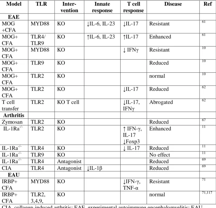
Table 1. Effect of TLR deficiency or inhibition on autoimmune diseases in animal models
Model TLR
Inter-vention
Innate response
T cell response
Disease Ref EAE
MOG +CFA
MYD88 KO ↓IL-6, IL-23 ↓IL-17 Resistant 61
MOG+ CFA
TLR4/ TLR9
KO ↑IL-6, IL-23 ↑IL-17 Enhanced 61
MOG+ CFA
MYD88 KO ↓ IFNγ Resistant 10
MOG+ CFA
TLR9 KO Reduced 10
MOG+ CFA
TLR2 KO normal 10
MOG+ CFA
TLR2 KO ↓IL-17 Reduced 62
T cell transfer
TLR2 KO T cell ↓IL-17,
IFNγ
Abrogated 62
Arthritis
Zymosan TLR2 KO Reduced 67
IL-1Ra-/- TLR2 KO ↑ IFN-γ,
IL-17 ↓Foxp3
Enhanced 11
IL-1Ra-/- TLR4 KO ↓ IL-17 Reduced 11
IL-1Ra-/- TLR9 KO No effect 11
IL-1Ra-/- TLR4 Antagonist Reduced 69
CIA TLR4 Antagonist ↓IL-1β Reduced 69
EAU IRBP+ CFA
MYD88 KO ↓IFN-γ,
TNF-α
Resistant 71
IRBP+ CFA
TLR2, 3,4,9,
KO normal 71,117
22
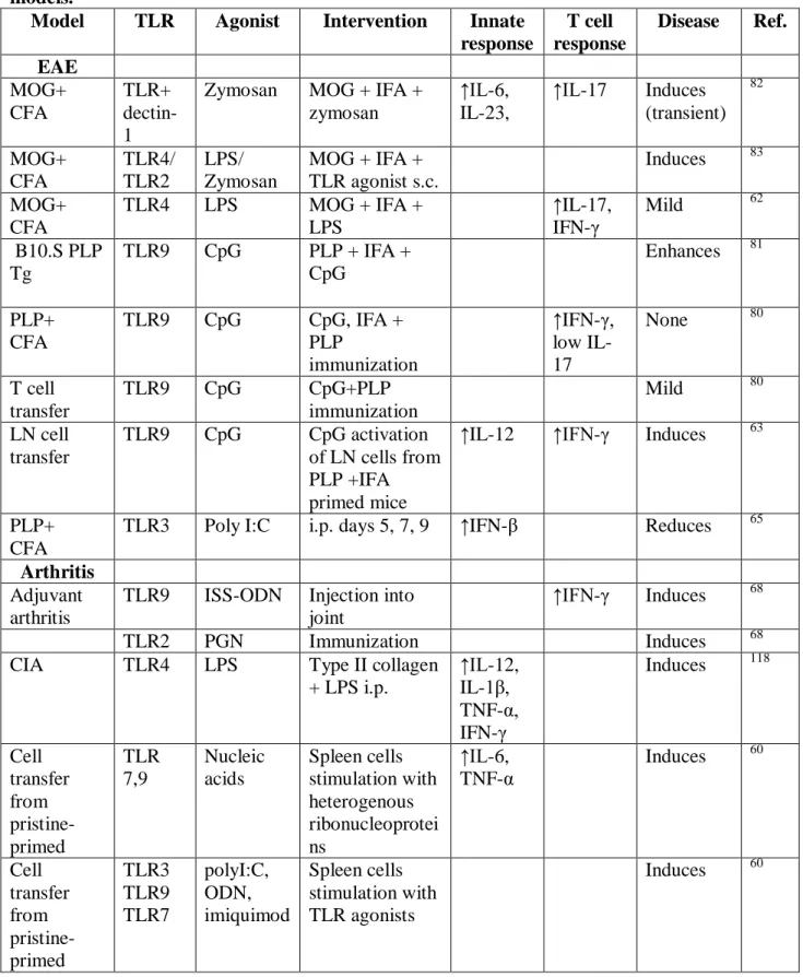
Table 2. Induction or modulation of autoimmune diseases by TLR agonists in animal models.
Model TLR Agonist Intervention Innate response
T cell response
Disease Ref. EAE MOG+ CFA TLR+ dectin-1
Zymosan MOG + IFA + zymosan
↑IL-6, IL-23,
↑IL-17 Induces (transient) 82 MOG+ CFA TLR4/ TLR2 LPS/ Zymosan
MOG + IFA + TLR agonist s.c.
Induces 83 MOG+
CFA
TLR4 LPS MOG + IFA +
LPS
↑IL-17, IFN-γ
Mild 62
B10.S PLP Tg
TLR9 CpG PLP + IFA +
CpG
Enhances 81
PLP+ CFA
TLR9 CpG CpG, IFA +
PLP
immunization
↑IFN-γ, low IL-17
None 80
T cell transfer
TLR9 CpG CpG+PLP
immunization
Mild 80
LN cell transfer
TLR9 CpG CpG activation of LN cells from PLP +IFA primed mice
↑IL-12 ↑IFN-γ Induces 63
PLP+ CFA
TLR3 Poly I:C i.p. days 5, 7, 9 ↑IFN-β Reduces 65 Arthritis
Adjuvant arthritis
TLR9 ISS-ODN Injection into joint
↑IFN-γ Induces 68
TLR2 PGN Immunization Induces 68
CIA TLR4 LPS Type II collagen
+ LPS i.p.
↑IL-12, IL-1β, TNF-α, IFN-γ
Induces 118
Cell transfer from pristine-primed TLR 7,9 Nucleic acids Spleen cells stimulation with heterogenous ribonucleoprotei ns ↑IL-6, TNF-α
Induces 60
Cell transfer from pristine-primed TLR3 TLR9 TLR7 polyI:C, ODN, imiquimod Spleen cells stimulation with TLR agonists
23
IL-ra-/- TLR4 LPS i.p. injection Enhanced 11
IL-ra-/- TLR2 Pam3Cys i.p. injection Enhanced 11
Other Antibody-associated vasculitis
TLR2 Pam3CSK4 Immunization
myeloperoxidas e + TLR agonist
↑IL-17 Induces 73
Antibody-associated vasculitis
TLR9 CpG Immunization myeloperoxidas e + TLR agonist
↑IFN-γ Induces 73
Psoriasis TLR7/ TLR8
Imiquimo d
Topical application
IL-23 ↑IL-17 Induces 74
EAU TLR4 LPS IRBP+CFA+LP
S
↑IL-6, IL-1β, TNF-α, IFN-γ,
↑IFN-γ, IL-17
Enhances 117
Myocarditis TLR9 CpG CpG i.p. PKC-θ
-
mice before/after immunization with
myosin+CFA
↑IL-17, TNF-α
Induces 90
24 Figure Legends
Figure 1. Bacteria-induced signaling in innate immune cells leading to T cell-promoting cytokine production. With the exception of Toll-like receptor 3 (TLR3), each of the TLRs employ myeloid differentiation primary response protein 88 (MYD88) as an adapter protein. In addition, TLR4 and TLR2 utilize MYD88-adapter-like (MAL), whereas Toll-interleukin-1 resistance (TIR) domain-containing adaptor-inducing interferon-β (TRIF) and TRIF-related adapter molecule (TRAM) are required for TLR3- and TLR4-mediated type I interferon (IFN) production. The figure illustrates some of the signalling pathways induced in a dendritic cell in response to a Gram-negative bacterium. Lipopolysaccharide (LPS) from the bacteria binds TLR4 in complex with MD2 and this complex initiates signalling by recruiting the adapter proteins MYD88, MAL, TRIF and TRAM. MYD88 associates with interleukin (IL)-1 receptor associated kinase 1(IRAK1) and IRAK4 and recruits tumor necrosis (TNF) receptor-associated factor 6 (TRAF6). This complex recruits transforming growth factor-β-activated kinase 1 (TAK1) leading to phosphorylation of IkB and activation of nuclear factor-κB (NF-κB) and consequent transcription of a range of genes coding for pro-inflammatory cytokines, including TNFα, interleukin 6 (IL-6), pro-IL-1β and pro-IL-18. In addition, TLR agonists activate the interferon regulatory factor (IRF) pathways, leading to IL-12p35 and type I IFN production. TLR-induced activation of TAK-1 also results in phosphorylation of mitogen activated protein kinases (MAPK), including p38 and ERK. Phosphorylation of ERK promotes transcription of IL-23p19 and IL-23 production. Phosphorylation of p38 activates cAMP response element-binding (CREB) leading to IL-10 production. Bacterial molecules, also bind intracellular nucleotide-binding oligomerization domain (NOD)-like receptor (NLRs), such as NOD-1 or NOD-2, which activate NF-κB and MAP kinases. Activation of NLRs, such as NLRP3, the associated inflammasome complex and caspase-1 facilitates the processing of pro-IL-1β and pro-IL-18 into mature cytokines. IL-12 promotes T helper 1 (TH1) cell differentiation, IL-10 induces regulatory
T (TReg) cells and IL-1, IL-6, IL-18 and IL-23 promote TH17 cell differentiation or expansion.
25
works with transforming growth factor-β (TGF-β) to promote differentiation of TH17 cells. TH17
cells are further activated by IL-1 and IL-23, but also by IL-17 and IL-21 produced early in the immune response by γδ T cells and other innate lymphoid cells (ILC). Interferon-γ (IFNγ) produced by TH1 cells activates macrophages, promoting release of inflammatory mediators,
such as IL-1β, tumor necrosis factor (TNF), matrix metalloproteinases (MMPs) and reactive oxygen species (ROS), which mediate tissue damage. IL-17 induces chemokine production, especially macrophage inflammatory protein (MIP)-2/IL-8, which recruits neutrophils to the site of inflammation. Finally, granulocyte-macrophage colony-stimulating factor (GM-CSF) produced by both TH1 and TH17 cells activates DC and macrophages and appears to be essential
for the development of autoimmunity. Biological response modifying drugs that are in use or in pre-clinical or clinical evaluation target many of the cytokines or the intracellular signaling molecules in innate cells or T cells.
Figure 3. Indirect and direct activation of T cells by TLR agonists. Pathogen-associated molecular patterns (PAMPs), bind to pathogen recognition receptors (PRRs), including Toll-like receptor (TLR), but also NOD-like receptors (NLR), C-type lectin receptors (CLR), and RIG-I-like receptors (RLR). TLR activation promotes co-stimulatory molecule (CD80 and CD86) and major histocompatability complex (MHC) expression (maturation) of immature dendritic cells (iDC) making them capable of activating naïve T cells. TLR-activated DC and macrophages (Mac) act as a source of immunomodulatory cytokines for directing differentiation of T helper 1 (TH1) or TH17 cells. DC and macrophage-derived interleukin 12 (IL-12) promotes differentiation
of naïve T cells into TH1 cells either directly or via interferon-γ (IFNγ) production from natural
killer (NK) cells. Activated TH1 cells secrete further IFNγ. IL-6 and transforming growth
factor-β (TGFfactor-β) promote differentiation of naïve T cells into TH17 cells. IL-1β, IL-1α or IL-18 in
synergy with IL-23 activate TH17 cells, inducing their proliferation and IL-17 secretion. γδ T
cells also secrete IL-17, IL-21 and IL-22 following stimulation with IL-1β, IL-1α or IL-18 with IL-23 released by PAMP activated DC or macrophages; this does not require association between the DC and γδ T cell or stimulation through the T cell receptor engagement. IL-21 and IL-17 produced by γδ T cells stimulate further differentiation of TH17 cells. There is also some
26
proliferation and cytokine production. However, in the case of TH17 cells, this appears to require
co-operation with IL-23.
27 References
1. Janeway, C.A., Jr. & Medzhitov, R. Innate immune recognition. Annu Rev Immunol 20, 197-216. (2002).
2. Mills, K.H. Induction, function and regulation of IL-17-producing T cells. Eur J Immunol 38, 2636-2649 (2008).
3. Wing, K. & Sakaguchi, S. Regulatory T cells exert checks and balances on self tolerance and autoimmunity. Nat Immunol 11, 7-13 (2010).
4. Akira, S., Uematsu, S. & Takeuchi, O. Pathogen recognition and innate immunity. Cell 124, 783-801 (2006).
5. Hennessy, E.J., Parker, A.E. & O'Neill, L.A. Targeting Toll-like receptors: emerging therapeutics? Nat Rev Drug Discov 9, 293-307 (2010).
6. Sutton, C.E., et al. Interleukin-1 and IL-23 Induce Innate IL-17 Production from gammadelta T Cells, Amplifying Th17 Responses and Autoimmunity. Immunity 31, 331-341 (2009).
The first report that γδ T cells promote autoimmune inflammation by providing a source of innate IL-17 and IL-21.
7. van Beelen, A.J., et al. Stimulation of the Intracellular Bacterial Sensor NOD2 Programs Dendritic Cells to Promote Interleukin-17 Production in Human Memory T Cells. Immunity 27, 660-669 (2007).
The first report that sensing of PAMPs though Nod-Like receptors promotes development of human Th17 cells.
8. Lalor, S.J., et al. Caspase-1-processed cytokines IL-1beta and IL-18 promote IL-17 production by gammadelta and CD4 T cells that mediate autoimmunity. J Immunol 186, 5738-5748 (2011).
Defined a role for PAMP-driven IL-18 as well as IL-1 in driving IL-17 production by CD4 and γδ T cells that are pathogenic in autoimmune diseases.
9. Ivanov, II, et al. Induction of intestinal Th17 cells by segmented filamentous bacteria. Cell 139, 485-498 (2009).
This study demonstrated that certain strains of commensal bacteria promote the induction of Th17 cells in the intestine, suggesting that mirobiata may precipitate autoimmunity.
10. Prinz, M., et al. Innate immunity mediated by TLR9 modulates pathogenicity in an animal model of multiple sclerosis. Journal of Clinical Investigation 116, 456-464 (2006).
This study demonstrated that signalling thorough MyD88 and TLR9 was required to promote the innate cytokines that drive the induction of Th17 cells in experimental autoimmunity.
11. Abdollahi-Roodsaz, S., et al. Stimulation of TLR2 and TLR4 differentially skews the balance of T cells in a mouse model of arthritis. J Clin Invest 118, 205-216 (2008).
This study demonstrated that TLR4 may be an important drug target for
rheumatoid arthritis; activation of TLR4 by microbial flora promoted T cell that were pathogenic in an arthritis model and disease was blocked using a TLR4 antagonist.
28
13. Jarnicki, A.G., et al. Attenuating regulatory T cell induction by TLR agonists through inhibition of p38 MAPK signaling in dendritic cells enhances their efficacy as vaccine adjuvants and cancer immunotherapeutics. J Immunol 180, 3797-3806 (2008).
14. Conroy, H., Marshall, N.A. & Mills, K.H. TLR ligand suppression or enhancement of Treg cells? A double-edged sword in immunity to tumours. Oncogene 27, 168-180 (2008).
15. Higgins, S.C. & Mills, K.H. TLR, NLR Agonists, and Other Immune Modulators as Infectious Disease Vaccine Adjuvants. Curr Infect Dis Rep 12, 4-12 (2010).
16. Bettelli, E., et al. Reciprocal developmental pathways for the generation of pathogenic effector TH17 and regulatory T cells. Nature 441, 235-238 (2006).
17. Mangan, P.R., et al. Transforming growth factor-beta induces development of the T(H)17 lineage. Nature 441, 231-234 (2006).
18. Veldhoen, M., Hocking, R.J., Atkins, C.J., Locksley, R.M. & Stockinger, B. TGFbeta in the context of an inflammatory cytokine milieu supports de novo differentiation of IL-17-producing T cells. Immunity 24, 179-189 (2006).
19. Sutton, C., Brereton, C., Keogh, B., Mills, K.H. & Lavelle, E.C. A crucial role for interleukin (IL)-1 in the induction of IL-17-producing T cells that mediate autoimmune encephalomyelitis. J Exp Med 203, 1685-1691 (2006).
20. Aggarwal, S., Ghilardi, N., Xie, M.H., de Sauvage, F.J. & Gurney, A.L. Interleukin-23 promotes a distinct CD4 T cell activation state characterized by the production of interleukin-17. J Biol Chem 278, 1910-1914 (2003).
21. Harrington, L.E., et al. Interleukin 17-producing CD4+ effector T cells develop via a lineage distinct from the T helper type 1 and 2 lineages. Nat Immunol 6, 1123-1132 (2005).
22. Park, H., et al. A distinct lineage of CD4 T cells regulates tissue inflammation by producing interleukin 17. Nat Immunol 6, 1133-1141 (2005).
23. Martin, B., Hirota, K., Cua, D.J., Stockinger, B. & Veldhoen, M. Interleukin-17-producing gammadelta T cells selectively expand in response to pathogen products and environmental signals. Immunity 31, 321-330 (2009).
24. Rachitskaya, A.V., et al. Cutting Edge: NKT Cells Constitutively Express IL-23 Receptor and ROR{gamma}t and Rapidly Produce IL-17 upon Receptor Ligation in an IL-6-Independent Fashion. J Immunol 180, 5167-5171 (2008).
25. Takatori, H., et al. Lymphoid tissue inducer-like cells are an innate source of IL-17 and IL-22. J Exp Med 206, 35-41 (2009).
26. Murphy, A.C., Lalor, S.J., Lynch, M.A. & Mills, K.H. Infiltration of Th1 and Th17 cells and activation of microglia in the CNS during the course of experimental autoimmune encephalomyelitis. Brain Behav Immun 24, 641-651 (2010).
27. Park, Y., Park, S., Yoo, E., Kim, D. & Shin, H. Association of the polymorphism for Toll-like receptor 2 with type 1 diabetes susceptibility. Ann N Y Acad Sci 1037, 170-174 (2004).
28. Hong, J., et al. TLR2, TLR4 and TLR9 polymorphisms and Crohn's disease in a New Zealand Caucasian cohort. J Gastroenterol Hepatol 22, 1760-1766 (2007).
29. van Heel, D.A., et al. Synergy between TLR9 and NOD2 innate immune responses is lost in genetic Crohn's disease. Gut 54, 1553-1557 (2005).
29
31. Serafini, B., et al. Dysregulated Epstein-Barr virus infection in the multiple sclerosis brain. J Exp Med 204, 2899-2912 (2007).
32. Buljevac, D., et al. Prospective study on the relationship between infections and multiple sclerosis exacerbations. Brain 125, 952-960 (2002).
33. Correale, J., Fiol, M. & Gilmore, W. The risk of relapses in multiple sclerosis during systemic infections. Neurology 67, 652-659 (2006).
34. Saal, J.G., et al. Persistence of B19 parvovirus in synovial membranes of patients with rheumatoid arthritis. Rheumatol Int 12, 147-151 (1992).
35. Saal, J.G., et al. Synovial Epstein-Barr virus infection increases the risk of rheumatoid arthritis in individuals with the shared HLA-DR4 epitope. Arthritis Rheum 42, 1485-1496 (1999).
36. Abraham, C. & Medzhitov, R. Interactions between the host innate immune system and microbes in inflammatory bowel disease. Gastroenterology 140, 1729-1737 (2011). 37. Wu, H.J., et al. Gut-residing segmented filamentous bacteria drive autoimmune arthritis
via T helper 17 cells. Immunity 32, 815-827 (2010).
38. Atarashi, K., et al. ATP drives lamina propria T(H)17 cell differentiation. Nature 455, 808-812 (2008).
39. Feng, T., Wang, L., Schoeb, T.R., Elson, C.O. & Cong, Y. Microbiota innate stimulation is a prerequisite for T cell spontaneous proliferation and induction of experimental colitis. J Exp Med 207, 1321-1332 (2010).
40. Herrmann, I., et al. Streptococcus pneumoniae Infection aggravates experimental autoimmune encephalomyelitis via Toll-like receptor 2. Infect Immun 74, 4841-4848 (2006).
41. Schrijver, I.A., Melief, M.J., Tak, P.P., Hazenberg, M.P. & Laman, J.D. Antigen-presenting cells containing bacterial peptidoglycan in synovial tissues of rheumatoid arthritis patients coexpress costimulatory molecules and cytokines. Arthritis Rheum 43, 2160-2168 (2000).
42. Klasen, I.S., et al. The presence of peptidoglycan-polysaccharide complexes in the bowel wall and the cellular responses to these complexes in Crohn's disease. Clin Immunol Immunopathol 71, 303-308 (1994).
43. Visser, L., et al. Phagocytes containing a disease-promoting Toll-like receptor/Nod ligand are present in the brain during demyelinating disease in primates. Am J Pathol 169, 1671-1685 (2006).
44. Schrijver, I.A., et al. Bacterial peptidoglycan and immune reactivity in the central nervous system in multiple sclerosis. Brain 124, 1544-1554 (2001).
45. Bsibsi, M., Ravid, R., Gveric, D. & van Noort, J.M. Broad expression of Toll-like receptors in the human central nervous system. J Neuropathol Exp Neurol 61, 1013-1021 (2002).
46. Rajan, N. & Langtry, J.A. Generalized exacerbation of psoriasis associated with imiquimod cream treatment of superficial basal cell carcinomas. Clin Exp Dermatol 31, 140-141 (2006).
47. Bach, J.F. The effect of infections on susceptibility to autoimmune and allergic diseases. N Engl J Med 347, 911-920 (2002).
30
49. Strachan, D.P. Family size, infection and atopy: the first decade of the "hygiene hypothesis". Thorax 55 Suppl 1, S2-10 (2000).
50. Walsh, K.P., Brady, M.T., Finlay, C.M., Boon, L. & Mills, K.H. Infection with a helminth parasite attenuates autoimmunity through TGF-beta-mediated suppression of Th17 and Th1 responses. J Immunol 183, 1577-1586 (2009).
51. Ochoa-Reparaz, J., et al. Central nervous system demyelinating disease protection by the human commensal Bacteroides fragilis depends on polysaccharide A expression. J Immunol 185, 4101-4108 (2010).
52. Seong, S.Y. & Matzinger, P. Hydrophobicity: an ancient damage-associated molecular pattern that initiates innate immune responses. Nat Rev Immunol 4, 469-478 (2004). 53. Oppenheim, J.J. & Yang, D. Alarmins: chemotactic activators of immune responses.
Curr Opin Immunol 17, 359-365 (2005).
54. Bianchi, M.E. DAMPs, PAMPs and alarmins: all we need to know about danger. J Leukoc Biol 81, 1-5 (2007).
55. Yanai, H., et al. HMGB proteins function as universal sentinels for nucleic-acid-mediated innate immune responses. Nature 462, 99-103 (2009).
56. Li, J., et al. Expression of high mobility group box chromosomal protein 1 and its modulating effects on downstream cytokines in systemic lupus erythematosus. J Rheumatol 37, 766-775 (2010).
57. Chen, C.J., et al. Identification of a key pathway required for the sterile inflammatory response triggered by dying cells. Nat Med 13, 851-856 (2007).
58. Farez, M.F., et al. Toll-like receptor 2 and poly(ADP-ribose) polymerase 1 promote central nervous system neuroinflammation in progressive EAE. Nat Immunol 10, 958-964 (2009).
59. Sacre, S.M., et al. The Toll-like receptor adaptor proteins MyD88 and Mal/TIRAP contribute to the inflammatory and destructive processes in a human model of rheumatoid arthritis. Am J Pathol 170, 518-525 (2007).
60. Hoffmann, M.H., et al. Nucleic acid-stimulated antigen-presenting cells trigger T cells to induce disease in a rat transfer model of inflammatory arthritis. J Autoimmun 36, 288-300 (2011).
61. Marta, M., Andersson, A., Isaksson, M., Kampe, O. & Lobell, A. Unexpected regulatory roles of TLR4 and TLR9 in experimental autoimmune encephalomyelitis. Eur J Immunol 38, 565-575 (2008).
62. Reynolds, J.M., et al. Toll-like Receptor 2 Signaling in CD4(+) T Lymphocytes Promotes T Helper 17 Responses and Regulates the Pathogenesis of Autoimmune Disease. Immunity 32, 692-702 (2010).
63. Ichikawa, H.T., Williams, L.P. & Segal, B.M. Activation of APCs through CD40 or Toll-like receptor 9 overcomes tolerance and precipitates autoimmune disease. J Immunol 169, 2781-2787 (2002).
This study demonstrated that innate immune cell activation and IL-12 induction through TLR9 promoted T cell that are pathogenic in experimental autoimmune
encephalomyelitis
31
65. Touil, T., Fitzgerald, D., Zhang, G.X., Rostami, A. & Gran, B. Cutting Edge: TLR3 stimulation suppresses experimental autoimmune encephalomyelitis by inducing endogenous IFN-beta. J Immunol 177, 7505-7509 (2006).
66. Onta, T., et al. Induction of acute arthritis in mice by peptidoglycan derived from gram-positive bacteria and its possible role in cytokine production. Microbiol Immunol 37, 573-582 (1993).
67. Frasnelli, M.E., Tarussio, D., Chobaz-Peclat, V., Busso, N. & So, A. TLR2 modulates inflammation in zymosan-induced arthritis in mice. Arthritis Res Ther 7, R370-379 (2005).
68. Ronaghy, A., et al. Immunostimulatory DNA sequences influence the course of adjuvant arthritis. J Immunol 168, 51-56 (2002).
69. Abdollahi-Roodsaz, S., et al. Inhibition of Toll-like receptor 4 breaks the inflammatory loop in autoimmune destructive arthritis. Arthritis Rheum 56, 2957-2967 (2007).
70. Sutmuller, R.P., et al. Toll-like receptor 2 controls expansion and function of regulatory T cells. J Clin Invest 116, 485-494 (2006).
71. Su, S.B., et al. Essential role of the MyD88 pathway, but nonessential roles of TLRs 2, 4, and 9, in the adjuvant effect promoting Th1-mediated autoimmunity. J Immunol 175, 6303-6310 (2005).
72. Kim, H.S., et al. Toll-like receptor 2 senses beta-cell death and contributes to the initiation of autoimmune diabetes. Immunity 27, 321-333 (2007).
73. Summers, S.A., et al. Toll-like receptor 2 induces Th17 myeloperoxidase autoimmunity while Toll-like receptor 9 drives Th1 autoimmunity in murine vasculitis. Arthritis Rheum 63, 1124-1135 (2011).
74. van der Fits, L., et al. Imiquimod-induced psoriasis-like skin inflammation in mice is mediated via the IL-23/IL-17 axis. J Immunol 182, 5836-5845 (2009).
75. Evans, H.G., Suddason, T., Jackson, I., Taams, L.S. & Lord, G.M. Optimal induction of T helper 17 cells in humans requires T cell receptor ligation in the context of Toll-like receptor-activated monocytes. Proc Natl Acad Sci U S A 104, 17034-17039 (2007). 76. Higgins, S.C., Jarnicki, A.G., Lavelle, E.C. & Mills, K.H. TLR4 mediates
vaccine-induced protective cellular immunity to Bordetella pertussis: role of IL-17-producing T cells. J Immunol 177, 7980-7989 (2006).
77. Acosta-Rodriguez, E.V., et al. Surface phenotype and antigenic specificity of human interleukin 17-producing T helper memory cells. Nat Immunol 8, 639-646 (2007).
78. LeibundGut-Landmann, S., et al. Syk- and CARD9-dependent coupling of innate immunity to the induction of T helper cells that produce interleukin 17. Nat Immunol 8, 630-638 (2007).
79. Ivanov, II, et al. Specific microbiota direct the differentiation of IL-17-producing T-helper cells in the mucosa of the small intestine. Cell Host Microbe 4, 337-349 (2008). 80. Tigno-Aranjuez, J.T., Jaini, R., Tuohy, V.K., Lehmann, P.V. & Tary-Lehmann, M.
Encephalitogenicity of complete Freund's adjuvant relative to CpG is linked to induction of Th17 cells. J Immunol 183, 5654-5661 (2009).
81. Waldner, H., Collins, M. & Kuchroo, V.K. Activation of antigen-presenting cells by microbial products breaks self tolerance and induces autoimmune disease. J Clin Invest 113, 990-997 (2004).