Copyright © 2016 Authors. This is an open access article distributed under the Creative Commons Attribution License, which permits unrestricted use, distribution, and reproduction in any medium, provided the original work is properly cited.
International Journal of Engineering & Technology
Website: www.sciencepubco.com/index.php/IJET doi:
Research paper, Short communication, Review, Technical paper
Medical Image Compression using Standard Deviation-Based
Wavelet Coefficients Thresholding Method
N.S.A.M Taujuddin1, R. Ibrahim2, S. Sari1
1Faculty of Electrical and Electronic Engineering, 2Faculty of Computer Science and Information Technology,
Universiti Tun Hussein Onn Malaysia, 86400 Parit Raja, Batu Pahat, Johor, Malaysia.
{shahidah,rosziati,suhailas}@uthm.edu.my
Abstract
In recent decades, digital images have become increasingly important. With many modern applications use image graphics extensively, it tends to burden both the storage and transmission process. Despite the technological advances in storage and transmission, the demands placed on storage and bandwidth capacities still exceeded its availability. Moreover, the compression process involves eliminating some data that degrades the image quality. Therefore, to overcome this problem, an improved thresholding and quantization techniques for image compression is proposed. Firstly, the generated wavelet coefficients obtained from the Discrete Wavelet Transform (DWT) pro-cess are thresholded by the proposed Standard Deviation-Based Wavelet Coefficients Threshold Estimation Algorithm. The proposed algorithm estimates the best threshold value at each detail subbands. This algorithm exploits the huge number of near-zero coefficients exist in detail subbands. For different images, the distribution of wavelet coefficients at each subband are substantially different. So, by calculating the standard deviation value of each subband, a better threshold value can be obtained. The results are then compared to the existing algorithms and it is found that the proposed compression algorithm shows double increase in compression ratio performance, produces higher image quality with PSNR value above 40dB.
Keywords: Image compression, thresholding, wavelets
1.
Introduction
Recently, there is a growing interest among researchers focusing on compression of various types of images and data. Advances over the past decade in many aspects of digital technology espe-cially devices for image acquisition, data storage, bitmapped print-ing and display have brought out many applications for digital imaging. With increasing demand on digital images, the uncom-pressed images requires considerable transmission bandwidth and storage capacity.
For example, reputable social media such as Flickr and Facebook allows their users to manage their daily digital photographs online in a very convenient way which led to enormous amounts of digi-tal images uploaded. Now, it already reach 3000 photo upload per minute for Flickr and 58000 photo upload per minute for Face-book [1] .
Hence, compression is one of the promising technique that can reduce the storage and network traffic requirements, therefore improving system efficiency [2].
2.
Literature Review
In the contemporary multimedia world, digital imaging plays an important role in social network, education, business, remote sens-ing, military as well as in the clinical environment. This is mainly motivated by the special features of digital imaging such that it
can be easily archived, stored and shared with another. Besides that, it can be used in more than one location at the same time. In addition, digital data also do not suffer from aging [3].
With advances in digital imaging application it offer broader range of images types and sizes. The recent image type named High Dynamic Range (HDR) image is an image with extended bit. It able to capture the full visible color of real world with details in highlights and shadows. Although it consume a large storage ca-pacity, HDR gaining its popularity in photography and 3D games [4], [5].
Image compression is a process of reducing the amount of data in an image by removing the redundant data without affecting the quality of the image by keeping the resolution and visual quality of the reconstructed image as close to the original image as possi-ble.
There are a huge number of approaches proposed in literature, mainly focusing on developing an algorithm able to compact as much as possible the original image in few coefficients. From the reading, it is identified that wavelet is the most prominent tool used in compression as proved by the amount of algorithms sug-gested [6]–[9].
Discrete Wavelet Transform (DWT) is an algorithm that sampling the small wave discretely. It can effectively separate the frequency of input into high frequency and low frequency subbands leading to a better resolution capability [10].
‘not-so-wanted’ details. Here, some coefficient values for pixels in an image are thrown out or set to zero. This is called the thresholding process and it give a smoothing effect to the image [11].
Paul in his work proposing a method that approximating the threshold value using Shannon Entropy [12]. Shannon Entropy is used to measure the randomness of image probabilistic partition, then by using this value, a set of threshold value is generated. This method provide less error or information loss because of multiple threshold value are used, but, it suffer with high computational time.
In a study conducted by Devi and Mini, they use the prediction concept in attaining the threshold value.The threshold value is predicted based on previous neighbouring sample [13]. They cal-culate the weighted sum of previous sample and predict the threshold value for the next sample using Auto Regression (AR) model. Because of the aim of this algorithm is to reduce the re-dundant info, it just suitable with image with high correlation such as medical images.
Later, Kim in his research extend this work by implementing it on RGBW (Red, Green, Blue, White) image [14].
In addition to the above method, Savic proposed a pixel value prediction threshold in the pprocessing stage [15]. In this re-search, the correlation between adjacent pixels is defined using histogram analysis. The information obtained in histogram of variances is then being used to decide the threshold value. This algorithm is suitable to be implemented in low bit rate system. However, prediction of this algorithm will be less accurate for less correlated image.
3.
Methodology
In this research, grey scale (8 bit) images are used for analysis purposes. Although the current digital images uses colour (24 bit or more) images, but for research and analysis purposes, grey scale image is frequently being used in developing algorithm to simplifies the algorithm and reduces computational requirements [9], [16].
This research uses medical images with different Region of Inter-est (ROI) and Region of Background (ROB) obtained from DI-COM (Digital Imaging and Communication in Medicine) Library [17]. This database provides real medical images for education and research purpose [18]–[21].
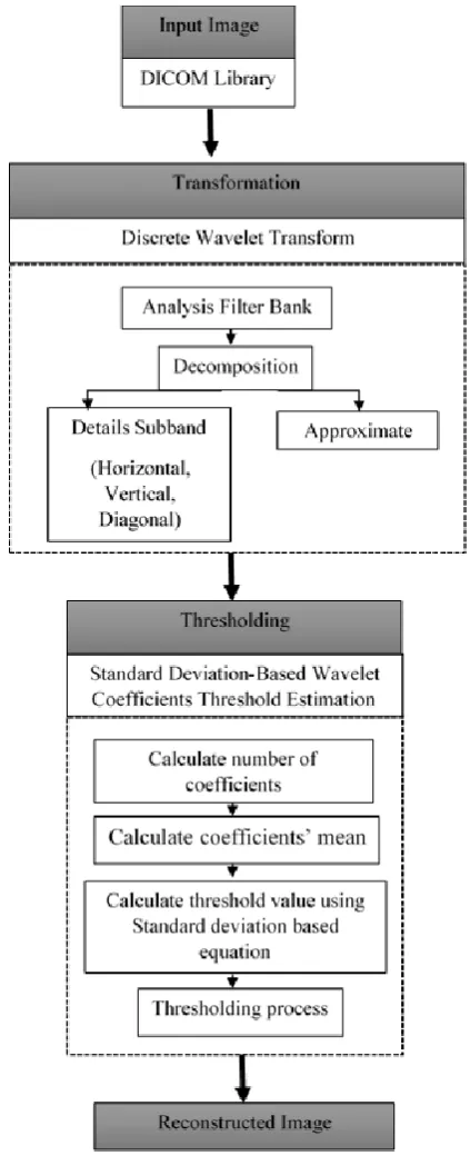
Figure 1 shows the framework of the proposed method. At the first phase, the original image was transformed into wavelet coef-ficients using Discrete Wavelet Transform (DWT). Discrete Wavelet Transform is used as transformation tool.It will divide the image into four different subbands. The approximate coef-ficients are placed in approximate subband (low resolution ap-proximate image) while the detail coefficients are placed in hori-zontal subband (intensity variation along column, horihori-zontal edge), vertical subband (intensity variation along row, vertical edge) and diagonal subband (intensity variation along diagonal).
Then, the wavelet coefficients will do through thresholding pro-cess where here the dispersion trend of the wavelet coefficients is measured and then the threshold value at detail subbands is esti-mated. The purpose of this process is to obtain a higher compres-sion ratio without harming the image quality.
[image:2.595.333.545.77.598.2]
The wavelet coefficients for approximate subband are represented by:
(1)
Where;
j0 is the wavelet scale;
N1 and N2 are the length and width of an image; S(n1 , n2) is the image function;
k1 and k2 are the index written from n1 and n2 respectively; Ø is the scaling function filter;
While the detail subbands are expressed as;
(2)
Where;
i is the details subband either Diagonal (D), Vertical (V) or Horizontal (H);
is the wavelet filter.
In the wavelet transform, the noise energy is distribute in all wave-let coefficients, while the original signal energy is found in some of the coefficients. Therefore, the signal energy is found much larger than noise energy. So, small coefficients can be considered as caused by noise while large coefficients are triggered by signif-icant signal features.
So, based on this idea, thresholding process where the removing of the small absolute coefficients value while retaining the large absolute coefficient value can be done. It will produce finer recon-struct signal. Since this method is taking the condition that the amplitude of wavelet transform coefficients signals are much larg-er than noises, so the unconsidlarg-ered noise will be removed while holding the significant.
Here, the thresholding value is calculated using the Standand De-viation concept.The standard deDe-viation can be defined as:
(3) Where,
(4)
Here, n is representing the amount of wavelet coefficient at the specific detail subband, while is wavelet coefficient value at a specific point at that subband.
By applying equation (2) into equation (3) and (4), the new de-tail subband threshold value can be calculated. After the new es-timated threshold value is obtained, the thresholding process will take place. At this point, each coefficient value that is lower than λv, will be discarded while the rest are remain.
4.
Result and Analysis
[image:3.595.302.548.121.479.2]This experiment was carried out on Matlab platform by using medical images obtained at DICOM Library (see Figure 2).
Figure 2: Medical Images Obtained from DICOM (Digital Imag-ing and Communication in Medicine) Library [17]
[image:3.595.316.551.640.758.2]To test the performance of the proposed method, it was compared with the prominent wavelet-based compression algorithm: Em-bedded Zerotree Wavelet (EZW), Set Partitioning in Hierarchical Trees (SPHIT), Wavelet Different Reduction (WDR) and Adap-tive Scanned Wavelet Different Reduction (ASWDR). The per-formance analysis is done on Peak Signal to Noise Ratio (PSNR) and Compression Ratio (CR).
Table 4.1: PSNR Value Comparison between Wavelet-Based Compression Algorithms with Proposed Method
Image EZW SPHIT WDR ASWDR Proposed
CT_Brain 36.52 36.52 36.52 36.52 48.11
CT_Chest 43.20 39.47 43.20 43.20 54.82
Xray_Kidney 39.24 34.47 39.24 39.24 47.03
Xray_Teeth 38.89 37.90 38.89 38.89 44.03
MR_Knee 35.77 35.08 35.77 35.77 46.04
MR_Brain 38.58 37.11 38.58 38.58 46.60
cannot virtually differentiate between the original and reconstruct-ed image [23]. This PSNR value also indicates no blurring or im-age quality degradations appear on reconstructed imim-age.
Table 4.2: Compression Ratio Comparison between Wavelet-Based Com-pression Algorithms with Proposed Method
Image EZW SPHIT WDR ASWDR Proposed
CT_Brain 8.63 5.54 9.41 9.10 6.47
CT_Chest 5.43 2.03 5.90 5.70 7.02
Xray_Kidney 4.17 1.51 4.34 4.23 10.03
Xray_Teeth 1.15 0.74 1.11 1.08 8.05
MR_Knee 7.23 4.80 7.66 7.53 9.04
MR_Brain 7.14 4.36 7.24 7.04 9.60
As for EZW, SPIHT, WDR and ASWDR algorithms, the thresh-old value was initially set based on significance of wavelet coeffi-cients [24]. Thus it does not exploiting the different characteristic at each subband. It also do not allow lossless reconstruction even on the retaining coefficients. Besides, the EZW, SPIHT, WDR and ASWDR algorithms use uniform quantization that partially fails to recognize the near zero coefficients that largely found in each subband.
On the other hand, the proposed Standard Deviation-Based Wave-let Coefficients Threshold method exploiting the different charac-teristic of subband by estimating the near zero coefficient for a satisfactory threshold value. By using this property, an efficient threshold estimation was obtained thus resulting in a better PSNR and compression ratio value as stated in this section.
5.
Conclusion
In this work, the Medical Image Compression using Standard Deviation-Based Wavelet Coefficients Thresholding Method is proposed. The proposed method can estimate the suitable thresh-old value by measuring the dispersion trend or characteristic of wavelet coefficients in each detail subband using standard devia-tion concept. Throughout the experiments done, it was found that the proposed Standard Deviation-Based Thresholding Algorithm benefited in reducing the image size as can be seen in higher com-pression ratio without harming the image quality as can be seen in higher PSNR value. As for the future work, the proposed method will be implemented in colour image by executing the proposed method in parallel.
References
[1] X. Xu, J. Ma, and L. Nie, “Weakly supervised image parsing via label propagation over discriminatively semantic graph,” J. Vis. Commun. Image Represent., vol. 40, pp. 808–815, 2016.
[2] A. J. Hussain, D. Al-Jumeily, N. Radi, and P. Lisboa, “Hybrid Neural Network Predictive-Wavelet Image Compression System,” Neurocomputing, vol. 151, pp. 975–984, Mar. 2015.
[3] M. M. Isaac and M. Wilscy, “Image Forgery Detection Based on Gabor Wavelets and Local Phase
Quantization,” Procedia Comput. Sci., vol. 58, pp. 76–83, 2015.
[4] R. Athilakshmi, J. Jayanthi, and B. Chithra, “HIGH QUALITY EDGE PRESERVATION USING
WAVELET BY LLSURE IMAGE FILTERING,” Int. J. Adv. Res. Comput. Sci. Electron. Eng., vol. 3, no. 4, pp. 236–241, 2014.
[5] M. Iwahashi, T. Yoshida, H. Kiya, and A. R. Reduction, “Range Reduction of HDR Images for Backward Compatibility with LDR Image Processing,” in APSIPA Annual Summit and Conference, 2014, pp. 2–5.
[6] S. D. Thepade and P. Bidwai, “Iris recognition using fractional coefficients of transforms, Wavelet Transforms and Hybrid Wavelet Transforms,” 2013 Int. Conf. Control. Comput. Commun. Mater. ICCCCM 2013, no. Iccccm, 2013.
[7] R. Loganathan and Y. S. Kumaraswamy, “Active Contour Based Medical Image Segmentation and Compression Using Biorthogonal Wavelet and Embedded Zerotree,” Indian J. Sci. Technol., vol. 6, no. April, pp. 4390–4395, 2013.
[8] D. a. Karras, “Improved Video Compression Schemes of Medical Image Sequences based on the Discrete Wavelet Transformation of Principal Textural Regions and Intelligent Restoration Techniques,” 2007 IEEE Int. Symp. Intell. Signal Process., pp. 1–6, 2007.
[9] J. Li, “An improved wavelet image lossless compression algorithm,” Opt. - Int. J. Light Electron Opt., vol. 124, no. 11, pp. 1041–1044, Jun. 2013.
[10] S. Liu and J. Chen, “A Fast Multi-focus Image Fusion Algorithm by DWT and Focused Region Decision Map,” in Asia-Pacific Signal and Information Processing Association Annual Summit and Conference (APSIPA), 2016, pp. 1–7.
[11] M. Vijay, L. S. Devi, M. Shankaravadivu, and M. Santhanamari, “Image Denoising Based On Adaptive Spatial and Wavelet Thresholding Methods,” in IEEE International Conference on Advances in Engineerinf, Science and Management, 2012.
[12] S. Paul and B. Bandyopadhyay, “A novel approach for image compression based on multi-level image thresholding using Shannon Entropy and Differential Evolution,” Proc. 2014 IEEE Students’ Technol. Symp., pp. 56–61, Feb. 2014.
[13] P. S. A. Devi and M. G. Mini, “Compression of Medical Images by Prediction on Wavelet Transform
Coefficients,” Bonfring Int. J. Adv. Image Process., vol. 2, no. 4, pp. 9–16, 2012.
[14] S. Kim, M. Kim, J. Kim, and H. Lee, “Fixed-Ratio Compression of an RGBW Image and Its Hardware Implementation,” IEEE J. Emerg. Sel. Top. Circuits Syst., vol. 6, no. 4, pp. 484–496, 2016.
[15] M. S. Savic, Z. H. Peric, and N. Simic, “Expert Systems with Applications Coding algorithm for grayscale images based on Linear Prediction and dual mode quantization,” Expert Syst. Appl., vol. 42, pp. 7285–7291, 2015.
[17] P. Suapang and K. Dejhan, “Medical Image Compression and DICOM-Format Image Archive,” pp. 1945–1949, 2009.
[18] H. Kaur, R. Kaur, and N. Kumar, “Lossless compression of DICOM images using genetic algorithm,” in 2015 1st International Conference on Next Generation Computing Technologies (NGCT), 2015, no. September, pp. 985–989.
[19] V. S. Nguyen, M. Ha, T. Hoang, and M. Quang, “A Research on 3D Model Construction from 2D DICOM,” in International Conference on Advance Computing and Applications, 2016, pp. 159–163.
[20] S. J. Pinto and J. P. Gawande, “Performance analysis of medical image compression techniques,” in 2012 Third Asian Himalayas International Conference on Internet, 2012, pp. 1–4.
[21] P. Somvanshi, “Tumor Preserving Medical Image Compression,” Int. J. Comput. Appl., vol. 54, no. 2, pp. 41–46, 2012.
[22] R. K. Yadav, S. P. Gangwar, and H. V Singh, “Study and analysis of wavelet based image compression
techniques,” Int. J. Eng. Sci. Technol., vol. 4, no. 1, pp. 1–7, 2012.
[23] G. Sreelekha and P. S. Sathidevi, “An HVS based adaptive quantization scheme for the compression of color images,” Digit. Signal Process., vol. 20, no. 4, pp. 1129–1149, Jul. 2010.
![Figure 2: Medical Images Obtained from DICOM (Digital Imag- ing and Communication in Medicine) Library [17]](https://thumb-us.123doks.com/thumbv2/123dok_us/8749967.891675/3.595.302.548.121.479/figure-medical-images-obtained-digital-communication-medicine-library.webp)
